

Research Article | DOI: https://doi.org/10.31579/2834-8427/028
Surveillance of genetic diversity and evolution in locally transmitted SARS-CoV-2 in Pakistan during the first wave of the COVID-19 pandemic
- Muhammad Shakeel 1
- Muhammad Irfan 1
- Zaibunnisa 1
- Muhammad Rashid 2
- Sabeeta Kanwal Ansari 2
- Ishtiaq Ahmad Khan 1*
1Jamil-ur-Rahman Center for Genome Research, Dr. Panjwani Center for Molecular Medicine and Drug Research, ICCBS, University of Karachi, Karachi-75270, Pakistan.
2National Institute of Virology, Dr. Panjwani Center for Molecular Medicine and Drug.
*Corresponding Author: Ishtiaq Ahmad Khan, Jamil-ur-Rahman Center for Genome Research, Dr. Panjwani Center for Molecular Medicine and Drug Research, ICCBS, University of Karachi, Karachi-75270, Pakistan.
Citation: Muhammad Shakee, Muhammad Irfan, Zaibunnisa, Muhammad Rashid, Ishtiaq A. Khan, et.al, (2024), Benefits of Timed Mating over Blind Mating of Mice to Get Timed Embryos, J Clinical Gynaecology and Breast, 3(3); DOI: 10.31579/2834-8427/028
Copyright: © 2024, Ishtiaq Ahmad Khan. This is an open access article distributed under the Creative Commons Attribution License, which permits unrestricted use, distribution, and reproduction in any medium, provided the original work is properly cited.
Received: 26 April 2024 | Accepted: 10 May 2024 | Published: 27 May 2024
Keywords: covid-19, genetic evolution, pandemic, sars-cov-2 lineages, spatio-temporal surveillance
Abstract
Mus Musculus has provided scientists a well-adapted animal for the developmental studies. There is one on one co-relation between higher mammalian genes to mice genes hence making them superior model to study expression and translation research. Albino strain of house mice were used in this study with comparison of random or blind matings in mice and timed pregnant mice. The working behind blind matings is minimal and maintenance time and required force was also minimal as compared to timed matings. The results of comparisons of weight gain with pregnancies was similar in two groups where some un-pregnant mice also showed average or above average weight gain with time. The accurate embryonic stages requirement for the two experiments were e13.5, e14.5 and e15.5 (peak of neurogenesis in mice). The timed matings showed to be superior regarding total pregnancies and having accurate stages and is recommended to be used in order to save trouble of not getting what you require and more sacrifice of mice then is needed.
Introduction
The scientific name of house mice is Mus Musculus and it belongs to kingdom animalia, phylum chordate, class mammalian and order rodenta. Mice are preferred in molecular research because they show great similarity with human with respect to genetic, physiological and anatomical properties (Bryda 2013). To investigate human health almost 59% animals used in biomedical research are mice (Taylor and Alvarez 2019). Short life cycle, short gestation period and high breeding efficiency make it excellent as a model organism in laboratory (Merlo, Altruda et al. 2012). More over mice are small, easy to handle and can be transported easily. They are used to study human diseases and human genetics because they are phylogenetically related to human (Perlman 2016). Gestation period of mice is between 18 to 22 days (Murray, Morgan et al. 2010). To understand developmental biology, it is necessary to carry out accurate staging of embryos that help to compare embryonic development of mice with same or different species or group of animals (Wong, van Eede et al. 2015). Almost 95% of the genes of human and mice are similar so it is excellent model to study transcriptional analysis (Simon 3, 5 et al. 2002). The estrous cycle of female mice consists of four stages namely proestrous, estrous, metestrous and diestrous (Byers, Wiles et al. 2012). There are various methods to evaluate the estrous cycle of mice that include visual observation (Auta and Hassan 2016), vaginal cytology (McLean, Valenzuela et al. 2012), histology of reproductive organs (Byers, Wiles et al. 2012), vaginal wall impedance (Rodriguez, Araki et al. 1997) and biochemistry of urine (Singletary, Kirsch et al. 2005). During the estrous stage of the estrous cycle female mice ovulate and breed with male mice (Bertolin and Murphy 2014). For investigating genetics of microcephaly and other developmental disorders mice is most suitable model (Moon 2006).
Material and Method
Housing of animals
We set up two experiments to get timed embryos at different embryonic days (from e13.5- e18.5) In first experiment we took 10 female’s mice in a separate cage and housed 5 male mice each in a separate cage to increase their sperm level, fertility and also to avoid aggressive behavior (Zidar, Weber et al. 2019). We used wooden cages that had proper fresh water supply. The pelleted food was given which was made up of ground ingredients. We used fine wooden scrap for bedding. The cages were placed in an air-conditioned room. In second experiment we took 8 female mice and 4 male mice under same conditions.
Experiment 1: Random or blind mating
• Initially female mice were labeled by using green and black permanent makers. One, two, three and four black bands on the tail of mice represent mice number 1, 2, 3 and 4 respectively while full black tail show mice number 5. Similarly, one, two, three and four green bands represent mice number 6, 7, 8 and 9 respectively. Further full green tail shows mice number 10.
• Body weight of all female mice was measured in the start of the experiment. Then all female mice were shifted male cages in this way that each male cage has two female mice. Vaginal plug was checked in next day early in the morning. The female mice in which vaginal plugs were observed separated from male and housed in a separate cage.
• Weight of all female mice was recorded on daily basis.
• At the desired stage the mice were dissected to collect the embryos.
Experiment 2: Timed mating
• Method of labeling mice was used as previous. Weight of each female mice was also recorded on daily basis.
• To evaluate estrous stage of mice we use vaginal cytology protocol (Byers, Wiles et al. 2012).
• Female mice that were in estrous and pro-estrous stage were housed with male mice in the evening.
• On the next day, in the morning, the female mice were separated from male mice.
• The remaining female mice were house with male mice when they reached estrous or pro-estrous stage.
Dissection of mice:
On the desired embryonic day, the mice were dissected to collect embryos. We preserved embryos in RNA latter, 4%PFA (paraformaldehyde), formalin and some embryos were snap freezed. We did this for further studies
Results
Weight gain was observed in each mouse, though it did not reflect true pregnancy (Table 1).
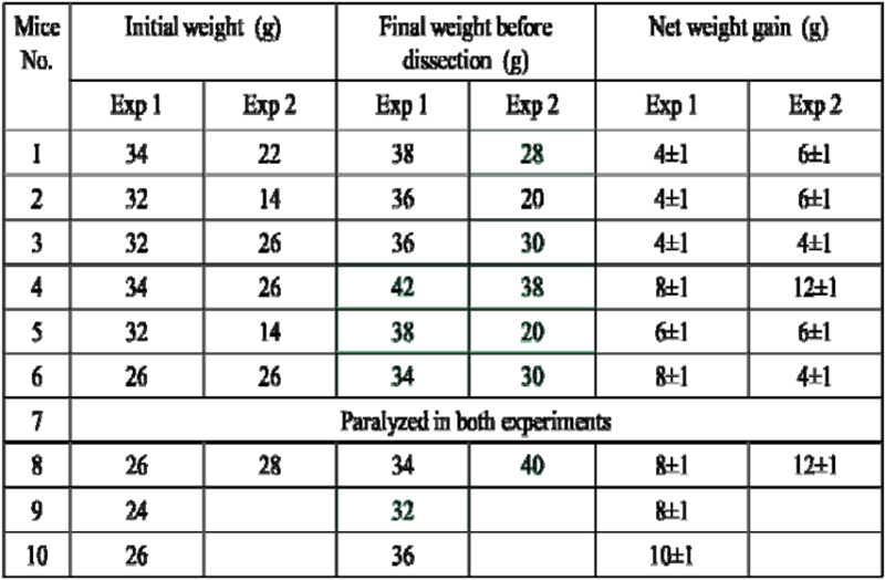
Table1: Correlation of mice with weight during the two experiments
The vaginal plug was observed once but it led to false pregnancy. In random mating we got 4 pregnant mice. Blind VS Timed matings in mice Blind matings 10 female with 5 stud males. Experiment took place from 16-05-22 to 20-06-22 (Table 2).
| MICE No. | Pregnancy induced after blind mating | Stages in days | Adult tissues collected | Embryonic brain collected | Rest of the embryos collected |
1 | | NA | | NA | NA |
2 | | NA | | NA | NA |
3 | | NA | | NA | NA |
4 | | E 6.5 | | (Wholeembryos) | |
5 | | E 12.5 | | | |
6 | | E 13.5 | | | |
7 | Died | NA | | NA | NA |
8 | | NA | | NA | NA |
9 | | E 18.5 | | | |
10 | Died | NA | | NA | NA |
Table 2: Random or blind matings and the success in pregnancies and collection of samples
Timed matings were set on 24-03-23 and finished in 10-05-23. Total of 8 female mice were mated with 4 stud male mice (Table 3).
| MICE No. | Pregnancy induced after blind mating | Stages in days | Adult tissues collected | Embryonic brain collected | Rest of the embryo collected |
1 | | E 14.5 | | | |
2 | | E 7.5 | | | |
3 | | E 13.5 | | | |
4 | | E 14.5 | | | |
5 | | E 15.5 | | | |
6 | | E 15.5 | | | |
7 | Died | NA | | | |
8 | | E 13.5 | | | |
Table 3: Timed pregnant mice results showing more pregnancies and accurate embryonic stages
Vaginal cytology of timed pregnant mice. The mice which were in the estrous and pro- estrous phase give accurate stage of embryo (Figure 1).
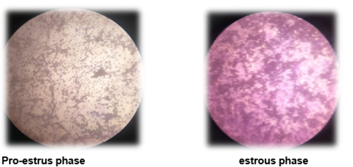
Figure 1: The stages visible after crystal violet dye staining vaginal smear which are superior to setup matings at in mice estrous cycle
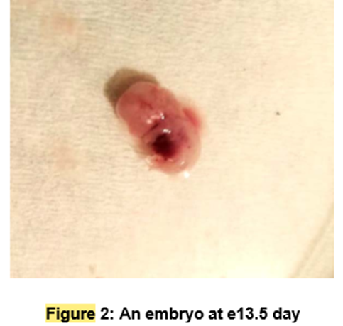
Embryonic day was identified by characteristic feature of embryos. Like the embryo of 13.5 stage has following. The foot plate is anterior and posterior distal borders are sectioned now. Hand plate has more cleared sectioned than foot plate. In hind limb only ankle region is identifiable while in forelimb elbow and wrist are distinguishable. Furthermore, pinna and whiskers are appearing.
At 14.5 embryonic stage. The separation of individual digits in the anterior footplate is a unique feature of this stage. Although there isn't yet any discernible separation between the growing toes, the rear footplate exhibits significant indentations.
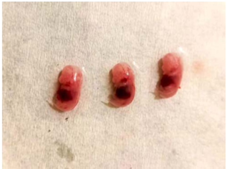
Figure 3: Three mice embryos recovered at e14.5 stage in mice development
At embryonic stage 15.5 Complete interdigital indentation is present on the hand and foot plates, and the vanishing of the webbing improves the delineation of each individual digit. The elbow and knee are obviously bent. Hair follicles can be found in the cephalic region, but not along the margin of the vibrissae. The anterior area of the back is gray, and the curvature of the back is markedly different from the currently straight post-cranial vertebral axis.
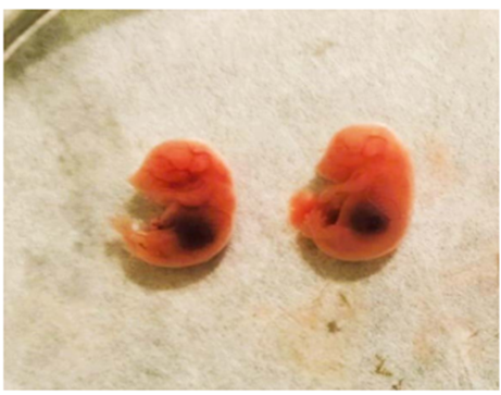
Figure 4: Two embryos at e15.5 day in mouse staging according to Theiler staging
Discussion
In the series of experiment done in this study we got contradicting results showing no/very little dependency on weight gain when talking about pregnancies. Average weight gain in random or blind matings in all females with time was 4.5+- 1g regardless of pregnancy. However, in the pregnant mice which were 4 out of 10 females’ average weight was 7.5+_1g. Which was a bit of difficult to judge correct stage of pregnancy as we got 6+-1g weight gain in the unpregnant mice only. We mated the mice every day and separated in the morning. From mice behaviour and other features, we separated females which looked pregnant. The true judgement numbers regarding pregnant to unpregnant mice was 4 and 5 respectively out of total 10 mice. Which is not very significant number. Moreover, we were only sure of staging after dissecting out embryos and didn’t always find the required range. This experiment was however not very lengthy and took four weeks to complete. The second experiment we ran was timed matings in mice. Total average weight gain in mice regardless of pregnancy was 6+-1g. The pregnant females gained average weight 7.3+-1g and unpregnant mice got average weight gain of around 5.5+-1. The values of weight gains in both experiments were almost comparable but just on the criterion of weight it is hard to judge the pregnancy and more so the exact stage we are looking for as was our requirement in our study. The time taken to finish the second experiment was however lengthy and took almost two months and required more housekeeping of mice and care for the animal requirements.
The second criterion was total number of pregnancies in both experiment in random or blind mating only 4 out of 10 females were found pregnant after dissection. In timed matings on the other hand 7 out of 8 mice got pregnant and apart from one mouse all six gave the accurate embryonic stages which were proven by the criterion of the Theilerr mice staging. However, we had only small number of animals available for these experiment as we don’t have a facility of animal house in our campus. We had to commute to different campus and used their facility under their approved animal use ethics and we respect it.
Conclusions
The timed matings gave us more pregnant mice at desired stages than mice with blind matings. As previously suggested, we confirm same results that although time taken for the whole experiment was more with timed matings and more afforts and housekeeping was required to get the desired results as can be seen from the dates of the experiments the desired outcome was statistically significant than blind matings.
Acknowledgements
We thank our whole research team and HEC, Pakistan for providing funds for this project. We especially thank Professor Qaisar Jabeen, Department of Pharmacy for allowing us to work in their animal house facility.
References
- Zidar, J., Weber, E. M., Ewaldsson, B., Tjäder, S., Lilja, J., Mount, J., . . . Törnqvist, E. J. (2019). Group and single housing of male mice: collected experiences from research facilities in Sweden. 9(12), 1010.
View at Publisher | View at Google Scholar - Auta, T. and A. Hassan (2016).
View at Publisher | View at Google Scholar - Bertolin, K. and B. D. Murphy (2014). Reproductive tract changes during the mouse estrous cycle. The guide to investigation of mouse pregnancy, Elsevier: 85-94.
View at Publisher | View at Google Scholar - Bryda, E. C. (2013).
View at Publisher | View at Google Scholar - Byers, S. L., M. V. Wiles, S. L. Dunn and R. A. Taft (2012).
View at Publisher | View at Google Scholar - Byers, S. L., M. V. Wiles, S. L. Dunn and R. A. J. P. o. Taft (2012).
View at Publisher | View at Google Scholar - McLean, A. C., N. Valenzuela, S. Fai and S. A. Bennett (2012).
View at Publisher | View at Google Scholar - Merlo, G. R., F. Altruda and V. Poli (2012).
View at Publisher | View at Google Scholar - Moon, A. M. (2006).
View at Publisher | View at Google Scholar - Murray, S. A., J. L. Morgan, C. Kane, Y. Sharma, C. S. Heffner, J. Lake and L. R. Donahue (2010).
View at Publisher | View at Google Scholar - Perlman, R. L. (2016).
View at Publisher | View at Google Scholar - Rodriguez, I., K. Araki, K. Khatib, J.-C. Martinou and P. Vassalli (1997).
View at Publisher | View at Google Scholar - Simon 3, E. B. I. B. E. G. N. K. A. M. E. R. A. G. S. G. S. A. U.-V. A. W., R. G. i. B. I. A. J. F. G. R. P. G. 5, B. A. P. 6, N. C. f. B. I. A. R. C. D. M. H. W. M. D. R. S. V. 7, D. o. M.M. P. L. 8, D. o. M. G. A. S. E. D. E. T. R. A. U. C. 9, C. f. B. Science and E. B. R. D. M. F. T. S. H. A. H. F. K. D. K. W. J. R. K. M. S. Matthias (2002).
View at Publisher | View at Google Scholar - Singletary, S. J., A. J. Kirsch, J. Watson, B. O. Karim, D. L. Huso, P. D. Hurn and S. J. Murphy (2005).
View at Publisher | View at Google Scholar - Taylor, K. and L. R. Alvarez (2019).
View at Publisher | View at Google Scholar - Wong, M. D., M. C. van Eede, S. Spring, S. Jevtic, J. C. Boughner, J. P. Lerch and R. M. Henkelman (2015).
View at Publisher | View at Google Scholar - Zidar, J., E. M. Weber, B. Ewaldsson, S. Tjäder, J. Lilja, J. Mount, C. I. Svensson, E. Svensk, E. Udén and E. J. A. Törnqvist (2019).
View at Publisher | View at Google Scholar
