

Clinical Image | DOI: https://doi.org/10.31579/2835-8325/017
Pearls in the Eye
1 Assistant Professor (Designated), Deptt. of Ophthalmology, Shri Lal Bahadur Shastri Government Medical College and Hospital, Nerchowk, Distt. Mandi, Himachal Pradesh, India.
2 Medical Officer (Specialist), Deptt. of Ophthalmology, Shri Lal Bahadur Shastri Government Medical College and Hospital, Nerchowk, Distt. Mandi, Himachal Pradesh, India.
3 Associate Professor, Deptt. of Ophthalmology, Shri Lal Bahadur Shastri Government Medical College and Hospital, Nerchowk, Distt. Mandi, Himachal Pradesh, India.
*Corresponding Author: Anubhav Chauhan, Assistant Professor (Designated), Deptt. of Ophthalmology, Shri Lal Bahadur Shastri Government Medical College and Hospital, Nerchowk, Distt. Mandi, Himachal Pradesh, India.
Citation: Anubhav Chauhan, Neha Chauhan, Deepak K. Sharma, (2023), Pearls in the Eye, Clinical Research and Clinical Reports, 2(3); 10.31579/2835-8325/017
Copyright: ©, 2023, Anubhav Chauhan. This is an open-access artic le distributed under the terms of the Creative Commons Attribution License, which permits unrestricted use, distribution, and reproduction in any medium, provided the original author and source are credited.
Received: 01 May 2023 | Accepted: 10 May 2023 | Published: 17 May 2023
Keywords: OPD; B scan; leprosy; ectropion uvea
Abstract
A 60-year-old female came to our OPD for pain and diminition of vision in her right eye for the past two months.
Ophthalmic images
A 60-year-old female came to our OPD for pain and diminition of vision in her right eye for the past two months. Her best corrected visual aquity was 6/60 in the right eye and 6/9 in the left eye. She was a known case of lepromatous leprosy and was on treatment for the past five months from the Department of dermatology. Slit lamp examination of the right eye revealed 1+ cells in the anterior chamber, ectropion uvea plus yellow-white pinhead sized lesions resembling beads i.e 'iris pearls' around the pupillary area (Figure1 and 1a) along with posterior subcapsular cataract. Fundus of the right eye could not be seen because of cataract and her B scan was normal. The only significant findings in her left eye were an immature senile cataract. She was started on topical steroids plus cycloplegics and was on a regular followup.
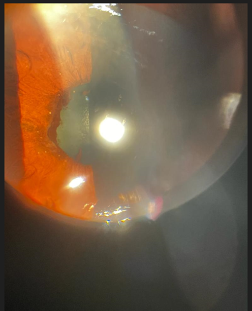
Figure 1
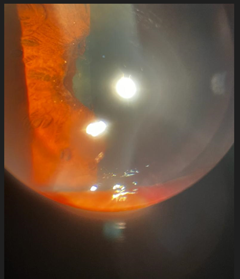
Figure 1a
The involvement of eye is seen more commonly in lepromatous leprosy form of the disease. [1] Iris pearls are pathognomonic findings of lepromatous leprosy and are a diagnostic uveal manifestation. They usually resemble a necklace or the beads of a rosary. [2] These are around 0.3 to 1.0 mm in diameter, dull yellow in colour, asymptomatic, and are mostly found around pupillary margins. These bacilli are likely to get lodged in the anterior segment of the eye, as this is the coolest part of the eye. It particularly involves the uveal tissue due to high vascularity of the area. [3] Infectious uveitis is an uncommon cause of iris nodules. Most cases of uveitis withiris nodules can be attributed to diseases such as sarcoidosis, Vogt-Koyanagi-Harada syndrome, multiple sclerosis, Fuchs heterochromic iridocyclitis and sarcoidosis. [4]
Source of Support-None
The paper being submitted has not been published,simultaneously submitted,or already accepted for publication elsewhere.
Conflicts of Interest
The authors declare that they have no competing interest.
Financial Disclosure(s)
The authors have no proprietary or commercial interest in any material discussed in this article.
References
- Ffytche TJ. (1981). Role of iris changes as a cause of blindness in lepromatous leprosy. Br J Ophthalmol, 65(4):231-239.
View at Publisher | View at Google Scholar - Ahluwalia NS, Choudhary P, Shakya R, Revankar A. (2022). Unmasking Hansen’s disease through an ophthalmologist’s eye. Indian J Ophthalmol 70:2671-2673.
View at Publisher | View at Google Scholar - Thool A, Mehta K. (2021). Typical iris pearls in lepromatous leprosy. J Evolution Med Dent Sci, 10(03):167-169.
View at Publisher | View at Google Scholar - Myers TD, Smith JR, Lauer AK, Rosenbaum JT. (2002). Iris nodules associated with infectious uveitis. Br J Ophthalmol, 86:969-974.
View at Publisher | View at Google Scholar
