

Research Article | DOI: https://doi.org/10.31579/2834-5029/017
Metabolic Liver Diseases: Hemochromatosis
- Rehan Haider *
Riggs pharmaceutical, Department of Pharmacy, University of Karachi, Pakistan.
*Corresponding Author: Rehan Haider, Riggs pharmaceutical, Department of Pharmacy, University of Karachi, Pakistan.
Citation: Haider R., (2023), Metabolic Liver Diseases: Hemochromatosis, International Journal of Biomed Research. 2(2): DOI:10.31579/2834-5029/017
Copyright: © 2023, Divia Paul A, This is an open access article distributed under the Creative Commons Attribution License, which permits unrestricted use, distribution, and reproduction in any medium, provided the original work is properly cited.
Received: 05 April 2023 | Accepted: 18 April 2023 | Published: 28 April 2023
Keywords: chronic disease; hemochromatosis; metabolism; hepcidin; metabolism; homeostasis; iron; metabolism; iron overload
Abstract
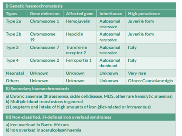
Hereditary hemochromatosis is classified into 4 subtypes (Table 1). Type 1 is the well-known form of iron overload due to an autosomal recessive genetic metabolic malfunction; the homozygous C282Y mutation of the HFE a gene on chromosome 6 bills for more than ninety% of clinical phenotypes in populations of Caucasian origin (Feder 1996) [1]. This mutation results in inadequate excessive intestinal iron absorption that, once a long time, may cause iron overload and harm to diverse organs (discern 1). sorts 2a and 2b of genetic hemochromatosis are juvenile types of iron overload that result in an intense outcome earlier than age 30, with cardiomyopathy and hypogonadism. The corresponding mutations are positioned within the hemojuvelin and hepcidin genes, respectively (Roetto 1999) [2]. type 3 has specifically been defined in Italian families and refers to a mutation in the transferrin receptor 2 genes (Girelli 2002) [3] Scientific results of type three hemochromatosis are much like type 1. Types 2 and three are autosomal recessive. Mutations in the autosomal dominant type of hemochromatosis are located within the gene coding for the basolateral iron transporter ferroportin 1 (Njajou 2001) [4]. In the evaluation of the alternative types, iron is gathered in type four, especially in macrophages. Ferritin levels are markedly elevated, although transferrin saturation is barely high, and secondary hemochromatosis is normally due to multiple blood transfusions in hemolytic anemia, thalassemia, sickle mobile anemia, and myelodysplasia syndrome. Iron first accumulates in RES macrophages and is then transferred to parenchymal cells. Iron may gather faster with common blood transfusions than with genetic hemochromatosis; iron overload often results in severe cardiomyopathy and liver cirrhosis, leading to a powerful diagnosis. therapy consists of iron chelators because phlebotomies cannot be completed due to underlying anemia. This evaluation will identify type 1 HFE hemochromatosis, Germany’s most established genetic shape. Most consequences of iron overload are similar, regardless of the reason. Accordingly, the pathophysiology of tissue and organ damage by using extra iron is best mentioned in detail for HFE hemochromatosis.
Introduction
Type 1 HFE hemochromatosis
History
The association between liver cirrhosis, pigment deposits in the liver, and diabetes mellitus was recognized over a century ago (Trousseau 1865, Troisier 1871, Hanot and Schachmann 1886) [5-7]. The term hemochromatosis was first introduced in the 19th century (Recklinghausen 1889) [8] but was not generally accepted until it was used as the title of a classic monograph (Sheldon 1935) [9]. The controversy over whether hemochromatosis was merely a form of alcoholic liver cirrhosis (MacDonald 1960) [10] or a genetic error of iron metabolism (Sheldon 1935, Crosby 1966) [11] lasted almost a century until the association between special HLA Haplotypes and hemochromatosis which recognized the genetic nature of the disease was described (Simon 1975) [12] The mode of inheritance was identified as an autosomal recessive disorder (Simon 1977) [13]. Finally, the major mutation on the HFE gene associated with clinical manifestations was identified (Feder 1996).
Epidemiology
Type 1 hemochromatosis is probably the most prevalent genetic metabolic error in Caucasian populations (Adams 2005) [14]. The prevalence of C282Y homozygotes is approximately 0.5% in central Europe and the Caucasian population of North America, and the prevalence of C282Y and H63D heterozygotes is approximately 40% in similar populations (Adams 2005). Phenotypic expression also depends on several non-genetic factors such as the amount of dietary iron and blood loss (Figure 2 For example, due to menses, females develop clinical consequences of Iron overload 5–8 times less frequently and 10–20 years later than males do. It is now widely accepted that not all C282Y homozygous men develop the full clinical manifestation of hemochromatosis. It also remains unclear how many men will show clinical disease during their lifetime and what factors determine that phenotype, the
homozygous C282Y mutation accounts for more than 90% of the clinical phenotype in Caucasian populations (Feder1996; Adams 2005) (Table 2). A point mutation in H63D is also frequently identified in the HFE gene, as well as other less frequent mutations. None of these gene alterations or polymorphisms, found in up to 40% of Caucasians, correlated with the phenotype. A subject with a C282Y variation on one allele and an H63D variation on the other is called a “compound heterozygote” (Table 2}Only a small percentage of such compound heterozygotes are at risk of clinical consequences of iron overload (Gallego, 2015)15.A recent meta-analysis showed a positive association between compound heterozygosity for C282Y/H63D and the risk of NAFLD and HCC but not liver cirrhosis (Ye et al. 2016){16}. C282Y and H63D heterozygotes are at no risk of iron overload. (Table 2). In non-Caucasian populations, other genes may also be involved in iron overload.
Etiology and Pathogenesis
intestinal iron absorption and loss are finely balanced under physiological conditions. Approximately 10% of the total daily iron intake (10–20 mg) is absorbed by the small intestine (1–2 mg). However, subjects with the homozygous C282Y mutation may absorb up to 20% of their iron intake, that is, up to 2–4 mg/day. Thus, homozygotes have an excessive iron intake of approximately 1 mg/day. It may therefore take several decades until iron. stores approach 10 g, above which organ damage is considered starting. Many patients at the clinical end stage of hemochromatosis, including liver cirrhosis and diabetes mellitus, have total body iron stores of 20–30g. Intestinal iron absorption is downregulated when iron stores increase in these patients, as in patients with genetic hemochromatosis. However, this down regulation occurs at an increased level compared to subjects without the HFE gene mutation. Correspondingly, intestinal iron absorption is significantly increased in patients with hemochromatosis when the iron stores are depleted by phlebotomy. It is important to continue phlebotomy after iron depletion to prevent re accumulation (see Table 4). However, these regulatory processes do not explain how HFE gene mutations cause an increase in intestinal iron absorption, since the HFE gene product is neither an iron transporter nor an iron reductase or oxidase. However, carriers and regulators of cellular iron uptake and release have also been identified (Pietrangelo 2002, Fleming 2002, Fletcher 2002) [17-19]. Some of these carriers also interact with the HFE gene product to regulate intestinal iron absorption (Pietrangelo 2002; Fleming 2002; Townsend 2002; Fletcher 2002), and the Nramp2 protein is the luminal iron carrier. Luminal iron reductase has also been identified as a Cytb protein. (Duodenal cytochrome B) (Fletcher 2002; Townsend 2002; Fleming 2002; Pietrangelo 2002). The basolateral iron transporter ferroportin 1 (also named Ireg1 or MTP1) has been identified (Donovan 2000, Abboud 2000) [20,21], as well as the basolateral iron oxidase hephaestin (Vulpe 1999). Mutations in some of these proteins are responsible for rare types 2–4 genetic hemochromatosis, although none of these genes are altered in type 1 hemochromatosis. Two other proteins have been shown to act as important iron-regulating proteins: transferrin receptor 2 and hepcidin (Pietrangelo 2002, Fletcher 2002, Fleming 2005) [22]. Mutations in the transferrin receptor 2 genes may lead to rare type 3 hemochromatosis, and mutations in the ferroportin 1 gene may lead to type 4 hemochromatosis. More recent studies have also indicated that hepcidin may be the most important regulator of iron metabolism and is involved in iron deficiency and overload. Hepcidin has been shown to down-regulate the basolateral iron carrier ferroportin. It has also been demonstrated that hepcidin itself is upregulated by HFE.Thus, an HFE mutation may reduce the upregulation of hepcidin, which then does not downregulated ferroportin. The corresponding increase in ferroportin expression finally causes an increase in intestinal iron uptake (DeDomenico, 2007) [23]. There may be further interactions among HFE, transferrin receptor 2, Nramp2, Dcytb, ferroportin, hephaestin, and hepcidin, all of which are currently being studied.
Diagnostic Laboratory tests
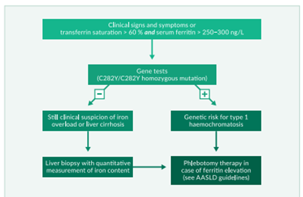
Any increase in serum iron level should start with the exclusion of hemochromatosis to avoid overlooking early disease. Normal serum iron, however, does not exclude hemochromatosis and increased infections and malignancies and thus has low specificity for indicating hemochromatosis (Niederau 1998) [24]. Ferritin levels increase, not due to genetic hemochromatosis, and are usually associated with normal or only slightly elevated transferrin saturation. Therefore, transferrin saturation should be measured to correctly interpret ferritin increase, and serum iron often occurs in the absence of hemochromatosis. Serum iron values are highly variable and should not be used for the diagnosis or screening of hemochromatosis. Determination of transferrin saturation is a better indicator of iron overload than serum iron. An increase in transferrin saturation usually precedes an increase in ferritin levels (Figure 1). Transferrin saturation is more sensitive and specific than serum ferritin for the detection of hemochromatosis. For screening, a threshold of 50% transferrin saturation may be optimal under fasting conditions. On the other hand, ferritin is a good indicator of increased iron stores and reliably indicates iron deficiency. It is less valuable for the early detection of hemochromatosis. In hemochromatosis, a slightly increased serum ferritin level (300–500 mg/mL) is usually accompanied by a transferrin saturation exceeding 80–90%. Unfortunately, serum ferritin is also increased, often in the presence of infections and malignancies, and thus has low specificity for indicating hemochromatosis (Niederau 1998). Ferritin increases not due to genetic hemochromatosis are usually associated with normal or only slightly elevated transferrin saturation. Therefore, transferrin saturation should be measured to correctly interpret ferritin levels.
Liver biopsy and resolution of liver iron concentration
Even though Simultaneous increases in serum ferritin and transferrin saturation strongly indicate a risk for hemochromatosis. Diagnosis should be made with the aid of genetic testing or liver biopsy with willpower. Iron content in the liver. Hepatic iron attention additionally increased with time in subjects with HFE gene mutations. To reap the “hepatic iron index,” liver iron concentrations were divided by the patient’s age. (Summers 1990) [25]. The semi-quantitative estimation of liver iron shops via Berlin blue coloration is much less touchy and unique than the chemical quantification of liver iron concentration. In the case of the homozygous C282Y gene, a liver biopsy is no longer required for the analysis of genetic hemochromatosis (parent three).There can also, but, be different motives to carry out a liver biopsy on iron overload: (1) subjects with biochemical or clinical evidence of iron overload within the absence of the homozygous C282Y mutation have to have a liver biopsy to verify iron overload; (2) in C282Y, homozygotes the danger of liver fibrosis and cirrhosis increase at ferritin values >one thousand mg/mL (Loreal1992) [26]; in the ones sufferers, liver biopsy is usually recommended because of the presence of liver cirrhosis markedly increases later hepatocellular carcinoma (HCC)threat and consequently warrants HCC screening.
Deferoxamine Testing and Ferro Kinetic Measurements
Determination of urinary excretion of iron after administration of deferoxamine lets in a few estimations of general frame iron shops. The deferoxamine check, however, is often the most effective indicator of pathological results, while serum ferritin and transferrin saturation are markedly accelerated and do not allow an analysis of early sickness. Ferro kinetic measurements these days are the simplest finished for medical research or in difficult diagnostic conditions.
Computed Tomography (CT), Magnetic Resonance Imaging (MRI), and Magnetometry
CT density measurements of the liver permit a semi quantitative estimation of iron awareness in the liver. However, this method is related to radiation and is, therefore, not allowed in many international locations where alternative methods are available. Alternatively, MRI permits reliable dimensions of liver iron content, supplied that special software is used and the equipment is calibrated for such a size. In clinical practice, the maximum MRIs do not fulfill these criteria. Bio-magnetometry permits the most accurate noninvasive measurement of liver iron awareness. However, this equipment is steeply-priced, and the most expensive allows the dimension of iron attention. Therefore, magnetometry is performed at only a few centers worldwide and is primarily used for scientific studies and not in daily clinical practice. With the availability of reliable and inexpensive genetic testing, CT and MRI magnetometry are not required for most patients.
Genetic Test:
Genetic tests in Caucasian populations have shown that the homozygous C282Y mutation accounts for more than 90% of patients with the clinical phenotype of type 1 hemochromatosis (Adams 2005, Erhardt 1999) [27]. Approximately 5% of patients with the clinical phenotype are C282Y/H63D compound heterozygotes, and the prevalence of C282Y or H63D heterozygosity in patients with the clinical hemochromatosis phenotype is considerably lower than that in the general population. Thus, a subject heterozygous for C282Y or H63D per se has no risk of iron overload. In subjects homozygous for C282Y, both serum ferritin and transferrin saturation are frequently increased; however, only male subjects have an increased risk for liver disease compared to subjects without HFE gene alterations in a recent large screening study. The number of C282Y homozygotes that will later develop clinical signs and symptoms due to iron overload is unknown. It is increasingly evident that only a minority of C282Y homozygotes progress to end-stage iron overload, with liver cirrhosis and diabetes mellitus. In subjects who are not C282Y homozygotes but have laboratory, histological, or clinical evidence of iron overload, further genes may be analyzed for mutations such as hemojuvelin, transferrin receptor 2, ferroportin 1, and hepcidin. executed only at some centers worldwide and is frequently used for clinical Early diagnosis and screening the prevalence of C282Y homozygotes is 0.5% in Caucasians (Adams 2005, Erhardt 1999). However, clinical manifestations are variable and depend on non-genetic factors such as dietary iron intake and blood loss. Until 1980, most patients with hemochromatosis had late irreversible complications, such as liver cirrhosis and diabetes mellitus. With a better understanding of the disease, the broad use of ferritin and transferrin saturation measurements, and the availability of reliable genetic tests, diagnostic efforts have concentrated on the detection of early disease before liver cirrhosis and diabetes mellitus. Several studies have shown that iron removal by phlebotomy is associated with a normal life expectancy in patients diagnosed early (Niederau 1985, Niederau 1996, Fargion 1992) [28] (Figure 4). Several other studies have focused on screening procedures to diagnose more patients with early disease (Edwards, 1988) [29]. These studies included populations with special risks, family members, and the general population (Niederau, 2002) [30]. It has been shown that an increasing number of patients are diagnosed early and that this trend increases survival (Figure 5). Many studies have shown that screening is useful for the detection of asymptomatic C282Y homozygotes using transferrin saturation and serum ferritin, as well as a genetic test for the C282Y mutation (Edwards 1988, Phatak 1998, Niederau 1998) [31]. Broad screening of the general population, however, is not recommended by the WHO and CDC, mainly because it is unknown how many asymptomatic C282Y homozygotes will later develop clinical disease (US Preventive Services Task Force 2007) [32]. The largest screening study analyzed HFE mutations in almost 100,000 North American subjects. In Caucasians, C282Y Homozygosity was 0.44%, a value similar to many previous studies in other populations with a similar background. In contrast, Asian or Black people rarely have an HFE gene mutation (Adams 2005). Among Caucasian C282Y homozygotes, only males showed a significant increase in liver disease compared to subjects without HFE gene variation (Adams 2005). Further prospective follow-up studies will determine how many asymptomatic C282Y homozygotes develop the clinical consequences of iron overload. It is also unknown whether ferritin levels during phlebotomy should be initiated in asymptomatic C282Y homozygotes (Table 4). The values recommended by the AASLD are based more on expert judgment than on solid data. The only solid data show that the risk of liver fibrosis and cirrhosis increases above the threshold of 1000 mg/mL for serum ferritin (Loreal, 1996). The value of screening for family members is obvious when a first-degree relative has clinical hemochromatosis. Such family screening is easy to perform using genetic testing. Heterozygous family members are not at risk for hemochromatosis unless they have other risk factors. The clinical phenotype of hemochromatosis is observed in 1–2% of patients with newly diagnosed diabetes mellitus and 3–15% of patients with liver cirrhosis (Niederau, 1999). These latter patients should be screened for iron overload, although such screening does not aim for a very early diagnosis. Nevertheless, cirrhotic and diabetic patients with hemochromatosis can benefit significantly from phlebotomy. Little is known about the prevalence of hemochromatosis in patients with arthropathy or cardiomyopathy of unclear etiology. Several smaller studies indicate that arthropathy may be an early clinical sign of iron overload, whereas cardiomyopathy usually occurs in late-stage iron overload. Complications such as iron overload, liver cirrhosis, diabetes mellitus, and increased skin pigmentation are the classical trios of genetic hemochromatosis. Cardiomyopathy, cardiac arrhythmia, and impotence are typical complications of advanced iron overload. In contrast, arthropathy may be an early sign of hemochromatosis, which may help with diagnosis in the pre-cirrhotic stage (Niederau 1996).
Liver disease
The liver is an organ that is affected by genetic iron overload early and heavily. In the early stages, excess iron stores are mainly found in peri-portal parenchymal cells, such as ferritin and hemosiderin. When excess iron further increases, lobular fibrosis develops, and iron stores are also found in the bile ducts and Kupffer cells. Septal fibrosis eventually progresses to cirrhosis. The stage of fibrosis is closely associated with the degree of excess iron. In many affected symptomatic patients with type 1 hemochromatosis, there are signs of liver disease at the time of diagnosis (Niederau 1985, Niederau 1996). Many nonspecific symptoms such as abdominal discomfort and fatigue may also be due to liver involvement. In asymptomatic patients diagnosed using screening procedures, signs of liver disease are infrequent. Complications due to cirrhosis such as ascites, jaundice, and portal hypertension are seen rarely and only in cases of advanced severe iron overload (Niederau 1985, Niederau 1996). The risk of liver cirrhosis increases with ferritin levels >1000 mg/mL (Loreal, 1996). Similar to insulin-dependent diabetes, liver cirrhosis cannot be reversed by iron removal (Niederau, 1996). However, less advanced stages, such as hepatic fibrosis and abnormalities in liver enzymes and function, respond well to iron removal (Niederau 1996) (Figure 5). Survival is significantly reduced in the presence of liver cirrhosis, whereas patients diagnosed at the pre-cirrhotic stage have a normal life expectancy when treated with phlebotomy(Niederau 1996) (Figure4) Association between hemochromatosis and other liver diseases. Some studies indicate that C282Y heterozygosity may aggravate the progression of concomitant liver diseases such as porphyria cutanea tarda, chronic hepatitis C, alcoholic hepatitis, and non-alcoholic steatohepatitis (NASH). In these latter patients, one might find slightly elevated liver iron and serum ferritin levels when they are C282Y heterozygotes; however, most studies have shown that these associations are. is of minor importance in the clinical course of the disease. To date, phlebotomy has only been proven meaningful in porphyria cutanea tarda because it can ameliorate cutaneous manifestations.
Liver carcinoma
Liver carcinoma develops in approximately 30% of patients with hemochromatosis and cirrhosis independent of iron depletion (Niederau 1996). The interval between complete iron depletion and the reported diagnosis of liver cancer is approximately nine years in large cohorts of German patients (Niederau 1985; Niederau 1996). The risk of liver cancer was 100-to 200-fold higher in patients with hemochromatosis than in the general population (Figure 6). Liver cancers, including hepatocellular carcinoma (HCC) and cholangiocellular carcinoma, develop in patients with cirrhosis. Thus, cancer screening using ultrasound and AFP (twice a year) is recommended only for patients with cirrhosis. Patients who develop liver cancer usually have the most cancer. of iron accumulation among various subgroups (Niederau 1996, Niederau1999)
Diabetes Mellitus
The prevalence of diabetes in hereditary hemochromatosis ranges from 20 to 50% (Niederau 1996, Adams 1991) [33]. The prevalence and stage of diabetes are related to the degree of pancreatic iron deposition. Patients with diabetes have two-fold higher mobilizable iron content than those without diabetes (Yaouanq, 1995) [34]. Investigations into the prevalence of unrecognized genetic hemochromatosis in diabetic patients show some variation in Europe vs. elsewhere; screening revealed a prevalence of 5–8 per 1000 unrecognized cases in Europe (Singh 1992) [35] and 9.6 per 1000 in Australia (Phelps 1989) [36]. Diabetes mellitus and impaired glucose tolerance are frequent features of several chronic liver diseases (Creutzfeldt 1970; Blei 1982) [37,38]. This study (Niederau, 1984) [39] showed hyperinsulinemia and insulin resistance without impaired glucose tolerance in non-cirrhotic hemochromatosis. The increase in circulating insulin concentration is likely due to a decrease in the diminished hepatic extraction of insulin. With the progression of iron overload and destruction of beta cells, insulin secretion is impaired (Dy mock 1972, Bierens de Haan 1973) [40,41].In end-stage hemochromatosis, insulin deficiency is associated with a severe reduction in beta cell mass (Rahier, 1987) [42]. Insulin resistance observed in early iron overload may be partially reversible after phlebotomy therapy (Niederau 1985, 1996), whereas insulin-dependent diabetes is irreversible (Niederau 1996). Survival is significantly reduced in patients with diabetes mellitus at diagnosis compared with that in patients without diabetes (Niederau 1996). The survival of non-diabetic patients was virtually identical to that of a matched normal population. Cardiomyopathy and cardiac arrhythmia are complications of hemochromatosis caused by iron deposition in the heart (Buja and Roberts 1971, Short 1981) [43,44]. Clinical or electrocardiographic signs of heart disease can be found in 20–35% of patients with HFE hemochromatosis (Niederau, 1985).Arrhythmia usually responds well to iron removal (Short 1981; Niederau 1996). In type 1, hemochromatosis cardiomyopathy is rare and usually associated with advanced iron overload and an older patient population. However, cardiomyopathy is a frequent cause of death, particularly in young patients who present with cardiac disease due to hemochromatosis (Finch 1966, Short 1981) [45]. It is also clear that young patients with severe cardiomyopathy may be affected by juvenile type 2 hemochromatosis, which may result in severe iron overload, hypogonadism, cardiomyopathy, liver cirrhosis, and amenorrhea by ages 15–24 years. Type 2-associated cardiomyopathy is often irreversible despite the initiation of phlebotomy or chelation therapy and may require immediate transplantation of the heart and potentially of the liver (von Herbay 1996, Jensen 1993) [46,47].
Arthropathy
Joint changes in genetic hemochromatosis may occur in two different ways (Schuhmacher 1964, Dy mock 1970, Niederau 1985, Niederau 1996) [48,49]. The most prevalent changes are seen in metacarpophalangeal joints II and III, in the form of cystic and sclerotic changes, cartilage damage, and narrowing of the intra-articular space. Occasionally, other joints of the hands and feet are affected. Large joints, that is, the knees and hips, may be affected by chondrocalcinosis. However, the pathogenesis of joint changes in hemochromatosis remains unclear. Arthropathy is one of the few complications that are not associated with the degree of iron overload. It has been speculated that iron may inhibit pyrophosphatase and thereby lead to the crystallization of calcium pyrophosphate. Alternatively, iron may have direct toxic effects on the joints. Arthropathy may be an early sign of hemochromatosis and may help in making a diagnosis at the pre-cirrhotic stage (Niederau 1996). Therefore, hemochromatosis should be considered in all patients with arthropathy of unknown etiology. Endocrine Abnormalities. Endocrine abnormalities are a late consequence of iron overload in contrast to the early onset of arthropathic changes. Sexual impotence and loss of libido may occur in up to 40% of male patients (Niederau 1985). Endocrine abnormalities in hemochromatosis are mainly, if not exclusively, caused by pituitary failure. This is in contrast to alcoholic cirrhosis, in which testicular failure is predominant (Kley 1985a, Kley 1985b) [50,51]. In contrast to alcoholic cirrhosis, in which estrogen levels are usually increased, estrogen levels decrease in hemochromatosis (Kley 1985a). Most endocrine changes are late and irreversible complications of genetic hemochromatosis and do not respond well to phlebotomy treatment (Niederau 1996). Iron overload infrequently affects other endocrine organs, such as the thyroid and adrenal glands. Severe hypogonadism with amenorrhea in young women and impotence in young men are thought to be due to type 2 hemochromatosis. Increased skin pigmentation is observed mainly in areas exposed to sunlight. A large part of the darkening of pigmentation is thought to be due to an increase in melanin and not due to excess iron, and the increase in skin pigmentation is reversible during iron removal (i.e., phlebotomy). Other potential complications. Iron overload has been speculated to aggravate atherosclerosis; however, evidence for this is rather weak (Niederau, 2000) [52]. There have also been reports that extra hepatic malignancies may be increased in HFE hemochromatosis (Amman 1980, Fracanzani 2001) while other studies have not found extra hepatic associations (Bain 1984, Niederau 1996 Elmberg 2003) [53.54], It is not clear whether HFE gene mutations are involved in the pathogenesis of Porphyria cutanea tarda since the prevalence of both risk factors varies greatly in different parts of the world, and associations between HFE gene mutations and porphyria have often been described in Southern Europe but not in Northern Europe (Toll 2006) [55].Polymorphisms beyond C282Y homozygosity. Recent studies have suggested that the C282Y and H63D polymorphisms in the HFE gene are associated with selection advantage. This selection may also explain the high frequency of up to 40% of these polymorphisms in Celtic populations (Adams 2005). These polymorphisms are almost exclusively found in individuals of Celtic descent. A French study recently showed that these polymorphisms are seen in 27% of the French general population (Hermine 2015) [56]. Interestingly, 80% of French winners in WM, EM, and Olympic sports events had one of these polymorphisms (Hermine 2015). A recent Swiss study showed that C282Y homozygotes are several centimeters taller than the reference population (Cippa 2013) [57], although these homozygotes are usually considered unhealthy. Indeed, the greater height and physical fitness of the Celts have already been mentioned by Julius Caesar in his work “De Bello Gallico “(Caesar 50 a.c.). Thus, subjects with heterozygous HFE polymorphisms are usually “very healthy” people without a major risk of iron overload and associated organ damage. HFE heterozygotes may have an increased risk of developing liver fibrosis only in the presence of other hepatotoxic factors, such as hepatitis C or fatty liver disease (Erhardt 2003).
Therapy
Phlebotomy treatment
Phlebotomy is the standard of care for removing iron from genetic hemochromatosis. One phlebotomy session removed approximately 250 mg of iron from the body. Since patients with the classical clinical phenotype may have an excess of 10–30 g iron, it may take 12–24 months to remove the iron overload when phlebotomies of 500 mL blood are performed weekly (Table 4). Phlebotomy is generally well tolerated, and hemoglobin level usually does not drop below 12 g/dl. Several studies have shown that liver iron is completely removed at such low ferritin levels; thus, the effect of therapy can be checked by ferritin measurements, and a control liver biopsy is not necessary. After the complete removal of excess iron, the intervals of phlebotomies may be increased to once every 2–3 months; serum ferritin should be kept in the lower normal range between 5 and 00 mg/mL. Phlebotomy should not be interrupted for longer intervals; however, there is a risk of iron accumulation due to genetic autosomal recessive metabolic malfunction.
Erythrocytapheresis
Three prospective, randomized studies have compared the advantages and disadvantages of erythrocyte apheresis compared to phlebotomy in patients with hereditary HFE hemochromatosis (Roustabout-Sestrienkova 2012, Sundic 2013, Rom-bout-Sestrienkova 2016) [58-60], erythrocyte apheresis can theoretically remove up to. three times more red blood cells per single procedure when compared with regular phlebotomy and thus may have clinical and economic benefits. In one of these studies, serum ferritin levels initially declined more rapidly in the apheresis group; however, the time to normalization of the ferritin level was equal in both groups (Sundic The cumulative costs for materials and technician times until achievement Of the desired ferritin levels were three-fold higher in the apheresis group (Sundic 2 In the other study, after adjustments for initial serum ferritin and body weight, the number of therapeutic procedures was lower for erythrocyte apheresis when compared with regular phlebotomy (0.43; 95% CI, 0.35–0.52; p <0>
The third study evaluated the effectiveness of erythrocyte apheresis over phlebotomy for maintenance therapy in patients with HFE hemochromatosis (Rombout-Sestrienkova, 2016). The two treatment arms, randomized, crossover clinical trial involved 46 patients who were treated for one year with either erythrocyte apheresis or phlebotomy to maintain a ferritin level < 50>
In summary, regular phlebotomy remains the gold standard for removing excess iron in hereditary hemochromatosis type 1 patient. It has fewer side effects and is more cost-effective than erythrocyte apheresis. Phlebotomy is usually monitored by repetitive measurements of serum-ferritin levels. According to the ESAL and AASLD guidelines, phlebotomies should be performed at frequent intervals until serum ferritin is reduced to low normal values of approximately 50–100 mg/mL (Bacon, 2011; EASL, 2010) [61,62]. Thereafter, the interval of phlebotomy can be prolonged to ensure that serum ferritin remains at 50–100 mg/mL. It is known that the liver and other organs do not contain excess iron when ferritin is within this range. On the other hand, it is also known that transferrin saturation may still be increased up to 70% at such ferritin levels in C282Y homozygotes. Recent studies have shown that serum concentrations of Non-Transferrin-Bound Iron (NTBI) and Labile Plasma-Iron (LPI) may increase. sharply beyond a transferrin saturation of 70–80% (Cabanchik 2014).{63} Such increases in NTBI and LPI may be associated with oxidative stress and risks for cell damage (Hershko 1978, Le Lan 2005, Pootrakul 2004, Hod 2011, Brissot 2012, Cabanchik 2014). [64-69] Therefore, there is a current debate on whether transferrin saturation should be used for monitoring long-term phlebotomy and whether transferrin saturation should be maintained below 50% (Cabanchik 2014, de Swart 2015). This means that there were a considerable number of patients. who would be at risk of becoming iron-deficient, which should be avoided according to the EASL and AASLD guidelines (Bacon 2011, EASL 2010). It is as well. reported that usual ferritin monitoring ensures a normal life expectancy in patients diagnosed without liver cirrhosis (Niederau 1985, Niederau 1996). Thus, monitoring of phlebotomy treatment should be based on serum ferritin, which should be maintained at 50–100 mg/mL (Bacon 2011, EASL 2010). Iron removal by chelators. Deferoxamine therapy for genetic hemochromatosis is not recommended because phlebotomy is more effective, with fewer side effects and lower costs. A phase 2/3 study proved the safety and effectiveness of the new oral iron chelator deferasirox in genetic HFE hemochromatosis (Phatak 2010). However, deferasirox is currently approved for the treatment of secondary hemochromatosis.
Diet
A diet low in iron is not recommended for patients with genetic hemochromatosis. One phlebotomy with 500 mL of blood removed approximately 250 mg of iron. A difficult-to-follow iron-restricted diet for a complete year would result in a single phlebotomy. It is therefore recommended that patients simply do not eat excessive amounts of food with very high iron content (such as the liver) and that they do not eat food to which iron has been added (Table 4).
Liver transplantation
Advanced liver cirrhosis and carcinoma may be indications for liver transplantation in hemochromatosis (Kowdley 1995; Brandhagen 2000) [70,71]. The prognosis of patients who have a liver transplant for hemochromatosis is markedly worse than that of patients with other liver diseases; a considerable number of patients with hemochromatosis die after transplantation due to infectious complications or heart failure (Brandhagen2000). Liver transplantation does not heal the original genetic defects.
Prognosis
Untreated hemochromatosis often has a poor prognosis in the presence of liver cirrhosis and diabetes mellitus. The prognosis was markedly worse in patients with cirrhosis than in those without cirrhosis at diagnosis (Figure 3); the same was true for diabetes mellitus. It is generally accepted that phlebotomy therapy improves prognosis. Patients diagnosed and treated in the early non-cirrhotic stage have a normal life expectancy (Figure 3) (Niederau 1985; Niederau 1996). Thus, early diagnosis markedly improved the prognosis (Figure 4). Iron removal by phlebotomy also improves outcomes in patients with liver cirrhosis. The prognosis of liver cirrhosis due to hemochromatosis is markedly better than that of other types of cirrhosis (Powell, 1971). Hepatomegaly and elevation of aminotransferases often regress after iron removal (Niederau 1985; Niederau 1996) (Figure 5). Insulin-dependent diabetes mellitus and hypogonadism are irreversible complications that occur despite complete iron removal (Niederau 1996) (Figure 5). However, early changes in glucose and insulin metabolism may be ameliorated by iron removal. For unknown reasons, arthropathy does not respond well to phlebotomy treatment, although it may be an early sign of iron overload (Figure 5). The AASLD consensus guidelines recommend starting phlebotomy treatment at ferritin values >300 mg/mL in men and >200 mg/mL in women. mg/mL in women. The risk of liver fibrosis and cirrhosis increased only at ferritin levels >1000 mg/mL. Further studies need to determine whether asymptomatic C282Y homozygotes with ferritin values of 300 and1000 mg/mL need to be treated or one might wait and monitor ferritin at that stage.
Juvenile hereditary hemochromatosis
Two genes have been associated with juvenile hemochromatosis:90% of cases are associated with mutations in hemojuvelin (HJV) (locus name HFE2A, which encodes HJV), while 10% of cases are associated with HAMP (locus name HFE2B, which encodes hepcidin). Despite the nomenclature for HFE2A and HFE2B, juvenile hemochromatosis is not associated with HFE mutations. To avoid confusion, most physicians used the terms type 2A (hemojuvelin mutations) and type 2 B (HAMP mutations). Mutations in hemojuvelin are associated with low levels of hepcidin in the urine, suggesting that hemojuvelin regulates hepcidin. Hepcidin is the key regulator of intestinal iron absorption and iron release from macrophages. Hepcidin facilitates ferroportin internalization and degradation. Hepcidin mutations may thereby lead to an increase in ferroportin and thus iron uptake from the intestine. Juvenile hemochromatosis is a rare condition. Clustering of HJV mutations can be observed in Italy and Greece, although few families account for this phenomenon. Mutations in HJV represent the majority of worldwide cases of juvenile hemochromatosis, and only a few patients have been diagnosed with HAMP-related juvenile hemochromatosis. Juvenile hemochromatosis is characterized by the onset of severe iron overload in the first two to three decades of life. Clinical features include hypogonadism, cardiomyopathy, and liver cirrhosis (Diamond 1989; Vaiopoulos 2003). The main cause of death is cardiomyopathy (De Gobbi 2002; Filali 2004). In contrast to HFE type 1 hemochromatosis, both sexes were affected equally. Mortality can be reduced in juvenile hemochromatosis when diagnosed early and properly treated. Phlebotomy is the standard treatment for juvenile hemochromatosis and is treated similarly to HFE hemochromatosis (Tavill, 2001). In patients with juvenile hemochromatosis and anemia or severe cardiac failure, the administration of chelators such as deferoxamine has been attempted to reduce mortality, and some case reports suggest that this might improve the left ventricular ejection fraction (Kelly 1998). Transferrin receptor 2 (TFR2)-related type 3 hemochromatosis TFR2-related hemochromatosis is defined as type 3 and is also known as HFE3. however, the term HFE3 should not be used because the HFE gene is not affected in type 3 hemochromatosis. TFR2-related hemochromatosis is inherited in an autosomal recessive manner. TFR2 is a type II 801-amino acid transmembrane glycoprotein expressed in hepatocytes and at low levels in Kupffer cells (Zhang 2004). A finely regulated interaction between TFR2, TFR1, and HFE is now thought to affect the hepcidin pathway, and consequently, iron homeostasis (Fleming 2005). Patients with homozygous TFR2 mutations have increased intestinal iron absorption, leading to iron overload. Hepcidin concentrations in urine are low in TFR2. hemochromatosis (Nemeth 2005). TFR2-related hemochromatosis is very rare, with only approximately 20 cases reported worldwide (Mattman, 2002). The age of onset in TFR2-related type 3 hemochromatosis occurs earlier than in HFE-associated type 1 hemochromatosis (Piperno, 2004; Girelli, 2002; Hattori, 2003). However, progression is slower than that in juvenile type 2 (De Gobbi, 2002; Roetto, 2001; Girelli, 2002). This phenotype was similar to that of type 1. Many patients present with fatigue, arthralgia, abdominal pain, decreased libido, or biochemical signs of iron overload (Roetto 2001, Girelli 2002, Hattori 2003). The complications of type 3 hemochromatosis include cirrhosis, hypogonadism, and arthropathy. Cardiomyopathies and diabetes mellitus are rare. Hepatocellular carcinoma has not been observed in a small number of cases. Most individuals with type 3 hemochromatosis have Italian or Japanese genetic backgrounds. Some Japanese males have liver cirrhosis at diagnosis (Hattori, 2003). Similar to type 1 hemochromatosis, the penetration of type 3 hemochromatosis is also considerably less than 100% (Roetto, 2001). Standard therapy is iron removal by weekly phlebotomy similar to the management of type 1 disease. Individuals with increased ferritin should be treated similarly to those with HFE hemochromatosis kind 4 hemochromatosis–Ferroportin disorder.Ferroportin-related iron overload (also called Ferroportin disorder)changed into first identified by way of Pietrangelo (1999), who defined an Italian own family with an autosomal dominant Non-Nocere hemochromatosis. Many households’ individuals had iron overload ensuing in liver fibrosis, diabetes, impotence, and cardiac arrhythmias. in addition to autosomal dominant inheritance, features distinguishing this from HFE hemochromatosis covered early iron accumulation in reticuloendothelial cells and a marked boom in ferritin in advance than what is seen in transferrin saturation (Pietrangelo 1999, Rivard 2003, Montosi 2001, Wallace 2004, Fleming 2001). numerous sufferers showed reduced tolerance to phlebotomy and have become anemic no matter accelerated ferritin (Pietrangelo 1999, Jouanolle 2003). In 2001, this shape of non-Nocere hemochromatosis was related to mutations of ferroportin (Montosi 2001) that had just been recognized as the basolateral iron transporter (Abboud 2000, Donovan 2000). considering that time, several mutations in the gene were implicated in patients from various ethnic origins with previously unexplained hemochromatosis. Iron overload sickness due to ferroportin mutations has been defined as a kind 4 hemochromatosis or Ferroportin ailment (for evaluation, see Pietrangelo 2004). Iron export is tightly regulated because each iron deficiency and iron extra are dangerous. the principal regulator of this mechanism is the peptide hepcidin which binds to ferroportin, and induces its internalization and degradation, thereby reducing iron efflux (Nemeth 2004). A boom in iron absorption may be brought about either by hepcidin deficiency or its useless interplay with ferroportin. All recent studies have shown that hepcidin deficiency appears to be the not unusual function of maximum sorts of genetic hemochromatosis (mutations in HFE, transferrin receptor 2, hemojuvelin, or hepcidin itself). The last instances of genetic iron overload are because of.heterozygous mutations in the hepcidin target, ferroportin. because of the moderate clinical penetrance of the genetic disorder, there had been doubts approximately the cause of iron removal remedy. but an extra recent look indicates that there can be clinically applicable iron overload with organ harm and liver cancer in patients carrying the A77D mutation of ferroportin (Corradini 2007). Treatment schemes are similar to those described for other types of genetic hemochromatosis. Secondary hemochromatosis Pathophysiology Most forms of secondary hemochromatosis are due to hemolytic anemia associated with poly transfusions such as thalassemia, sickle cell disease, and myelodysplastic syndromes (MDS). Most of these patients need blood transfusions regularly for survival. However, in the long run, multiple blood transfusions often lead to iron overload if patients are not treated with iron chelators. In general, iron overload due to blood transfusions is similar to genetic hemochromatosis; however, secondary iron overload develops much faster than the genetic forms (McLaren 1983), sometimes as soon as after 10–12 blood transfusions (Porter 2001). Subsequently, secondary iron overload can result in more rapid organ damage when compared with genetic hemochromatosis. Secondary iron overload cannot be treated by phlebotomy because marked anemia is the clinical marker of the disease. Secondary iron overload often limits the prognosis of patients with thalassemia; life expectancy deteriorates with increasing iron concentrations in the liver (Telfer, 2000). Therapy with an iron chelator may reduce the transfusional iron burden if the frequency of transfusion is not too high. The development of HFE versus secondary hemochromatosis not only differs in terms of the speed of iron accumulation but also the type of organ damage; in secondary hemochromatosis, cardiomyopathy is often the complication that limits the prognosis (Liu 1994). It is interesting that interestingly, very frequently in juvenile genetic hemochromatosis, where there is also rapid iron accumulation. In general, serum ferritin values closely reflect the liver iron concentration and may be used as an indication of the timing of therapy as well as to check the effects of iron chelation.For many years, deferoxamine was the only iron chelator available in most countries but in some countries, deferiprone is also approved for patients who do not tolerate deferoxamine (Hoff brand, 2003). The clinical use of deferiprone is limited due to side effects such as agranulocytosis and neutropenia (Refaie 1995). Long-term data prove that deferoxamine can reduce iron overload and its organ complications (Olivieri, 1994, Cohen, 1981).
Deferoxamine, however, needs to be given daily subcutaneously or by IV infusion for several hours. Thus, patients with thalassemia often report that deferoxamine treatment is worse than thalassemia itself (Gold beck,2000). Therefore, adherence problems often limit the beneficial effects of this iron chelator (Cohen 1989). Without iron chelation, children with thalassemia often develop severe cardiomyopathy before age 15 (Cohen, 1987). After that age, liver cirrhosis is also a significant complication in secondary iron overload due to thalassemia (Zurlo 1992). Iron chelation should start early to prevent complications of iron overload. By the ages of 3–5, liver iron concentration may reach values associated with a significant risk for liver fibrosis in severe thalassemia (Angelucci 1995). Children younger than 5 should therefore be cautiously treated with chelators if they have received transfusions for more than a year (Olivieri, 1997). Deferoxamine can reduce the incidence and. ameliorate the course of iron-associated cardiomyopathy (Olivieri, 1994, Brittenham 1994, Miskin 2003). Deferasirox is an oral iron chelator with high selectivity for iron III (Nick 2003). Deferasirox binds to iron in a 2:1 proportion with a high affinity and increases biliary iron excretion (Nick 2003). This chelator can reduce iron overload in hepatocytes and cardiomyocytes (Nick, 2003, Hershko, 2001). Due to its half-life of 11–18 hours, it needs to be taken only once daily (Nisbet-Brown 2003). Deferasirox exerted similar iron chelation when compared with deferoxamine in patients with thalassemia; the effect of 40 mg/kg deferoxamine was similar to that of 20 mg/kg deferasirox (Piga 2006). Both in adults and children 20–30 mg/kg/day deferasirox significantly reduced liver iron concentration and serum ferritin. (Cappellini 2006). Magnetic resonance imaging showed that 10–30 mg/kg/day of deferasirox may also reduce iron concentration in the heart within one year of maintenance therapy. Deferasirox may cause minor increases in serum creatinine as well as gastrointestinal discomfort and skin exanthema which are usually self-limiting. Considering the compliance problems with deferoxamine, deferasirox has a better cost-effectiveness ratio (Vichinsky2005). Deferasirox is defined as standard therapy both in the guidelines of the National Comprehensive Cancer Network (NCCN) (USA) and in the international guidelines on MDS (Green berg 2006, Gattermann 2005) Use of blood from patients with HFE hemochromatosis (type 1) for blood donation For some decades, it has been debated whether blood phlebotomized from patients with HFE hemochromatosis may be used for blood transfusions (Nouel 1991, Barton 1999, Conry-Cantilena 2001, De Buck 2012, Leitmann 2013). In many countries, blood from hemochromatosis patients is still not used for blood transfusion because of several arguments and precautions: For a long time, such blood has not been accepted by many blood banks because there was a hypothesis that such blood may be associated with increased risk for the recipient. Indeed, excess iron may increase the risk of bacterial and viral infections (Walker, 2000, Khan, 2007, Drake Smith, 2008). In particular, there were some hints that hydrophilic bacteria, including Vibrio sp., Salmonella sp., and Yersinia sp. grow particularly well in iron-overloaded blood (Nouel 1991, Cauchie 1987, Boelaert 1987, Piroth 1997). There have also been reports that Yersinia enterocolitica is responsible for post-transfusion, sepsis, and death (Leclercq, 2005). In vitro, there is a significantly decreased antibacterial activity against S. Typhimurium LT2and better survival of Vibrio vulnificus in blood from iron-overloaded HFE patients when compared with healthy subjects (Jolivet-Gougeon, 2007, Jolivet-Gougeon 2008, Bullen 1991). In contrast, such risks were not present in blood from iron-depleted patients with HFE hemochromatosis (Jolivet-Gougeon 2008, Bullen 1991). A further study showed that the presence of anti-Yersinia antibodies was similar in the blood of uncomplicated HFE hemochromatosis patients when compared to blood from control donors (Jolivet-Gougeon, 2007). Based. on screening tests for antibodies to hepatitis B core antigen, syphilis, human immunodeficiency virus, hepatitis C virus, hepatitis B surface antigen, and Human T-lymphotropic virus, no statistically significant difference could be found for HFE donors versus regular donors (Leitman 2003, Sanchez 2001). It has in addition been argued that blood donation by hemochromatosis patients is not voluntary because they benefit from the donation (Conry-Cantilena 2001, De Gonzalez 2007, Pennings 2005). Also, phlebotomies from hemochromatosis patients do not require financial compensation and may thus provide a financial advantage for the physician (Leitman 2013). The latter argument needs to be discussed considering that the management of hemochromatosis patients as well as the use of their blood varies between industrialized countries (Butzeck 2011, Leitman 2013). In any case, it has been proposed that all phlebotomies should be free to hemochromatosis patients to eliminate any financial incentives and the non-voluntary character of the donation (Leitman 2013). In general, blood banks need to observe rigorously that their criteria for hemochromatosis patients are also applicable to other donors. In a cohort of 130 subjects with HFE polymorphisms referred to a blood center for management, 76% met all eligibility criteria for allogeneic blood donation and 55% had previously been blood donors before being made aware of their HFE diagnosis (Leitmann, 2003). In the latter study, HFE donors were documented to more regularly observing their donation appointments more than non-Nocere donors and they were less likely to have low-screening hemoglobin of < 12>
Conclusion
Hemochromatosis is referred to know to be an iron-storage ailment with genetic heterogeneity, however, a completely closing common metabolic pathway resulting in inappropriately low production of the hormone hepcidin. This results in a growth in intestinal absorption and deposition of excessive quantities of iron in parenchymal cells, which in turn affects eventual tissue damage and organ failure. a systematic enigma has been the variable scientific expression, with some patients offering hepatic cirrhosis at a young age and others almost asymptomatic for life. research is unraveling this puzzle utilizing figuring out environmental elements—especially alcohol intake—and associated enhancing genes that modulate phenotypic expression. An excessive index of suspicion is wanted for early prognosis, but this will result in presymptomatic remedy and an ordinary life expectancy. Venesection (phlebotomy) remedy remains the mainstay of remedy, however, possible treatments are the scenario of cutting-edge-day studies.
Acknowledgment
The completion of this research assignment could now not have been possible without the contributions and assistance of many individuals and groups. We’re. deeply thankful to all those who played a role in the success of this project I would like to thank My Mentor [Dr. Naweed Imam Syed Prof branch of mobile Biology at the University of Calgary for their useful input and guidance for the duration of the research system. Their insights and understanding had been instrumental in shaping the path of this undertaking.
Authors’ Contribution
I would like to increase our sincere way to all the members of our take a look at, who generously shared their time, studies, and insights with us. Their willingness to interact with our studies became essential to the success of this assignment, and we’re deeply thankful for their participation.
Funding:
The authors received no financial support for the research, authorship, and/or publication of this article.
Conflict of Interest:
The authors declare no conflict-of-interest.
References
- Feder JN, Gnirke A, Thomas W, et al. (1996). A novel MCH class I-like gene is mutated in patients with hereditary hemochromatosis. Nature Genetics; 13:399-407.
View at Publisher | View at Google Scholar - Roetto A, Totaro A, Piperno A, et al. (2001). new mutations inactivating transferrin receptor 2 in hemochromatosis type 3. Blood; 97:2555-2260.
View at Publisher | View at Google Scholar - Girelli D, Bozzini C, Roetto A. (2002). Clinical and pathological finding in hemochromatosis type 3 due to a novel mutation in transferrin receptor 2 gene. Gastroenterology; 122:1295-1302.
View at Publisher | View at Google Scholar - Njajou OT, Vaessen N, Joosse M, et al. (2001). A mutation in SLC11A3 is associated with autosomal dominant hemochromatosis. Nat Genet; 28:213-214.
View at Publisher | View at Google Scholar - Trosseau A. (1865). Glucosurie: Diabetes sucre. Bull Soc Anatom (Paris); 2:663.
View at Publisher | View at Google Scholar - Troisier M. (1871). Diabete sucre. Bull Soc Anatom (Paris); 16:231-235.
View at Publisher | View at Google Scholar - Hanot V, Schachmann M. (1886). Sur la cirrhose pigmentaire dans le diabe`te. sucré. Arch Physiol Norm Pathol; 7:50-72.
View at Publisher | View at Google Scholar - Recklinghausen von FD. (1898). Über Hämochromatose. Berl Klin Wochenschr; 26:925.
View at Publisher | View at Google Scholar - Sheldon JH. (1935). Hemochromatosis. Oxford University Press, London.
View at Publisher | View at Google Scholar - Short EM, Winkle RA, Billingham ME. (1979). Myocardial involvement in idiopathic hemochromatosis. Am J Med; 70:1275-1279.
View at Publisher | View at Google Scholar - MacDonald RA, Mallory GK. (1960). Hemochromatosis and hemosiderosis. Study in 211 autopsied cases. Arch Intern Med; 105:686-700.
View at Publisher | View at Google Scholar - Mattman A, Huntsman D, Lockitch G, et al. (2002). Transferrin receptor 2. (TfR2) and HFE mutational analysis in non-C282Y iron overload: identification of a novel TfR2 mutation. Blood; 100:1075-1077.
View at Publisher | View at Google Scholar - Crosby WH. (1966). Hereditary hemochromatosis. In: Ingelfinger FJ (Ed) Controversy in internal medicine. Saunders, Philadelphia: 261-270.
View at Publisher | View at Google Scholar - De Domenico I, Diane M, Ward DM, et al. (2007). Hepcidin regulation: ironing out the details. J Clin Invest; 117:1755-1758.
View at Publisher | View at Google Scholar - Simon M, Pawlotsky Y, Bourel M, et al. Hemochromatose idiopathique. Maladie associee a l’antigen HLA-A3? Nouv Press Med 1075; 4:1432.
View at Publisher | View at Google Scholar - Simon M, Bourel M, Genetet B. (1977). Idiopathic hemochromatosis demonstrates recessive transmission and early detection by family HLA typing. N Engl J Med 1977; 297:1017-1021.
View at Publisher | View at Google Scholar - Adams PC, Reboussin DM, Barton JC, et al. (2005). Hemochromatosis and iron-overload screening in a racially diverse population. N Engl J Med 2005; 352:1769-1778.
View at Publisher | View at Google Scholar - Gallego CJ, Burt A, Sundaresan AS, et al. (2015). Penetrance of hemochromatosis in HFE genotypes resulting in p. Cys282Tyr and p. [Cys282Tyr]; [His63Asp] in the emerging network. Am J Hum Genet; 97:512-520.
View at Publisher | View at Google Scholar - Ye Q, Qian BX, Yin WL, Wang FM, Han T. (2016). Association between the HFE C282Y, H63D polymorphisms and the risks of non-alcoholic fatty liver disease, liver cirrhosis, and hepatocellular carcinoma: An updated systematic review and meta-analysis of 5,758 cases and 14,741 controls. PLOS One;11(9): e0163423.
View at Publisher | View at Google Scholar - Pietrangelo A. (2000). Physiology of iron transport and the hemochromatosis gene. Am J Physiol Gastrointest Liver Physiol;282: G403-G414.
View at Publisher | View at Google Scholar - Pietrangelo A. (2004). Hereditary hemochromatosis -a new look at an old disease. N Engl J Med; 350:2383-2397.
View at Publisher | View at Google Scholar - Fleming RE, Sly WS. (2002). Mechanisms of iron accumulation in hereditary hemochromatosis. Ann Rev Physiol; 4:663-680.
View at Publisher | View at Google Scholar - Fletcher LM, Holiday JW. (2002). Hemochromatosis: Understanding the mechanism of disease and implications for diagnosis and patient management following the recent cloning of novel genes involved in iron metabolism. J Intern Med; 251:181-192.
View at Publisher | View at Google Scholar - Donovan A, Brownlie A, Zhou Y, et al. (2000). Positional cloning of zebra fish ferroportin1 identifies a conserved vertebrate iron exporter. Nature; 403:778-781.
View at Publisher | View at Google Scholar - Abboud S, Haile DJ. (2000). A novel mammalian iron-regulated protein involved in intracellular iron metabolism. J Biol Chem; 275:19906-19912.
View at Publisher | View at Google Scholar - Adams PC, Speech ley M, Kertesz AE. (1991). Long-term survival analysis in hereditary hemochromatosis. Gastroenterology; 101:368-372.
View at Publisher | View at Google Scholar - Fleming RE, Sly WS. (2002). Mechanisms of iron accumulation in hereditary hemochromatosis. Ann Rev Physiol; 4:663-680.
View at Publisher | View at Google Scholar - De Domenico I, Diane M, Ward DM, et al. (2007). Hepcidin regulation: ironing out the details. J Clin Invest; 117:1755-1758.
View at Publisher | View at Google Scholar - Niederau C, Niederau CM, Littauer A, et al. (1998). Screening for iron overload and iron deficiency. Ann Int Med; 128:337-345.
View at Publisher | View at Google Scholar - Summers KM, Holiday JW, Powell LW. (1990). Identification of homozygous hemochromatosis subjects by measurement of the hepatic iron index. Hepatology; 12:20-25.
View at Publisher | View at Google Scholar - Loreal O, Deugnier Y, Moirand R. (1992). Liver fibrosis in genetic hemochromatosis. Respective roles of iron and non-iron related factors in 127 homozygous patients. J Hepatol; 16:122-127.
View at Publisher | View at Google Scholar - Erhardt A, Niederau C, Osman Y, et al. (1999). Demonstration of HFE polymorphism in German patients with hereditary hemochromatosis. Dtsch Med Wochenschr; 124:1448-1452.
View at Publisher | View at Google Scholar - Fargion S, Mandelli C, Piperno A, et al. (1992). Survival and prognostic factors in 212 Italian patients with genetic hemochromatosis. Hepatology; 15:655-659.
View at Publisher | View at Google Scholar - Edwards CQ, Griffen LM, Goldgar D, et al. (1988). Prevalence of hemochromatosis among 11,065 presumably healthy blood donors. N Engl J Med; 318:1355-1362.
View at Publisher | View at Google Scholar - Niederau C. (2000). Iron overload and atherosclerosis. Hepatology; 32:672-674.
View at Publisher | View at Google Scholar - Phatak P, Brissot P, Wurster M, et al. (2010). A Phase 1/2, dose-escalation trial of deferasirox for the treatment of iron overload in HFE-related hereditary hemochromatosis. Hepatology; 52:1671-1779.
View at Publisher | View at Google Scholar - U.S. Preventive Services Task Force. (2007). Screening for Hemochromatosis: Recommendation Statement. Am Fam Phys; 75:11-21.
View at Publisher | View at Google Scholar - Vaiopoulos G, Papanikolaou G, Politou M, et al. (2003). Arthropathy in juvenile hemochromatosis. Arthritis Rheum; 48:227-230.
View at Publisher | View at Google Scholar - Adams PC, Speech ley M, Kertesz AE. (1991). Long-term survival analysis in hereditary hemochromatosis. Gastroenterology; 101:368-372.
View at Publisher | View at Google Scholar - Yaouanq JM. (1995). Diabetes and hemochromatosis: current concepts, management, and prevention. Diabete et metabolism; 21:319-329.
View at Publisher | View at Google Scholar - Singh BM, Grunewald RA, Press M, et al. (1992). Prevalence of hemochromatosis amongst patients with diabetes mellitus. Diabet Med; 9:730-731.
View at Publisher | View at Google Scholar - Phelps G, Chapman I, Hall P, et al. (1989). Prevalence of genetic hemochromatosis among diabetic patients. Lancet; 2:233-234.
View at Publisher | View at Google Scholar - Creutzfeldt W, Frerichs H, Sickinger K. (1970). Liver diseases and diabetes mellitus. Prog Liver Dis; 3:371-407.
View at Publisher | View at Google Scholar - Blei AT, Robbins DC, Drobny E, et al. (1982). Insulin resistance and insulin receptors in hepatic cirrhosis. Gastroenterology; 83:1313-1318.
View at Publisher | View at Google Scholar - Niederau C, Berger M, Stremmel W, et al. (1984). Hyperinsulinemia in non-cirrhotic hemochromatosis: impaired hepatic insulin degradation? Diabetologia; 26:441-444.
View at Publisher | View at Google Scholar - Dymock W, Cassar J, Pyke DA, et al. (1972). Observations on the pathogenesis, complications, and treatment of diabetes in 115 cases of hemochromatosis. Am J Med; 52:203-210.
View at Publisher | View at Google Scholar - Bierens deHaan B, Scherrer JR, Stauffacher W, et al. (1973). Iron excess, early glucose intolerance, and impaired insulin secretion in idiopathic hemochromatosis. Eur J Clin Invest; 3:179-87.
View at Publisher | View at Google Scholar - Rahier J, Loozen S, Goebbels RM, et al. (1987). The hemochromatosis human pancreas: a quantitative immuno chemical and ultra structural study. Diabetologia; 30:5-12.
View at Publisher | View at Google Scholar - Buja L, Roberts W. (1971). C Iron in the heart. Am J Med; 51:209-221.
View at Publisher | View at Google Scholar - Buring ML. (2002). Hemochromatosis: red cross blood service policy. Med J Aust; 176:564.
View at Publisher | View at Google Scholar - Short EM, Winkle RA, Billingham ME. (1979). Myocardial involvement in idiopathic hemochromatosis. Am J Med; 70:1275-1279.
View at Publisher | View at Google Scholar - Finch SC, Finch CA. (1996). Idiopathic hemochromatosis, an iron storage disease. Medicine; 34:381-430.
View at Publisher | View at Google Scholar - Herbay von A, Niederau C, Pelichowska M, et al. (1996). Kardiomyopathie als Todesursache bei genetischer Hämochromatose. Z Gastroenterol; 34:178-182.
View at Publisher | View at Google Scholar - Jensen PD, Bagger JP, Jensen FT, et al. (1993). Heart transplantation in a case of juvenile hereditary hemochromatosis followed up by MRI and endomyocardial biopsies. Eur J Haematol; 51:199-205.
View at Publisher | View at Google Scholar - Schuhmacher HR. (1964). Hemochromatosis and arthritis. Arthr Rheum; 7:41-50.
View at Publisher | View at Google Scholar - Dymock W, Hamilton EBD, Laws JW, et al. (1970). Arthropathy of hemochromatosis: clinical and radiological analysis of 73 patients with iron overload. Ann Rheum Dis; 29:469-476.
View at Publisher | View at Google Scholar - Kley HK, Stremmel W, Niederau C, et al. (1985). an Androgen and estrogen response to adrenal and gonadal stimulation in idiopathic hemochromatosis evidence for decreased estrogen formation. Hepatology: 251-256.
View at Publisher | View at Google Scholar - Kley HK, Niederau C, Stremmel W, et al. (1985). Conversion of androgens to estrogens in idiopathic hemochromatosis: comparison with alcoholic cirrhosis. J Clin Endocrinol Metabol; 61:1-6.
View at Publisher | View at Google Scholar - Niederau C. (2000). Iron overload and atherosclerosis. Hepatology; 32:672-674.
View at Publisher | View at Google Scholar - Bain C, Brad bear R, Siskind V, et al. (1984). A cohort study of the risk of malignancy in hemochromatosis and other nonalcoholic liver diseases. Hepatology;4: A1020.
View at Publisher | View at Google Scholar - Elmberg M, Hultcrantz R, Ekbom A, et al. (2003). Cancer risk in patients with hereditary hemochromatosis and their first-degree relatives. Gastroenterology; 125:1733-1741.
View at Publisher | View at Google Scholar - Toll A, Celis R, Ozalla MD, et al. (2006). The prevalence of HFE C282Y gene mutation is increased in Spanish patients with porphyria cutanea tarda without hepatitis C virus infection. J Eur Acad Dermatol Venereol; 20:1201-1206.
View at Publisher | View at Google Scholar - Hermine O, Dine G, Genty V, et al. (2015). Eighty percent of French sports winners in Olympic, World, and European competitions have mutations in the hemochromatosis HFE gene. Biochimie; 119:1-5.
View at Publisher | View at Google Scholar - Cippà PE, Krayenbuehl P. (2013). Increased height in HFE hemochromatosis. N Engl J Med; 369:785-786.
View at Publisher | View at Google Scholar - Rombout-Sestrienkova E, Nieman FH, et al. (2012). Erythrocytapheresis versus phlebotomy in the initial treatment of HFE hemochromatosis patients: results from a randomized trial. Transfusion; 52:470-477.
View at Publisher | View at Google Scholar - Sundic T, Hervig T, Hannisdal S, et al. (2014). Erythrocytapheresis compared with whole blood phlebotomy for the treatment of hereditary hemochromatosis. Blood Transfus;12: S84-89.
View at Publisher | View at Google Scholar - Rombout-Sestrienkova E, Winkens B, et al. (2016). Erythrocytapheresis versus phlebotomy in the maintenance treatment of HFE hemochromatosis patients: results from a randomized crossover trial. Transfusion; 56:261-270.
View at Publisher | View at Google Scholar - Bacon BR, Adams PC, Kowdley KV, Powell LW, Tavill AS. (2011). Diagnosis and management of hemochromatosis: 2011 practice guideline by the American Association for the Study of Liver Diseases. Hepatology; 54:328-343.
View at Publisher | View at Google Scholar - EASL. (2010). EASL clinical practice guidelines for HFE hemochromatosis. J Hepatol; 53:3–22.
View at Publisher | View at Google Scholar - Cabantchik ZI. (2014). Labile iron in cells and body fluids: physiology, pathology, and pharmacology. Frontiers Pharmacol; 5:1-11.
View at Publisher | View at Google Scholar - Hershko C, Graham G, Bates GW, Rachmilewitz EA. (1978). Non-specific serum iron in thalassemia: an abnormal serum iron fraction of potential toxicity. Br J Haematol; 40:255-263.
View at Publisher | View at Google Scholar - Le Lan C, Loreal O, Cohen T, et al. (2005). Redox-active plasma iron in C282Y/C282Y hemochromatosis. Blood; 105:4527-4531.
View at Publisher | View at Google Scholar - Pootrakul P, Breuer W, Sametband M, et al. (2004). Labile plasma iron (LPI) as an indicator of chelatable plasma redox activity in iron-overloaded beta-thalassemia/HbE patients treated with an oral chelator. Blood; 104:1504-1510.
View at Publisher | View at Google Scholar - Hod EA, Britten ham GM, Billote GB, et al. (2011). Transfusion of human volunteers with older, stored red blood cells produces extra vascular hemolysis and circulating non-transferrin-bound iron. blood; 118:6675-6682.
View at Publisher | View at Google Scholar - Brissot P, Ropert M, Le Lan C, Loreal O. (2012). Non-transferrin bound iron: a key role in iron overload and iron toxicity. Biochim Biophys Acta; 1820:403-410.
View at Publisher | View at Google Scholar - Cabanchik ZI. (2014). Labile iron in cells and body fluids: physiology, pathology, and pharmacology. Frontiers Pharmacol; 5:1-11.
View at Publisher | View at Google Scholar - Kowdley K, Hassanein T, Kaur S, et al. (1995). Primary liver cancer and survival in patients undergoing liver transplantation for hemochromatosis. Liver Transpl Surg; 1:237-241.
View at Publisher | View at Google Scholar - Brandhagen DJ, Alvarez W, Therneau TM, et al. (2000). Iron overload in cirrhosis-HFE genotypes and outcome after liver transplantation. Hepatology; 31:456-460.
View at Publisher | View at Google Scholar
