

Case Report | DOI: https://doi.org/10.31579/2834-5029/013
Foetal Visceral Maturation as A Contributing Tool for The Evaluation of Covid-19 Vertical Maternal Transmission
1 Department of Anatomy, Fr Muller Medical College, Kankanady, Mangalore, Karnataka, India.
2 Department of Pathology, Indira Gandhi Medical college and Research Institute, Kathirkamam, Puducherry, India.
3 Clinical Pathology, Life Nity International Laboratory, 104 Mermaid building, Sheik Zayed 1st Street, Abu Dhabi, U.A.E.
4 Department of Anatomy, All India Institute of Medical Sciences, Bhubaneswar – 751019, Odisha State, India.
*Corresponding Author: Divia Paul A, Department of Anatomy, Fr Muller Medical College, Kankanady, Mangalore, Karnataka, India.
Citation: Divia Paul A., Banushree CS., Shankar Narayan S.R., Manisha R. Gaikwad, (2023) Foetal Visceral Maturation as A Contributing Tool for The Evaluation of Covid-19 Vertical Maternal Transmission, International Journal of Biomed Research. 2(2): DOI:10.31579/2834-5029/013
Copyright: © 2023, Divia Paul A, This is an open access article distributed under the Creative Commons Attribution License, which permits unrestricted use, distribution, and reproduction in any medium, provided the original work is properly cited.
Received: 09 February 2023 | Accepted: 23 February 2023 | Published: 03 March 2023
Keywords: maternal infection with SARS-CoV-2; impaired fetal organ maturation; vertical infection
Abstract
In recent times, the histology of the embryological development of various organs has been studied and helps to correlate with the gestational age (GA). Ultrasound data were obtained from the medical records department and possible genetical abnormalities were excluded and causes of death were identified as in utero death or spontaneous. In viral defense mechanisms during pregnancy, syncytiotrophoblast of placenta possesses high rates of basal autophagy.
Introduction
Following the identification of an outbreak of novel coronavirus infection (SARSCoV-2), in Wuhan, China, in December 2019, there was concern for the potential effects of the illness on pregnant women which can impair the visceral maturation of foetus by vertical infection [1-2]. In recent times, the histology of the embryological development of various organs has been studied and helps to correlate with the gestational age (GA) [3]. Thymus is a lymphoepithelial organ and the key regulator of cellular immunity of the body [4]. Meanwhile, largest accumulation of lymphoid tissues in the body in spleen serves as defense against microorganisms that penetrate the circulation [5]. The human kidney develops through a complex process termed 'branching morphogenesis between 22- and 36-weeks’ gestation. This creates a radial glomerular pattern [6]. The histology of lung can also be a reliable parameter, [7] and radial alveolar count requires a pleural section parallel to the bronchiolar tree. The radial alveolar count described by Emery and Mithal is the number of alveoli crossed by an imaginary straight line drawn from the center of a terminal bronchiole to the nearest pleural surface [8]. The case series helped to find the relationship between the SARSCoV-2 occurring during pregnancy with the nature and extent of impaired organ maturation in foetus. It also aided to characterize the foetal organ pathology findings and to identify pathological risk can impair visceral maturation of foetus. Gestational age was calculated based on maternal data (last menses: Naegele’s rule) [9]. Ultrasound data were obtained from the medical records department and possible genetical abnormalities were excluded and causes of death were identified as in utero death or spontaneous.
In viral defense mechanisms during pregnancy, syncytiotrophoblast of placenta possesses high rates of basal autophagy [10-11]. This has critical role in the maternal-fetal interface and the destruction of the trophoblast may serve as a potential mechanism for a pathogenic virus to penetrate the chorionic villi and reach the fetal circulation results in programmed cell death [11]. The information on the effects of SARS-CoV-2 infection in pregnancy is limited [12-13]. Thymic microstructure has specialized anatomical organization which is directly propotional to their function [14]. The medulla represents a site where each single positive thymocytes accumulate prior to their exit into the periphery. Subsets of medullary thymic epithelial cells are involved in multiple aspects of T -cell development and thymic migration. Medullary heterogeneity provides a better understanding of the mechanisms controlling α and β T-cell development especially in innate and adaptive immune systems. [15-16]. In severe chronic forms of viral diseases splenic tissues exhibits white pulp atrophy, to the degree that secondary lymphoid follicles completely disappear [17]. Changes in the structure of the spleen with splenic or lymphoid stromal hyperplasia, may be followed by lymphoid atrophy and disorganized compartments of the spleen. The development of human kidney is a complex process. The definitive and morphologically distinctive sequential developmental pattern of the glomerulus, commencing as early as 7th–8th week of gestation and continuing up to 35–36th weeks of gestation, makes the fetal kidneys excellent viscera for estimation of period of gestation [6]. The histology of lung can also be a reliable parameter and radial alveolar count requires a pleural section parallel to the bronchiolar tree [7-8]. Development of lung is a continuous process till 8 years and by 20th week type 1 pneumocyte differentiates. Pneumocytes when infected early, can led to recruitment of leukocytes into the pulmonary interstitium, production of pro-inflammatory cytokines, injury to parenchymal cells, collapse of the alveolar space which compromise of gas exchange and could cause hypercapnia [18].
Case Series
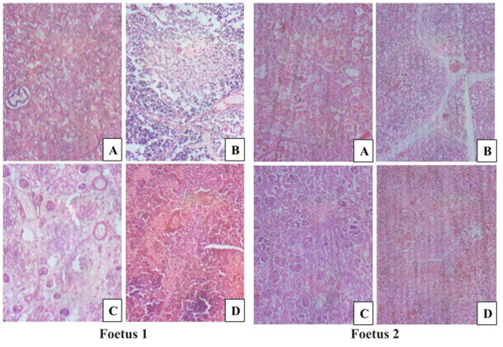
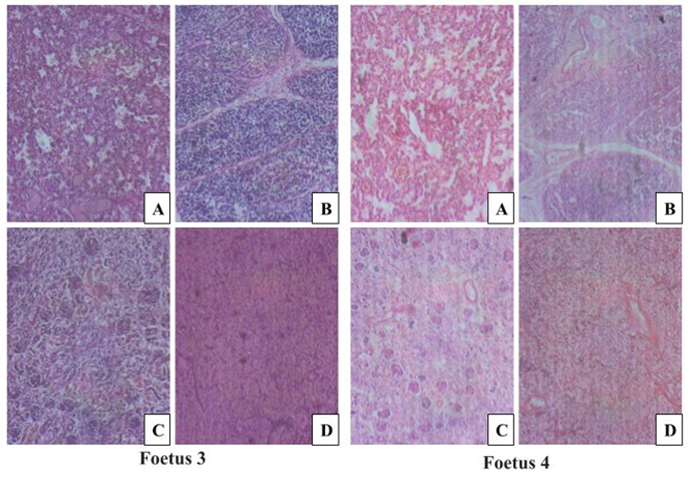
Four different histological samples namely, thymus, spleen, lungs and kidneys were processed. All samples were fixed in 10% formalin and embedded in paraffin; 5 μm sections were stained with hematoxylin-eosin for light microscopy. The microscopic examinations were done to evaluate the degree of visceral maturation based on knowledge of the developmental chronology of foetal tissue. The GA with histological development of the foetal thymus maturation and spleen were noted. For the kidneys, gestational age was estimated by counting the rows of glomeruli between two well-oriented columns of Bertin running from the arcuate artery to the nephrogenic zone or with sequential development of glomerulus by counting the average radial glomerular count in cortical zones. Lung development was determined by the different stages of development based on the maturation.
History of covid-19 infection was during the first trimester of pregnancy for mothers of foetus number 1 (33 wks) and 4 (37 wks) whereas it was during the last trimester for mothers of foetus number 2(38 wks) and 3(27 wks). No history of gestational diabetes as well as pregnancy related complication for all mothers. Antenatal scans were showing normal growth for foetus number 2(38 wks) and 3 (27 wks) where as it was impaired for foetus number 1 and 4 during the routine USG scans of fifth month.
Foetus No.1 of 33 weeks weight and 1.26 Kg had the following features that the kidney section studied from first image showed radial glomerular count (RGC) of 15. Second image show 17 and third image showed one glomerulus. Thymus section show thymic parenchyma with well-formed trabeculae. Parenchyma composed of loosely arranged mesenchymal cells and lymphocytes. Cortex and medulla cannot be distinguished. No hassle’s corpuscles seen. Spleen section showed predominantly red pulp and indistinct white pulp. No trabeculae or capsule noted. Lung section showed extensive area of haemorrhage with small foci and congested blood vessels, which are suggestive of intravascular coagulation. Lungs are in saccular stage of lung development.
Foetus No.2 of 38 weeks weight and 2.5 Kg had the following features that the kidney section studied from first image showed RGC of 2. Second image show 27 and third image is not showing any glomeruli. Thymus had well-formed capsule and trabeculae dividing into lobules which is composed of lymphocytes seen. Two hassle’s corpuscles were noted. Spleen showed well-formed trabeculae with red pulp. White pulp not seen in the image provided. Lungs is in alveolar stage of lung development. There was no hyaline membrane in Alveoli.
Foetus No.3 of 27 weeks weight and 900 gms had the following features that the kidney section studied from first image showed RGC of 17. Second image RGC is 3. Thymus had thin capsule and trabeculae dividing lymphocytes into lobules. No Hassle’s corpuscles were noted. Spleen showed trabeculae with red pulp. No white pulp seen. Lungs are in saccular stage of lung development.
Foetus No.4 of 37 weeks weight and 2 Kg had the following features that the kidney section studied from first image showed radial glomerular count of 27 and second image show 0. Thymus had thin capsule and trabeculae dividing lymphocytes into lobules. No Hassle’s corpuscles were noted. Spleen showed trabeculae with red pulp. No white pulp seen. Lung section showed extensive area of haemorrhage with small foci and congested blood vessels, which are suggestive of intravascular coagulation. Lungs are in saccular stage of lung development. Written informed consent of the concerned families was obtained for all the four cases.
| Sl.no | G.A. (In Wks) | Birth Wt. (In K.g.) | Thymus | Spleen | Kidney | Lung |
| Foetus 1 | 33 Wks
| 1.26 Kg | Thymus group IV corresponding to 18-24 weeks. So, there is delayed development of spleen. | Spleen group IV corresponding to 18-24 weeks. So, there is delayed development of spleen. | Kidney average RGC is 11 corresponding to 24 weeks of gestation. so, there is impaired glomerulogenesis | Lungs show primitive alveoli form corresponding to 26-32 weeks of gestation so there is delayed structural development of lung. |
| Foetus 2 | 38 Wks | 2.5 Kg | Thymus development corresponds to 25-38(group V) weeks of gestation suggestive of normal development. | Spleen group IV and corresponds to 18-24 weeks. So, there is delayed development of spleen. | Kidney RGC is 14.5 corresponding to 28 weeks of gestation. So, there is delayed glomerulogenesis | Lung development corresponds to 32-38 weeks of gestation suggestive of normal development. |
| Foetus 3 | 27 Wks | 900 gms | Thymus corresponds to 18-24 weeks (group IV). So, there is mild delayed development of spleen. | Spleen development corresponds to 18-24 week (group IV) suggestive of impaired development. | Kidney radial glomerular count is 10 corresponding to 22 weeks of gestation. So, there is delayed glomerulogenesis | Lung development corresponds to 26-32 weeks of gestation suggestive of normal development. |
| Foetus 4 | 37 Wks | 2kg | Thymus corresponds to 18-24 weeks (group IV). So, there is delayed development of spleen. | Spleen development corresponds to 18-24 week (group IV) suggestive of impaired development. | Kidney, radial glomerular count is 13.5 corresponding to 27 weeks of gestation. So impaired glomerulogenesis | Lung development corresponds to 26-32 weeks of gestation suggestive of impaired development. |
Table 1: Foetal organs and impaired maturation
GA-Gestational age, Wks-Weeks, Wt.-Weight, K.g.-Kilogram
Discussion
Congenital transmission of SARS-CoV-2 was detected during the first trimester in the placental cells, amniotic fluid and also in the fetal membrane. However, the evidences were inadequate to establish foetal tissue involvement due to lack of samples for autopsy to study the virus particles [19]. In the present case series, we had two still birth foetus was the mother’s diagnosed with SARS-CoV-2 during the first trimester of pregnancy. All organs which were studied showed delayed maturation which implies that the disease during the first trimester is causing more developmental issues on foetus as it is a period of organogenesis. The hypoxia induced on mothers due to the disease can also have an impact on foetoplacental circulation. However, an extensive study with a greater number of samples is needed to draw a conclusion. Fetal tissues like the liver, heart, lungs and hematopoietic cells also express ACE2 which indicates that the presence of the virus in the amniotic fluid will cause the fetal infection [20]. In the present case series, we had two foetuses obtained from mothers who were affected by SARS-CoV-2 during the last trimester. It has been noticed that lung and thymus development appeared to be normal when the infection was in the last trimester, where spleen and kidney development was impaired irrespective of trimester of pregnancy and viral infection.
The thymus is an organ commonly targeted by infectious pathogens such as viruses, bacteria, and fungi. Alterations of proliferation, secretion, migration, differentiation and death of thymocytes can be induced by these infections due to phenotypic and functional changes within the thymus. The behavior of mature, peripheral T-lymphocytes can be equally affected [21]. In the present case series two samples were the mother’s diagnosed with SARS-CoV-2 during the first trimester of pregnancy showed delayed maturation of thymus, where which implies that the disease during the first trimester is causing more developmental issues on foetus as it is a period of organogenesis.
Irrespective of the trimester all cases had impaired growth in the spleen in the present case series. Splenic tissue of human fetuses develops at 14th to 24th week of gestation. The function of spleen in the foetus is heamotopoiesis and it continues from the fetal period till the child birth. So, the red and white pulp attains maturation throughout the embryonic period though the maturation is complete. The viruses must have caused impaired function of the spelenic cords due to hypoxia induced to the mother. Mild to moderate disorganization of the white pulp with indistinct regions and severe congestion and hemorrhage, and proliferation of megakaryocytes of red pulp was observed in the case series. An obvious distinction between white and red pulp is not always proper and several plasma cell aggregates replace the populations of normal resident cell of the red pulp was observed [22].
Kidneys have the advantage of revealing clearly visible structural changes. The count of the glomerular zone extends from the top of the superficial definitive glomerulus to the bottom of the deepest glomerulus, at the junction with the medullas which are still recognizable in cases of advanced necrosis and frequently encountered in forensic practice [23]. In the case series all the samples have delayed and impaired glomerulogenesis. This indicates SARS-CoV-2 can cross the placental barrier. The SARS-CoV-2 can cross the placental barrier, as the viral RNA was detected in the amniotic fluid and the S proteins were detected in the fetal membrane [19].
Development of lung is a continuous process till 8 years and by 20th week there is differentiation of the type 1 pneumocyte. The histopathological changes we observed in the infected lungs of K18-hACE2 mice correlate with the impaired pulmonary function [18]. It has been noticed in the case series that lung development appeared to be normal when the infection was in the last trimester. The pseudoglandular period in which most of the lung elements develop except alveoli must have likely to be affected by the viral disease which resulted in impaired growth of lungs of foetus born from mothers affected with Covid-19 during the first trimester.
Conclusion:
The case series outlooks the impaired maturation of foetal vital viscera’s can be related to SARS-CoV-2 virus invasion and infection during the pregnancy and how and under what circumstances the foetus can get infected. The report helps us to find out the possible mechanisms behind it as well as the nature of the maternal and fetal response to COVID-19 occurring during pregnancy.
Limitations:
We couldn’t perform a placental or viseral immunohistochemical study with SARS-CoV-2 nucelocapsid-specific or cytokeratin-7 specific monoclonal antibody to detect SARS-CoV-2 antigen.
Acknowledgements:
All authors appreciate the great effort of histology lab technicians of Yenepoya Medical college, Mangalore for their timely help and assistance in the conduction of this study.
Declarations
Ethical Approval
We have obtained consents from parents of the still births involved in the case series study.
Competing interests
No competing interests of a financial or personal nature is evolved for the study.
Authors' contributions
All authors have equal contribution towards the study.
Contributions:
Dr Divia Paul A executed the project with proposal writing, data collection, dissection, sample processing, block making and H& E slide preparation and arranging photo microscopic image coupling.
Dr. Banushree CS, Dr. Shankar Narayan SR – analyzed and verified the slides and reported the foetal visceral maturation by double blinding method.
Dr Manisha R. Gaikwad- Helped in data coupling, and writing part of the manuscript.
Funding
No funding received for the study.
Availability of data and materials
With the permission of corresponding author datasets used can be accessed.
Author declaration
All authors declare no conflict of interest.
References
- Yang X, Yu Y, Xu J, Shu H, Liu H, et al. (2020). Clinical course and outcomes of critically ill patients with SARS-CoV-2 pneumonia in Wuhan, China: a single-centered, retrospective, observational study. The Lancet Respiratory Medicine. 8(5):475-481.
View at Publisher | View at Google Scholar - Allotey J, Stallings E, Bonet M, Yap M, Chatterjee S, et al. (2020). Clinical manifestations, risk factors, and maternal and perinatal outcomes of coronavirus disease 2019 in pregnancy: living systematic review and meta-analysis. BMJ. 370.
View at Publisher | View at Google Scholar - Piercecchi-Marti MD, Adalian P, Liprandi A, Figarella-Branger D, Dutour O, et al. (2004). Fetal visceral maturation: a useful contribution to gestational age estimation in human fetuses. Journal of Forensic Science. 49(5): JFS2004027-5.
View at Publisher | View at Google Scholar - Ajita RK, Naranbabu Singh T H, Ibochouba Singh Y, Chandramani Singh L. (2006). An insight into the structure of the thymus in human foetus – a histological approach. J. Anat. Soc. India. 55 (1): 45-49.
View at Publisher | View at Google Scholar - Dubey A, Jethani SL, Singh D. (2018). Gestational age estimation in human fetuses from histogenesis of the spleen. SRHU MEDICAL JOURNAL. 1(2):76-79.
View at Publisher | View at Google Scholar - Daković-Bjelaković M, Vlajković S, Cukuranović R, Antić S, Bjelaković G, et al. (2005). Quantitative analysis of the nephron during human fetal kidney development. Vojnosanit Pregl, 62:281-286.
View at Publisher | View at Google Scholar - Emery JL, Mithal A. (1960). The number of alveoli in the terminal respiratory unit of man during late intrauterine life and childhood. Arch Dis Child, 35:544–547.
View at Publisher | View at Google Scholar - Gaultier CL. (1988). Physiologie et physiopathologie du developpement et ´ de la maturation du poumon antenatal. Rev Mal Resp, 5: 213–222.
View at Publisher | View at Google Scholar - Loytved CA, Fleming V. (2016). Naegele's rule revisited. Sexual & Reproductive Healthcare. 8:100-101.
View at Publisher | View at Google Scholar - Ander SE, Diamond MS, Coyne CB. (2019). Immune responses at the maternal-fetal interface. Science immunology. 4(31).
View at Publisher | View at Google Scholar - Cornish EF, Filipovic I, Åsenius F, Williams DJ, McDonnell T. (2020). Innate immune responses to acute viral infection during pregnancy. Frontiers in Immunology. 11:2404.
View at Publisher | View at Google Scholar - Allotey J, Stallings E, Bonet M, Yap M, Chatterjee S, et al. (2020). Clinical manifestations, risk factors, and maternal and perinatal outcomes of coronavirus disease 2019 in pregnancy: living systematic review and meta-analysis. BMJ. 370.
View at Publisher | View at Google Scholar - Gajbhiye RK, Modi DN, Mahale SD. (2020). Pregnancy outcomes, newborn complications and maternal-fetal transmission of SARS-CoV-2 in women with COVID-19: a systematic review of 441 cases. MEDRXIV.
View at Publisher | View at Google Scholar - Alves NL, Takahama Y, Ohigashi I, Ribeiro AR, Baik S, et al. (2014). Serial progression of cortical and medullary thymic epithelial microenvironments. Eur. J. Immunol. 44(1):16-22.
View at Publisher | View at Google Scholar - Anderson G, Takahama Y. (2012). Thymic epithelial cells: working class heroes for T cell development and repertoire selection. Trends Immunol. 33(6):256-263.
View at Publisher | View at Google Scholar - Perry JS, Lio CW, Kau AL, Nutsch K, Yang Z, et al. (2014). Distinct contributions of Aire and antigen-presenting-cell subsets to the generation of self-tolerance in the thymus. Immunity. 41(3):414-426.
View at Publisher | View at Google Scholar - Djokic V, Akoolo L, Parveen N. (2018). Babesia microti infection changes host spleen architecture and is cleared by a Th1 immune response. Frontiers in Microbiol. 9:85.
View at Publisher | View at Google Scholar - Pan F, Ye T, Sun P, Gui S, Liang B, et al. (2020). Time course of lung changes at chest CT during recovery from coronavirus disease 2019 (COVID-19). Radiology. 295(3):715-721.
View at Publisher | View at Google Scholar - Shende P, Gaikwad P, Gandhewar M, Ukey P, Bhide A, et al. (2021). Persistence of SARS-CoV-2 in the first trimester placenta leading to transplacental transmission and fetal demise from an asymptomatic mother. Human Reproduction. 36(4):899-906.
View at Publisher | View at Google Scholar - Li M, Chen L, Zhang J, Xiong C, Li X. (2020). The SARS-CoV-2 receptor ACE2 expression of maternal-fetal interface and fetal organs by single-cell transcriptome study. PLoS One. 15(4):e0230295.
View at Publisher | View at Google Scholar - Albano F, Vecchio E, Renna M, Iaccino E, Mimmi S, et al. (2019). Insights into thymus development and viral thymic infections. Viruses. 11(9):836.
View at Publisher | View at Google Scholar - Santos SO, Fontes JL, Laranjeira DF, Vassallo J, Barrouin-Melo SM, et al. (2016). A minimally invasive approach to spleen histopathology in dogs: A new method for follow-up studies of spleen changes in the course of Leishmania infantum infection. Comparat. Immunol., Microbiol. and infect. Dis. 48:87-92.
View at Publisher | View at Google Scholar - Singer DB, Sung CJ, Wigglesworth JS. (1991). Fetal growth and maturation: with standards for body and organ development. In: Wigglesworth JS, Sung CJ, editors. Textbook of fetal and perinatal pathology. Cambridge: Blackwell Scientific, 11–47.
View at Publisher | View at Google Scholar
