

Research Article | DOI: https://doi.org/10.31579/2835-8295/146
Evaluation of Morphological Changes in Neurons of the Parietal Lobe Cortex of the Brain in Rats with Acute Respiratory Failure of Obstructive Genesis
- N.Ye. Maksimovich,
- M.A. Feduto,
- E.I. Bon, *
- S.M. Zimatkin,
- S.A. Sedinevskaya
Grodno State Medical University, Grodno, Republic of Belarus.
*Corresponding Author: E.I. Bon, Grodno State Medical University, Grodno, Republic of Belarus.
Citation: N.Ye. Maksimovich, M.A. Feduto, E.I. Bon, S.M. Zimatkin, S.A. Sedinevskaya, (2026), Evaluation of Morphological Changes in Neurons of the Parietal Lobe Cortex of The Brain in Rats with Acute Respiratory Failure of Obstructive Genesis, International Journal of Clinical Reports and Studies, 5(1); DOI:10.31579/2835-8295/146
Copyright: © 2026, E.I. Bon. This is an open-access artic le distributed under the terms of the Creative Commons Attribution License, which permits unrestricted use, distribution, and reproduction in any medium, provided the original author and source are credited.
Received: 11 December 2025 | Accepted: 16 January 2026 | Published: 06 February 2026
Keywords: obstruction; hypoxia; neurons; parietal cortex
Abstract
Acute respiratory failure, which can be caused by airway obstruction due to various reasons, has a negative effect on vital organs and, first of all, on the brain, reducing functional capabilities and leading to structural damage. When studying the cortex of the parietal lobe of the brain in rats under conditions of total and partial obstructive respiratory failure, structural changes were revealed after 30 and 60 minutes, which were manifested in a change in the size and shape of neurons, the degree of staining of their cytoplasm. For total obstructive respiratory failure lasting 30 minutes, a change in the shape of neurons is characteristic in the form of a loss of sphericity with an increase in elongation, and for a 60-minute period, a decrease in the area (by 35%, p < 0,05) of neurons is characteristic against the background of a significant increase in the number of hyperchromic wrinkled neurons in both time intervals to 75% and 80%, respectively. At the same time, partial obstructive respiratory failure in both periods studied was manifested by an increase in the area of neurons (by 24% and 45%, respectively, p < 0,05) without changing their shape against the background of an increase in the number of hypochromic neurons with signs of swelling and shadow cells (up to 80% and up to 95%, respectively). These differences are due to the different severity of acute respiratory failure.
Introduction
The main task of external respiration is to ensure adequate gas exchange with the environment to meet the metabolic needs of the body. Various reasons can lead to impaired external respiration and the development of acute respiratory failure [1]. In particular, obstruction of the airways can occur due to their spasm, edema, inflammatory infiltration, obstruction with sputum, mucus, foreign body, aspiration of gastric contents, blood, etc. (obstructive respiratory failure). The development of acute respiratory failure is based on alveolar hypoventilation. Low oxygen levels in the blood negatively affect the body as a whole, but primarily the vital organs and, first of all, the brain, reducing functionality and leading to structural damage [2, 3]. The severity of oxygen starvation of the brain depends on the degree of narrowing of the airways and the duration of the hypoxic period. Particular attention should be paid to the parietal cortex of the brain, damage to which leads to impaired response to sensory stimuli and spatial orientation [4].
Previously conducted studies of the parietal cortex of the brain under conditions of total tracheal obstruction revealed the presence of structural changes in neurons in the form of a decrease in area and a change in shape (loss of sphericity and an increase in elongation) of cells, as well as a change in the degree of chromatophilia, which was manifested by a decrease in the number of normochromic neurons with a simultaneous increase in the number of hyperchromic wrinkled neurons [5]. However, the processes of damage to neurons of the parietal cortex of the brain due to acute respiratory failure caused by partial obstruction (stenosis) of the airways have not been sufficiently studied. The aim is to evaluate morphological changes in neurons of the parietal cortex of the brain in rats with acute respiratory failure of obstructive genesis.
Materials and methods
The study was conducted on outbred white rats (30 males, weight 240±20 g), divided into 5 groups (n=6) in compliance with the requirements of Directive of the European Parliament and of the Council No. 2010/63/EU of 22.09.2010 on the protection of animals used for scientific purposes. In rats of the experimental groups, obstructive respiratory failure was modeled by total (groups 2, 3) or partial (groups 4, 5) compression of the trachea under intravenous thiopental anesthesia (40 mg/kg). Total obstructive respiratory failure was modeled by ligating the trachea below the cricoid cartilage of the larynx with a ligature for 30 minutes (group 2) and 60 minutes (group 3). Partial obstructive respiratory failure was modeled by placing a 1,5 mm diameter plastic wire on the trachea below the cricoid cartilage of the larynx with a ligature of the trachea in this area (narrowing of the lumen reached 65%) and subsequent removal of the wire with collection of material after 30 minutes (group 4) and 60 minutes (group 5).
The control group consisted of sham-operated rats with reproduction of all stages without tracheal stenosis (group 1).
The brain was quickly removed in the cold and fixed in Carnoy's fluid, followed by dehydration of the brain sections in increasing concentrations of ethanol and embedding them in paraffin. Frontal paraffin sections of the parietal lobe were then prepared with a thickness of 7 μm and stained using the Nissl method. The location of the parietal cortex was established using a stereotaxic atlas [6]. In each animal, 30 neurons of the fifth layer of the parietal lobe cortex were studied, determining their size and shape. Changes in the area and shape (form factor, elongation factor) of neurons were assessed using the ImageWarp image analysis program (Bitflow, USA). In histological preparations, different types of neurons were determined by the degree of staining of their cytoplasm and their percentage content.The obtained quantitative continuous data were processed using nonparametric statistics methods, the licensed computer program Statistica 10.0 for Windows (StatSoft, Inc., USA). The data are presented as Me (LQ; UQ), where Me is the median, LQ is the value of the lower quartile; UQ is the value of the upper quartile. Differences between the indicators of the control and experimental groups were considered reliable at p<0>
Results.
In the control group, the area of neurons in the parietal lobe cortex of the rat brain was 186,6 (180,2; 191,0) µm2. They had a rounded shape (form factor – 0,9 (0,9; 0,9) units, elongation factor – 1,2 (1,2; 1,3) units), distinct smooth contours of the cellular and nuclear membranes. In rats of the experimental groups, changes in the size and shape of neurons (Table 1) and the degree of staining of their cytoplasm occurred.
| Groups | Indicators | ||
| area (µm2) | form factor (units) | elongation factor (units) | |
| control | 186,6 (180,2; 191,0) | 0,9 (0,9; 0,9) | 1,2 (1,2; 1,3) |
| total obstruction 30 min | 180,6 (167,5; 188,7) | 0,7* (0,7; 0,7) | 2,1* (2,0; 2,2) |
| total obstruction 60 min | 121,5*# (118,6; 133,4) | 0,6* (0,6; 0,7) | 2,2* (2,1; 2,3) |
| partial obstruction 30 min | 231,4* (221,2; 240,5) | 0,9 (0,9; 0,9) | 1,2 (1,2; 1,3) |
| partial obstruction 60 min | 270,6 *& (261,3; 280,4) | 0,9 (0,9; 0,9) | 1,2 (1,2; 1,3) |
Note: – * – the differences are significant compared to the control group (р<0>
– # – the differences are significant compared to the “total obstruction 30 minutes” group (р<0>
– & – the differences are significant compared to the “partial obstruction 30 minutes” group (р<0>
Table 1 – Indicators of the size and shape of neurons in the parietal cortex of the brain of rats with total and partial obstructive respiratory failure (Me (LQ; UQ
After 30 minutes of total tracheal obstruction, there was no change in the area of neurons in the parietal lobe cortex (p>0,05). At the same time, a change in the shape of neurons was noted in the form of a loss of sphericity with an increase in the elongation of the perikarya. This demonstrates a decrease in the form factor by 23% (p<0>0,05), which indicates the preservation of the shape of neurons, in contrast to changes in total tracheal obstruction. By the 60-minute period of total tracheal obstruction, the area of neurons in the parietal lobe cortex decreased by 35% compared to the control group (p<0>0,05).
After 60 minutes of partial tracheal obstruction, the area of neurons increased by 45% compared to the control group (p<0>0,05) and the elongation factor (р>0,05). In the control group, up to 95% of the population of neurons in the parietal cortex of the brain were normochromic cells, and the remaining neurons were – hypochromic (4%) and hyperchromic (1%) cells (Fig. 1).
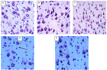
Figure. 1. Neurons of the parietal cortex of rats with total and partial obstructive respiratory failure. Digital micrograph. Nissl staining. Magnifying lens. х 20.
А – control group (normochromic neurons);
В – after 30 minutes of total obstruction (hyperchromic shrunken neurons);
С – after 60 minutes of total obstruction (hyperchromic shrunken neurons);
D – after 30 minutes of partial obstruction (hypochromic neurons with signs of swelling and shadow cells (indicated by arrow));
E – after 60 minutes of partial obstruction (hypochromic neurons with signs of swelling and shadow cells (indicated by arrow)).
In contrast to the control group, in the experimental groups with total tracheal obstruction in both study periods, hyperchromic shrunken neurons predominated: up to 75% in the group of rats with 30-minute obstruction (p<0>
Thus, in total and partial obstructive respiratory failure, opposite changes in the size and shape of neurons in the parietal cortex of the brain, and the degree of staining of their cytoplasm were noted. For total obstructive respiratory failure lasting 30 minutes, a change in the shape of neurons is characteristic in the form of a loss of sphericity with an increase in elongation, and for a 60-minute period, a decrease in the area of neurons is characteristic. At the same time, a significant increase in the number of hyperchromic wrinkled neurons was observed in both time intervals. At the same time, partial obstructive respiratory failure with a residual tracheal patency of 35% in both studied periods were manifested by an increase in the area of neurons without changing their shape with a simultaneous increase in the number of hypochromic neurons with signs of swelling and shadow cells. These differences are due to different rates of increase in acute oxygen deficiency.
References
- Tobin J. Martin, Horacio J. Androgue. (1997). Respiratory Failure. 576.
View at Publisher | View at Google Scholar - Guo, M. F. J. Z. Yu, C. G. Ma. (2011). Mechanisms related to neuron injury and death in cerebral hypoxic ischaemia. Folia Neuropathol. 49 (2).78–87.
View at Publisher | View at Google Scholar - Romano, A.D. G. Serviddio, F. de Matthaeis et al. (2010). Oxidative stress and aging. J. Nephrol. 23 (15). 29–33.
View at Publisher | View at Google Scholar - Bon, E. I. N. Ye. Maksimovich, S. M. Zimatkin. (2020). Histological changes in neurons of the parietal cortex of the brain of rats with subtotal and total ischemia. Bulletin of the Smolensk State Medical Academy. 23-17.
View at Publisher | View at Google Scholar - Feduto, M.A. N. Ye. Maksimovich, E.I. Bon et al. (2023). Modeling of Cerebral Anoxia of Respiratory Genesis in Rats. Archives of Urology and Nephrology. 2(1).
View at Publisher | View at Google Scholar - Paxinos, G. C. Watson. (1998). The Rat Brain in stereotaxis coordinates. Academic Press, Australia. 242.
View at Publisher | View at Google Scholar - Brown, B.M., R.G. Newcombe, Y. Zhao. (2009) non-null semi-parametric inference for the Mann-Whitney measure. J. of Nonparametric Statistics. 21 (6). 743– 755.
View at Publisher | View at Google Scholar - N.Y. MAKSIMOVICH – Head of the Department of Pathological Physiology named after D.A. Maslakov, Educational Institution “GrSMU”, Doctor of Medical Sciences,
View at Publisher | View at Google Scholar - M.A. FEDUTO – assistant at the Department of Pathological Anatomy (Forensic Medicine) of the Educational Institution “GrSMU”
View at Publisher | View at Google Scholar - E.I. BON - Associate Professor of the Department of Pathological Physiology named after D.A. Maslakov, Educational Institution
View at Publisher | View at Google Scholar - S.M. ZIMATKIN – Head of the Department of Histology, Cytology and Embryology, Educational Institution “GrSMU”, Doctor of Biological Sciences,
View at Publisher | View at Google Scholar - S.A. SEDINEVSKAYA – student, Educational Institution “GrSMU”
View at Publisher | View at Google Scholar
