

Case Report | DOI: https://doi.org/10.31579/2834-5010/027
Co-Occurrence of Wilson Disease and auto-Immune Hepatitis in 14-Year-old Female: A Case Report*
1Department of Pharmacy, Al-Kitab University, Kirkuk, Iraq.
2Department of Pharmacology and Toxicology, College of Pharmacy, University of Al-Mustanseriah, Baghdad, Iraq.
3Department of Clinical Pharmacy, College of Pharmacy, University of Al-Mustanseriah, Baghdad, Iraq.
4Department Pediatric, College of Medicine, Al-Nahrain University, Baghdad, Iraq.
*Corresponding Author: Fadwa Ghassan Hameed, Department of Pharmacy, Al-Kitab University, Kirkuk, Iraq.
Citation: Fadwa G. Hameed, Inam S. Arif, Mohammed M. Mohammed, Hala S. Arif, (2025), Co-Occurrence of Wilson Disease and auto-Immune Hepatitis in 14-Year-old Female: A Case Report, 2024, International Journal of Clinical Therapeutics, 4(5); DOI:10.31579/2834-5010/027
Copyright: © 2025, Fadwa Ghassan Hameed. This is an open access article distributed under the Creative Commons Attribution License, which permits unrestricted use, distribution, and reproduction in any medium, provided the original work is properly cited.
Received: 26 September 2025 | Accepted: 10 October 2025 | Published: 21 October 2025
Keywords: autoimmune liver disease; histopathology; wilson’s disease; kayser–fleischer rings
Abstract
Unusual cases of coexistence between Wilson's disease and autoimmune hepatitis have occurred. There are characteristics of both diseases in this community of patients, and laboratory and histo pathological findings can be misleading. Wilson disease's clinical appearance can differ widely; thus, there is not always an easy diagnosis. In addition to being childhood and young adult illnesses, Wilson's disease can also be triggered at any age. Liver disease and cirrhosis, neuropsychiatric disorders, Kayser-Fleischer (KF.) rings, and acute hemolysis events are the primary characteristics of Wilson's disease, frequently in combination with acute liver failure. Diagnosis is extremely difficult for children and adults with active liver disease. None of the latest Wilson's disease laboratory tests are optimal and may not be specific. Therefore, by taking into account acute hepatitis similar to Wilson's disease and autoimmune hepatitis, concomitant treatment with immunosuppression and penicillamine may have a superior impact.
Introduction
In Autoimmune Hepatitis, clinical, serological, histological and radiological characteristics overlap, but adequate diagnosis remains hampered by a lack of standardized diagnostic criteria. With regard to the risks and benefits of treatment, patients should be carefully consulted, taking into account the lack of randomized and monitored outcome evidence for medical interventions[1]. The coexistence of Wilson disease (WD) and autoimmune liver disease (ALD) in the same patients is a rare entity[2,3]. Necrosis of the hepatocytes and exposure of the intracellular antigen to the immune system was observed in WD, leading to the development of low-titer autoantibody[4]. WD screening is therefore highly recommended in patients classified as AIH, especially when the response to immunosuppressive drug treatment is disappointing. In this case, mixed steroid and d-penicillamine treatment may be successful. The main causes of acute and chronic hepatitis are Wilson's disease (WD) and autoimmune hepatitis (AIH). At the same time, the coexistence of these illnesses in one patient is rare. Hepatocyte necrosis and intracellular antigen exposure to the immune system is seen in WD. This observation is a misleading excuse to differentiate AIH from WD [5]. There is no proof of dermatological indicators of autoimmune disorders and levels of serum immunoglobulin are not elevated in this group of WD patients. WD screening is highly recommended in patients classified as AIH, Specifically, when immunosuppressive therapies have a poor response. Combined steroid and d-penicillamine therapy could be successful in this situation[6]. Autoimmune hepatitis has an emerging complexity that has posed many diagnostic and management challenges[7].These challenges present difficulties in understanding the many clinical phenotypes, improving current corticosteroid regimens, detecting depressed patients early, integrating new treatment options into safe and efficient management strategies, and developing new therapies specific to molecular and cellular sites[7,8]. Acute non-viral hepatitis, acute-on-chronic liver failure and acute liver failure can be included in WD. WD, autoimmune hepatitis (AIH), liver damage (DILI) triggered by medication and indeterminate causes, drug-induced liver injury (DILI) and causes that are indefinite should be included in the differential diagnosis before the viral etiology of acute hepatitis has been eliminated. WD has not historically been diagnosed in several WD patients who present with acute liver failure. The underlying, but unrecognized, cirrhosis is usually present. In addition to the conclusive characteristics of acute l-phosphatase, Classic Wilsonian Acute Liver Failure (ALF-WD)-Coombs-Classic Wilsonian Acute Liver Failure (ALF-WD)-Coombs-negative intravascular hemolysis, elevated serum copper and urinary copper excretion, moderate serum aminotransferase elevations compared to other etiologies of acute liver failure, and subnormal serum alkaline phosphatase are sometimes differentiated clinically in these patients. Any acute liver failure caused by WD is a medical emergency(as well as the classic Wilsonian acute liver failure). These patients should be relocated immediately to the liver transplantation and urgent liver transplantation management center[9]. Acute-on-chronic liver failure precipitated by infection, hypotension, or acute worsening of WD is a risk in patients with pre-existing serious liver damage caused by WD. Acute hepatic or acute-on-chronic liver failure is a risk for patients who have declined medically-advised WD care.
Case Presentation
In the Rare Diseases Clinic in AL-Imamen AL-kadhemiain medical hospital/Iraq-Baghdad, (June-2015), a case of a 14-year-old (wt.26 kg.) female patient has been reported as Acute Hepatitis with a negative prior history of ant other diseases but due to G6PD deficiency for a blood transfusion at age 5-years old. Patient lab data: ALT. (78 U/L) ,AST (100 U/L), ALP (420 U/L), TSB. (105 mg/dl.), Bilirubin (78.8 mg/dl) (Jaundice),INR.(0.9 sec.), Ceruloplasmin (17.9 mg/dl.),U. copper (105 mcg/dl.), U. copper/24hr.(1580 mg/dl.) and No Kayser-Fleischer (KF) ring . Patient start on Penicillamine full dose, but continue to have increase in Bilirubin and liver transaminases levels. After three months on Penicillamine (250 mg *2) meanwhile, she developed Tea colored urine, normal HBC and her liver enzymes improved in Oct.2015. Until Dec.2015,her lab data were normal as : ALT (55 U/L), AST (47 U/L) and ALP (255 U/L).So, she was shifted to zinc Sulfate 150mg/D, due to absence of Zinc Acetate (not available at the hospital and the market). In July.2016, U.copper varied from (32 to 84 mcg/dl.),there was another exacerbation of hepatitis by double elevation of transaminases and ALP (486 U/L) (remarkable elevation). So, she was resumed on Penicillamin to 1000mg/D divided in two daily doses, U.copper 945mcg/dl.) , which indicate Zinc Sulfate failure. On Sep.2016, Renal function tests were normal, Transaminases normalized, ALP(404 U/L) (more than 4 times upper the limit) so ,she was on Penicillamine + B6 assuming it’s a sort of Cholengitis). Early in 2017, patient shifted to Zinc Sulfate (150 mg/D), U. copper below (60 mcg/dl.) In April 2017,Zinc Acetate started (150mg*2 ), U. copper (64mcg/dl.) & ALP continue on a high level. July 2017: TSB. (1.44 mg/dl.), ALT (406 U/L), ALP (625 U/L),U.copper(58 mcg/dl..) Aug.2017: ALT (55 U/L), ALP(472 U/L), Lkm (+ve) ,Gamma Protein (159 g/L) mild elevation and U. copper (120 mcg/dl.).She was doing well on Zinc Acetate. In 2018:U.copper (88 mcg/dl),Gamma Globuline(19.7 g/L.), Liver Kidney Microsomes(LKM.) ( + ve(.
Patient stop the treatment:
March 2018: Gamma Globulin(21 g/L.), U. copper (172 mcg/dl.),s. copper (9 mcg/dl.), ALP (237 U/L), ALT (78 U/L.).
Patient maintained on Zinc Acetate.
Aug.2019, patient started:
Prednisolone (5 mg.) | Azathioprine (50 mg.) | Zinc Acetate (150 mg /D.) |
1*3 for 1 week | 1*1 | 1*1 |
1*2 for 1 week |
| |
1*1 for 1 week | ||
On Feb.2020:
Patient on Azathioprine (50mg/D) and Zinc Acetate (150mg/ D):
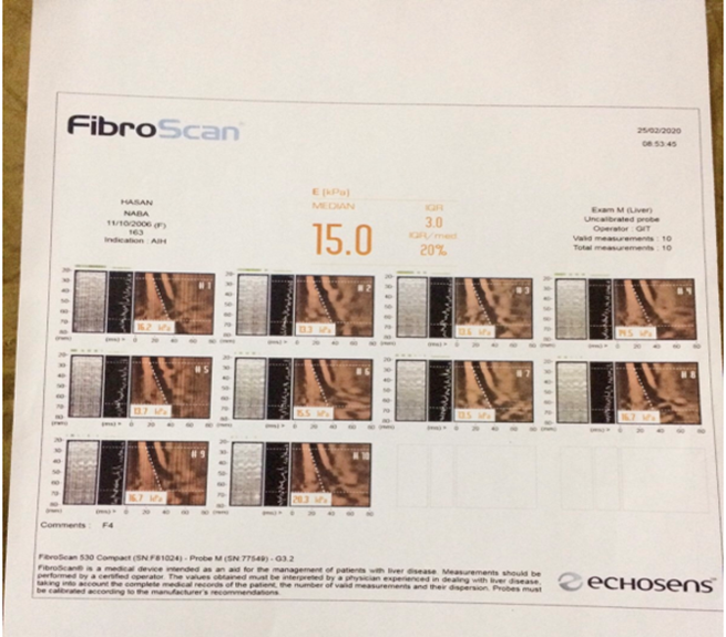
-U.copper.24hr.(34 mg/dl), ALT(73 U/L), AST (77 U/L), ALP (347 U/L) and the Fibro scan demonstrate stage 4 (F4): advanced liver scarring (cirrhosis).
Markers | On admission | After three months of Azathioprine and Zinc Acetate | Reference values |
Alanin transaminase (ALT.) | 100 | 73 | 10-49 U/L |
Aspartate transaminase (AST.) | 87 | 77 | <34> |
Alkaline phosphatase (ALP.) | 420 | 347 | 46-116 U/L |
Gamma globuline (GGT.) | 15.9 | 19.7 | <15> |
Bilirubin | 2.9 | 1.4 | <1> |
Urinary copper per day (U. Copper /D) | 105 | 38 | <40> |
S. Ceruloplamine | 17.9 | 23 | 22-58 mg/dl. |
International normalized ratio(INR.) | 0.9 | - | <1> |
Liver kidney microsome (LKM.) | + ve /post 2years. | + ve. |
|
Immunoglobulin G (IgG.) | 11.8 | 13.5 | 7-16 mg/dl. |
Table 1: Laboratory findings for the patients on admission versus three months of treatment with Azathioprine and Zinc acetate.
Discussion
Wilson Disease is a serious disease that can conduct to multiple hepatic outcomes, including active chronic hepatitis. Owing to the absence or malfunction of P-type ATPase ATP7B transporting copper, its etiology relies on the defective incorporation of copper into apo ceruloplasmin. Symptoms differ, usually either hepatic or neurological at the time of diagnosis, depending on the main affected organs. The epidemiological profile consists of patients between the ages of 5 and 35, although there have been records of younger and older cases. Low ceruloplasmine levels are seen in most patients with neurologic WD, but about half of Wilson's patients with liver disease may be outside the normal range[10]. The presence of Kayser-Fleischer rings is found at diagnosis in about 50 percent of cases[11]. Also, the elevation of urinary copper considered as an effective marker of diagnosis. When the etiology is still uncertain, liver biopsy is considered. AIH, on the other hand, is a chronic disease causing progressive destruction of liver parenchyma. The pathological mechanism is still unclear, and cirrhosis normally results without appropriate care[12]. The AIH diagnostic criteria are the presence of autoantibodies, hyper gamma globulinaemia, Aminotransferase and negative viral marker elevation. Unlike WD, AIH patients typically respond well to immune suppressor therapy, which precludes liver transplantation [13]. In literature, a few instances of similarities have been identified between these two diseases. Cases of WD and positive autoantibodies of two patients with liver disease have been documented by Milkiewicz et al. In their research, patients had a partial initial response to prednisolone therapy [14], unlike our case. Dara et al also mentioned a case where both AIH and WD characteristics were present. Their patient reached the scores for these diseases and was treated for all of them with a positive answer (azathioprine, prednisolone and d-penicillamine)[15]. AIH markers were also reported for acute hepatitis caused by WD, with generally bad results associated with delay in diagnosis. The role of autoantibodies in WD is still unknown; due to hepatocyte necrosis or even a concurrent display of both entities, it may be an early feature of its pathological process[16,17].For this reason, when faced with insufficient response to initial therapy, assessing AIH patients for WD can be life-saving[18]. In conclusion, whether it is due to primary Auto Immune Hepatitis and Wilson disease or Pencillamine sensitivity, further investigations are required. Lack of adherence was obvious especially when the patients start Zinc sulfate on Dec.2015, Due to Gastro intestinal side effects, poor adherence from the parents sides, especially a remarkable elevations in ALP levels was noticed and lead to Hepatitis exacerbations. When starting chelation therapy (D-penicillamine) on july 2016, there was a minimum decrease in ALP levels and cholangitis were observed. During 2017,patient start Zinc Acetate, she was doing well. Later in 2018, there was a significant worsening in ALP levels and increase in the Gamma globuline levels and LKM (+ve). Patient started Prednisolone (5 mg.),Azathioprine(50 mg.) Zinc Acetate (150 mg /D.) later on 2019, a cirrohis were shown on Feb.2020.Patients non adherent on medications or dietary copper contents leads to worsening in clinical presentations and even death. Therefore, adherence has an extremely important role for the long- term success of treatment. Failure to adhere to lifelong therapy can lead to substantial progression of WD- associated liver disease and/or liver failure, the latter requiring liver transplantation. A significant clinical appearance closely resembles autoimmune hepatitis, children, but also young adults, in particular (AIH). This is a chronic inflammatory liver disease characterized by increased serum aminotransferases, hyper gammaglobulinemia, non-specific involvement of autoantibodies (microsomal antibody of the liver kidney, LKM ,anti-nuclear antibody, ANA; smooth muscle antibody, SMA;), hepatitis interface histology, and immunosuppression response[19]. As there is no single pathognomonic function, a rating system that has been revised and modified allows the diagnosis of AIH. Classic AIH can have a chronic or acute appearance[20,21].Significantly, WD's obvious simple acute hepatitis proved to be close to acute autoimmune hepatitis. Several studies record a pediatric WD clinical appearance close to that of AIH[25-28,29,30]. Because of the early cases of WD that clinically mimic autoimmune hepatitis[22-24]. In others, WD was misdiagnosed as AIH, and treated, albeit inadequately, as AIH. For example, in adult girls, an 8-month history of jaundice, ascites, hyperglobulinemia, positive ANA, hepatitis device, and liver biopsy cirrhosis demonstrated improvement in corticosteroid therapy .But WD screening findings a month later showed positive results with low serum ceruloplasmin, increased urinary copper and 385 μg/g dry weight liver copper. D-penicillamine was initiated and corticosteroids, she becomes clinically stable and tapered off [25]. ANA-positivity (type 1 AIH) is more common than LKM-positivity(type 2 AIH) in AIH-like WD, but this latter pattern may occur. Since acute liver failure due to AIH can be difficult to diagnose, acute liver failure presents major diagnostic problems. A similarly non-descriptive clinical picture of acute liver failure may be present in pediatric patients with WD, not the traditional ALF-WD picture. Initially, a 17-year-old girl with acute liver failure initiated a corticosteroid investigation focused on clinical and histological features of AIH in the midst of widespread IgG and negative autoantibodies. Clinical decline was responsible for urgent liver transplantation. Explant histopathology was consistent with WD; baseline 24-hour urinary copper excretion was diagnosed with 10,322 μg/24 hours(162.6 μmol/24 hours)of urinary copper, with findings available only after transplantation[28]. There was a liver biopsy. A 15-year-old girl with acute liver failure had ANA 1:320, SMA 1:160, hyper gamma globulinemia, and plasma cell infiltration, but she also had low serum ceruloplasmin, Kayser Fleischer rings, and high concentrations of liver copper. She started off with both penicillamine and corticosteroids, but as her condition progressed, she underwent liver transplantation. She had WD, confirmed genetically[31].Another problem is that, as a consequence, some people with WD might get autoimmune hepatitis. In such cases, the disease mechanism may be similar to the situation in which a person with pre symptomatic WD experiences acute liver failure due to an inter current viral infection[32] .The affected individual develops AIH in this rare case, Possibly, but not certainly, due to damage to the hepatocellular plasma membrane caused by copper. In children with WD with prominent autoimmune traits, ulcerative colitis can occur[19,32]; however, patients on chelating (Trientine) has also been reported as an adverse occurrence. Some children have a genetic makeup that may be associated with ATP7B mutations and that predisposes them to autoimmune disease. In any case, the presence of each disease needs to be completely identified or denied[33]. Diagnostic complications may be primary sclerosing cholangitis(Roberts EA, unpublished observations)or, in particular, autoimmune sclerosing cholangitis(ASC), as stainless copper is used in liver biopsies[34].In these conditions and WD, the copper distribution pattern in the liver seems to be distinct.
Conclusion
The presence of autoimmune liver disease and Wilson's disease is considered as uncommon entity. The problem in diagnosing is that, clinicians should have a high degree of suspicion. A high degree of understanding and knowledge is required for this situation. Therefore, it is prudent to recognize this form of hepatitis at the same time in rare patients with dominant disease features and to initiate medical treatment for both of them.
References
- Trivedi PJ, Hirschfield GM. overlap syndromes and autoimmune liver disease. Alimentary Pharmacol Ther. 2012 Sep;36(6):517-33.
View at Publisher | View at Google Scholar - Dara N, Imanzadeh F, Sayyari AA, Nasri P, Hosseini AH. Simultaneous presentation of Wilson’s disease and autoimmune hepatitis; a case report and review of literature. Hepatitis monthly. 2015 Jun;15(6).
View at Publisher | View at Google Scholar - Yener S, Akarsu M, Karacanci C, Sengul B, Topalak O, Biberoglu K, et al. Wilson's disease with coexisting autoimmune hepatitis. J Gastroenterol Hepatol. 2004;19(1):114-6.
View at Publisher | View at Google Scholar - Milkiewicz P, Saksena S, Hubscher SG, Elias E. Wilson's disease with superimposed autoimmune features: report of two cases and review. J Gastroenterol Hepatol. 2000;15(5):570-4.
View at Publisher | View at Google Scholar - Czaja AJ.Diverse manifestations and evolving treatments of autoimmune hepatitis. Minerva Gastroenterol Dietol. 2005;51:313–333.
View at Publisher | View at Google Scholar - Czaja AJ. Special clinical challenges in autoimmune hepatitis: the elderly, males, pregnancy, mild disease, fulminant onset, and nonwhite patients. Semin Liver Dis. 2009;29:315–330. doi: 10.1055/s-0029-1233530.
View at Publisher | View at Google Scholar - Czaja AJ, Bianchi FB, Carpenter HA, et al. Treatment challenges and investigational opportunities in autoimmune hepatitis. Hepatology.2005;41:207–15.
View at Publisher | View at Google Scholar - Czaja AJ. Difficult treatment decisions in autoimmune hepatitis.World J Gastroenterol.2010;16:934–47.
View at Publisher | View at Google Scholar - Ahmad A, Torrazza-Perez E, Schilsky ML. Liver transplantation for Wilson disease. Handb Clin Neurol 2017;142:193-204.
View at Publisher | View at Google Scholar - European Association for Study of Liver. EASL Clinical Practice Guidelines: Wilson’sdisease. J Hepatol 2012;56.
View at Publisher | View at Google Scholar - Stremmel W, Meyerrose KW, Niederau C, et al. Wilson disease: clinical presentation,treatment, and survival. Ann Intern Med 1991;115:720.
View at Publisher | View at Google Scholar - Couto C, Bittencourt P. Controvérsias no diagnóstico e tratamento da hepatite autoimune.In: Savassi-Rocha P, Coelho L, Sanches M, eds. Tópicos em Gastroenterologia14 - Controvérsias. Rio de Janeiro: Guanabara Koogan, 2015.
View at Publisher | View at Google Scholar - Santos RG, Alissa F, Reyes J, et al. Fulminant hepatic failure: Wilson’s disease or autoimmune hepatitis? implications for transplantation. Pediatr Transplant 2005;9:112–6.
View at Publisher | View at Google Scholar - Milkiewicz P, Saksena S, Hubscher SG, et al. Wilson’s disease with superimposed autoimmune features: report of two cases and review. J Gastroenterol Hepatol 2000;15:570–4.
View at Publisher | View at Google Scholar - Dara N, Imanzadeh F, Sayyari AA, et al. Simultaneous presentation of Wilson’s disease and autoimmune hepatitis; a case report and review of literature. Hepat Mon 2015;15:e29043.
View at Publisher | View at Google Scholar - Loudianos G, Zappu A, Lepori MB, et al. Acute liver failure because of Wilson diseasewith overlapping autoimmune hepatitis features: the coexistence of two diseases? JPediatr Gastroenterol Nutr 2016;63:e23–4.
View at Publisher | View at Google Scholar - Yener S, Akarsu M, Karacanci C, et al. Wilson’s disease with coexisting autoimmune hepatitis. J Gastroenterol Hepatol 2004;19:114–6.
View at Publisher | View at Google Scholar - Deutsch M, Emmanuel T, Koskinas J. Autoimmune hepatitis or Wilson’s disease, aclinical dilemma. Hepat Mon 2013;13:e7872
View at Publisher | View at Google Scholar - Jang HJ, Jang JY, Kim KM. Appendiceal orifice inflammation in an 8-year-old girl with ulcerative colitis complicating Wilson’s disease. Gut Liver 2010;4:126.
View at Publisher | View at Google Scholar - Alvarez F, Berg PA, Bianchi FB, Bianchi L, Burroughs AK, Cancado EL, et al. International Autoimmune Hepatitis Group Report: review of criteria for diagnosis of autoimmune hepatitis. J Hepatol 1999;31:92938.
View at Publisher | View at Google Scholar - Ebbeson RL, Schreiber RA. Diagnosing autoimmune hepatitis in children: is the International Autoimmune Hepatitis Group scoring system useful?Clin Gastroenterol Hepatol 2004;2:935.
View at Publisher | View at Google Scholar - Archer GJ, Monie RD. Wilson’s disease and chronic active hepatitis. Lancet 1977;i:486.
View at Publisher | View at Google Scholar - Scott J, Gollan JL, Samourian S, Sherlock S. Wilson’s disease, presenting as chronic active hepatitis. Gastroenterology 1978;74:645.
View at Publisher | View at Google Scholar - Perman JA, Werlin SL, Grand RJ, Watkins JB. Laboratory measures of copper metabolism in the differentiation of chronic active hepatitis and Wilson disease in children. J Pediatr 1979;94:5648.
View at Publisher | View at Google Scholar - Milkiewicz P, Saksena S, Hubscher SG, Elias E. Wilson’s disease with superimposed autoimmune features: report of two cases and review. JGastroenterol Hepatol 2000;15:570.
View at Publisher | View at Google Scholar - Yener S, Akarsu M, Karacanci C, Sengul B, Topalak O, Biberoglu K, et al. Wilson’s disease with coexisting autoimmune hepatitis. J Gastroenterol Hepatol 2004;19:114.
View at Publisher | View at Google Scholar - Deutsch M, Emmanuel T, Koskinas J. Autoimmune hepatitis or Wilson’s disease, a clinical dilemma. Hepat Mon 2013;13:e7872.
View at Publisher | View at Google Scholar - Bruno Campos Santos, Ludmila Resende Guedes, Luciana Costa Faria and Claudia Alves Couto. Case report:Wilson’s disease presentation resembling autoimmune hepatitis.BMJ case report. October 25, 2019.
View at Publisher | View at Google Scholar - Dara N, Imanzadeh F, Sayyari AA, Nasri P, Hosseini AH. Simultaneous presentation of Wilson’s disease and autoimmune hepatitis; a case report and review of literature. Hepat Mon 2015;15:e29043.
View at Publisher | View at Google Scholar - Loudianos G, Zappu A, Lepori MB, Dessi V, Mameli E, Orru S, et al. Acute liver failure because of Wilson disease with overlapping autoimmune hepatitis features: the coexistence of two diseases? J Pediatr Gastroenterol Nutr 2016;63:e23.
View at Publisher | View at Google Scholar - Sallie R, Chiyende J, Tan KC, Bradley D, Portmann B, Williams R, et al. Fulminant hepatic failure resulting from coexistent Wilson’s disease and hepatitis E. Gut 1994;35:849.
View at Publisher | View at Google Scholar - Nery FG, Marques I, Magalhaes M, Miranda HP. Wilson’s disease and ulcerative colitis in the same patient: just a coincidence? A case report and literature review. Gastroenterology Res 2010;3:287.
View at Publisher | View at Google Scholar - Boga S, Jain D, Schilsky ML. Trientine induced colitis during therapy for Wilson disease: a case report and review of the literature. BMC Pharmacol Toxicol 2015;16:30.
View at Publisher | View at Google Scholar - Sood V, Rawat D, Khanna R, Alam S. Cholestatic liver disease masquerading as Wilson disease. Indian J Gastroenterol 2015;34:174.
View at Publisher | View at Google Scholar
