

Research Article | DOI: https://doi.org/10.31579/2835-8295/138
Detection of Storage Fungi Associated with Sunflower (helianthus annus l.) Seeds and Its Chemical Management
1University of Agriculture, Faisalabad, Pakistan
2National Agricultural Research Center, Islamabad, Pakistan.
*Corresponding Author: Muhammad Arshad Ullah, National Agricultural Research Center, Islamabad, Pakistan.
Citation: Sajid Waseem, Amer Habib and Muhammad A. Ullah, (2025), Detection of Storage Fungi Associated with Sunflower (helianthus annus l.) Seeds and Its Chemical Management, International Journal of Clinical Reports and Studies, 4(6); DOI:10.31579/2835-8295/138
Copyright: © 2025, Muhammad Arshad Ullah. This is an open-access artic le distributed under the terms of the Creative Commons Attribution License, which permits unrestricted use, distribution, and reproduction in any medium, provided the original author and source are credited.
Received: 29 July 2025 | Accepted: 06 November 2025 | Published: 13 November 2025
Keywords: macrophomina phaseolina; rhizopus arrhizal; sclerotium rolfsi; helianthus annus; aspergillus flavus and helminthosporium
Abstract
Sunflower (Helianthus annus L.) is one of the Pakistan most significant oilseed crop. Currently, sunflower facing several challenges including various foliar diseases as well as storage seed associated fungi. A variety of mycoflora have been associated to sunflower seeds in storage. So, it is critical to perform a thorough investigation of this issue. For this purpose, a survey of several locations in Vehari and Bahawalpur districtswas conducted to collect the seed samples during the sunflower harvesting stages. The samples were brought to Seed Health Testing Lab. Department of Plant Pathology UAF for further processing and mycological evaluation. The incidence and frequency percentage of seed-borne fungus, as well as their influence on seed germination and seedling vigor, were observed and documented. The research study was done under CRD. The results of this investigation revealed that the impact of fungicides on seed germination varies substantially. Tazolen and Alert plus were more effective and this might be attributable to the molecules swift entrance into the fungal mycelia rapid contact andpossible of destruction of the fungus protoplasm. These two fungicides chemical components may stop fungus propagules from sprouting. It can be confidently inferred and suggested that fungicidal seed treatment is extremely successful cost efficient and simple to use since it may minimise seed-borne mycoflora promote seed germination and protect seedlings for a considerable amount of time period. According to the present research work it is concluded that if seed treatment of sunflower seeds with proper fungicides is done before sowing of seeds, this is not only helpful for good germination but also helpful for controlling different field diseases like Charcoal rot (Macrophomina Phaseolina), Head rot (Aspergillus spp., Rhizopus arrhiza), Seedling wilt (Fusarium spp.) and Sclerotial wilt (Sclerotium rolfsi).
Introduction
Sunflower (Helianthus annus L.), a part of the family asteraceae, is one of the largest and most important oil seed crops. Sunflower is a spring crop and have originated in North America. Fats and oils are essential components of human nutrition. Seeds and fruits of many crops and plants are used to produce vegetable oil (Butt and Ali, 2005). Ukraine, Russia, Europe, and Argentina are among the countries that grow it commercially. Ukraine ranked as top producer of sunflower seeds during the year 2019-20 (16.5 million metric tons). With a production capacity of 15.3 million metric tons in 2019/2020. Russia is also a major producer of sunflower seeds of the worldwide. The sunflower seed fruit is a green capsule with several immature white colour seeds. The sunflower crop in Pakistan is grown on 151 thousand acres with a total seed output of 87 thousand metric tons and a maximum oil output of 33 thousand tonnes (Gop. 2021).
Fungi, bacteria, nematodes and viruses affect sunflowers causing a variety of disease. Alternaria helianthi and Septoria helianth were produce of leaf spot, which one of the most frequent of fungal foliar infections. Albugo tragopogonis, and Plasmopara halstedii are the organisms that generate brown and grey patches. Two of the most frequent fungi are white rust and downy mildew (Masirevic and Jasnic, 2006a, Achbani et al., 2000). Several of these fungi have been discovered to the seed-borne. Sunflower seeds are also highly affected by fungus, which attacks the plants at different stages of growth as well as during harvesting and storage. In Pakistan, Sharfun- Nahar et al. (2005), found a large number of fungi associated with sunflower seeds. Important oilseed crop and beautiful plant sunflower (Helianthus annuus L.) may thrive in the presence of sustained biotic and abiotic stress, such as drought, pathogen infection, and high temperatures (Badouin et al., 2017). EST databases were used by Giacomelliet al., 2010), to identify the 97 sunflower Genes encoding members. Sunflower has 112 Gene encoding genes that have recently been found, however research on Gene encoding proteins in oil crop species and their role in disease resistance in domesticated sunflower is still rare. 119 Ha Gene encoding proteins were found in the sunflower genome using bioinformatic methods whereas 741 Genes encoding members from other species of oil crop were collected and updated from earlier research (Zou et al., 2016). While preserving seeds in deposits storage fungus has occasionally had a significant negative effect. It may finally get to the point where germination is so low that it below the minimum limit at which time the lot is fully destroyed and the loss is irreparable.Additionally the impacts of storage fungal on germinating seeds in the field produced in weak plants that were more susceptible to disease (Zala et al., 2010). Sunflower is mostly grown in the winter season in eastern India specifically in West Bengal. Sunflower seed storage has so become a common method among sunflower producers. Sunflower seed quality degrades rapidly during storage in eastern Indian states due to poor climatic conditions particularly high temperature and humidity. Seed quality is degraded as a result of the additional dangerous pathogenic diseases (Saha and Mandal, 2016). Because one of the most essential areas for achieving a use of high-quality seeds for spreading results in a crop high yield fast development, and high plant density seed quality during storage requires special attention. Seed invigoration is one of several methods for maintaining and improving the quality of seeds. Different dry powder exposures are used to monitor the loss of seed quality in different crop seeds have been described (Bhattacharya et al., 2015). The benefits of seed vigoration employing a variety of ingredients (chemicals raw plant materials medicinal powders etc.) on different crop seed and seedling quality have been demonstrated by recent research characteristics carried out by researchers throughout the world (Basra et al., 2003). Storage time is another major aspect that influences seed quality. To rearrange cropping patterns and maintain optimal crop performance in the field a decision on the upper limit of seed storage time without losing quality is essential. In light of these factors the current study was created to investigate sunflower seed and seedling quality metrics under various storage temperature and seedling vigour conditions. Control of the seed-borne fungi is an important aspect hence their control by suitable storage conditions in seed dressing fungicidesof biological and physical methods, and growing resistant varieties are briefly dealt with (Vaidehi. 2006). New special form of fungus (Macrophomina phaseolina) was identified to be a cause of the pre and post-emergence of rotting sunflower plants in a study of sunflower (Helianthus annuus L.) pre and post emergence rotting (Vaidehi, 2002), and studies have revealed that is a new seed-borne disease of sunflower was most prevalent in alkaline soils. Macrophomina phaseolina, a pathogen, has been identified as the cause (Morar et al., 2004). The inhibitory impact of extracts from four plants was investigated. Fungal organisms have a crucial role in infected seeds, reducing their quality and longevity during storage (Afzal et al., 2010). Analysis performed on four sunflowers revealed the occurrence of several pathogenic and saprophytic fungi belonging to the following genera (Kakde et al., 2012). In the majority of seed nations major advances have been made in identifying chemical micromycetes and calculating the risk that aflatoxins offer (Kononenko et al., 2008, 2009). In India, Pakistan, Tanzania Malaysia, Iran and Egypt aflatoxins were evaluated as having a very high toxicity level in seeds. This infection was also present in seed processed products for oil, but with a higher prediction performance and a more intense accumulation of carcinogenic compounds (Abdullah et al., 2010). Experimental proof has been gained in a number of studies that when seeds are stored especially under high temperatures and moisture conditions the buildup of mycotoxins increases dramatically (Jeswal et al., 2013). Therefore, a research work was carried out to investigate the pathogens associated with sunflower seeds in storage condition.
Materials and Methods
Sunflower seeds were collected from various stores and farmers in various areas in the districts Vehari and Bahawalpur (ISTA,1976). Seed Health Testing Lab., Department of Plant Pathology, University of Agriculture, Faisalabad, was received samples that have been properly labeled and packed. The seeds were maintained in polythene bags and kept in the refrigerator for isolation identification and purification of associated pathogens. According to International Rules for Seed Testing Association, the blotter method was utilized to determine the seed borne pathogen linked with the seeds in the seed sample (ISTA, 2001).
The fungal presence on sunflower seeds was determined using the agar plate techniques followed by Neergaard (1979). For the test 400 seeds were randomly selected from each of the five kinds. After soaking half of the seeds in a sodium hypochlorite solution (1% NaOCl) for five minutes, they were washed and rinsed three times with distilled sterile water. A culture media of Potato Dextrose Agar was produced and placed onto 9cm Petri plates supplemented with Chloramphenicol (0.5 gl-1) as a bacteriostatic agent. The seeds were placed in Petri plates to grow (every Petri dish contained 20 seeds). Petri plates were next be incubated for 5-8 days at 26-29 °C with 12/h cycles of desolation and near solar radiation. Following incubation slides were made and examined under a light microscope to identify seed borne fungus. All of the Petri dishes were set up in a completely random order (100 seeds per rep.). Assessment of infection frequency of fungal pathogens was determined by the formula of Inhibition (%) as below
Inhibition (%) = Diameter of colony in control- diameter of colony in fungicide treated/ Diameter of colony of control x 100 (Dhingra and Sinclair., 1993)
Fungal species was identified primarily via microscopic inspection of the fungus to assess morphology culture pigmentation septations color conidia appearance conidiophores form mycelium growth spore masses structures and fruiting bodies. The growing on test experiment was taken place in the greenhouse for two months during the winter to measure seed germination plant survival and seedling vigor. 10 seeds from each sample were sown in each pot. The pots were put in a glasshouse with an average temperature of 25 °C ranging from 20 to 35°C and were watered to field capacity. Growth duration rates were observed during the first four weeks and percentage survival rates from the fifth and eighth weeks from the sunflower types under examination. Four fungicides were employed to suppress seed infection including Ayzoxistrobin, Difenoconazole, Thiophanate methyl and Pyraclostrobin. Each fungicide was applied twice as foliar spraying following seedling appearance from the second week to the last week of the experiment. At regular intervals the number of growing plants will be counted. Eight weeks before weekly data.
Vigor Index = (mean shoot length+ mean root length) x germination seed percentage [Baki and Andersen 1972; ISTA, 1979 and Haque et al., 2007].
Starting to make use of the seed component manufacturing method a sample of sunflower seeds was further analysed to determine the presence of seed-borne fungus in various regions of the seeds. Twenty-five seeds from each sample were immersed in sterilized distilled water in test tubes for 24 hours before being dissected aseptically into seed coat (testa and tegmen), embryo and endosperm. At 27±2°C, these components were platted on blotter paper for 7 days. After 7 days, the number of fungus linked with these seed components was counted (Habib et al., 2007). For this purpose,84 seeds were sown in 12 Petri plate @ 7 seeds per plate. These 12 Petri plates were divided into 4 sets. These plates were kept under 4 different incubation temperature i.e, 15°C, 20°C, 25°C and 30°C individually. These plates were kept as such for 10 days and then data was recorded.
The pathogenicity of various fungi was validated using spore suspensions collected from 15 day old colony development on PDA. The HgCl2 treated seeds were steeped for 12 hours in these spore solutions. These fungi were cultured in Petri plates (9cm) on PDA medium for 15 days at 15±1°C in an incubated shaken, and crushed to create the fungal suspension. Each period the suspension was then diluted in 200 cc of sterilized sterile water added to provide a uniform suspension for seed treatment. Prior to infestation the well-known sunflower seeds were surface sterilized for 2 minutes with a 2 % sodium hypochlorite (NaOCl) solution. These seeds were sowed individually in trays with sterilized sand as 24 seeds per tray infected with 15 days old culture of corresponding fungus. As a control non, infested seeds from this batch were planted. During the experiment trays were maintained at 27°C in vitro and sprayed with sterilized water at regular intervals to keep the trays at the appropriate moisture content. In pathogenicity trials healthy seed of sunflower before being contaminated with isolates of the most harmful organisms for sunflower oil flavus, Aspergillusniger, Alternariaalternata, Fusariummoniliforme, and Macrophomina accurately record were surface sterilized with 2 % sodium hypochlorite. At a rate of 2g/kg of seed four fungicides were used as Acrobat, Alert plus, Tazolen, and Trimiltox forte. After 24 hours of infection with isolates of test fungus these surface sterilized and artificially infected seedlings were treated with the four fungicides listed above. These infected and treated seeds were placed in Petri plates in groups of seven seeds each. Fungus infested seeds that had not been treated with fungul give out as a control direct. All of Petri dishes were maintained in a constant temperature of 25 degrees Celsius in an incubator. After 10 days data on seed germination was collected and statistically examined. To establish the presence of fungi, dead seedling pieces were plated on PDA and plates were examined in binocular modethen insepction were noted that and statistically evaluated (CRD design).To compare means, the data were statistically assessed using ANOVA and the Least Significant Difference test (Steel et al., 1997).
Results and Discussion
According to (ISTA, 1993), seed samples of four kinds of sunflower (Helianthus annuus L.), A-88, A-133, G-10, and G-134, were obtained from the department of Plant Breeding and Genetics U.A.F. Except for the damaged seeds which were removed 400 seeds of each kind were selected at random. Eighty-four seeds were placed in sterilized Petri dishes with two sterilized blotter papers as is (without any treatment), and eighty-four seeds were placed in the same dishes after being treated with a 2 % sodium hypochlorite (NaOCl) solution for two minutes and being rinsed with three washes of sterilized water. Similar to this two further sets of 28 seeds were plated on PDA media after being treated with 2% sodium hypochlorite (NaOCl).In the case of the blotter paper method the extent of seed-borne fungus was recorded after 8 days and after 4 to 6 days in the case of the PDA medium approach.
Aspergillus flavus was most detected and recorded (88.08 %) in case of A-88 variety followed by Aspergillus niger (78.55 %), Alternaria alternate (69.02 %) while Penicilium spp., was detected and recorded (19.07 %) in the lowest number in both blotter paper and ager plate technique. PDA media method was the best for detection of seed-borne fungi as compared to blotter paper technique because high number of fungi (62.58 %) was recorded on PDA medium as compared to fungi (50.33 %) recorded on blotter paper indicated in tables 1 and 2 Aspergilus flavus was most detected and recorded (90.47 %) in case of A-133 variety folGrowth of Aspergillus flavus on seedslowed by Aspergilus niger (83.32 %), Alternaria alternate (78.55 %) while Pencilium spp. and was detected and recorded (21.45 %) in the lowest number in both blotter paper and ager plate techniques. PDA media method was the best for the detection of seed-borne fungi as compared to blotter paper technique because high number of fungi (64.62 %) was recorded on PDA medium as compared to fungi (55.10 %) recorded on blotter paper (Tables 3 and 4)
Tables 3 and 4 mentioned that Aspergilus flavuswas most detected and recorded (92.85 %) in case of G-10 variety followed by Aspergilus niger (80.93 %), Alternaria alternate (76.17 %) while Pencilium spp. and was detected and recorded (21.45 %) in the lowest number in both blotter paper and ager plate techniques. PDA media method was the best for the detection of seed-borne fungi as compared to blotter paper technique because high number of fungi (68.02 %) was recorded on PDA medium as compared to fungi (51.70 %) recorded on blotter paper (Tables 5 and 6). The results indicated in tables 7 and 8 revealed that the Aspergilus flavus was most detected and recorded (92.85 %) in case of G-134 variety followed by Aspergilus niger (80.93 %), Alternaria alternate (76.17 %) while Pencilium spp. and was detected and recorded (69.02 %) in the lowest number in both blotter paper and ager plate techniques. PDA media method was the best for the detection of seed-borne fungi as compared to blotter paper technique because high number of fungi (54.42 %) was recorded on PDA medium as compared to fungi (44.90 %) recorded on blotter paper.
Fungi isolates from four sunflower samples were purified and identified to a particular level. Lactophenol was used to make their semi permanent slides. The species discovered are listed below and showed in figures.
- Aspergilus flavus: On the PDA, there are colonies. Conidiophores arose from substrate hypha, broad encircle, gross roughened the cell that grow these tiny fake feet likewise wide septate 202-2263 x 5.6-16.8 μ. Conidial heads are generally circular when young radiate, separating into multiple weakly defined columns, and are 133.6-389 μ in width. Conidia elliptical when juvenile, normally circular or subglobose, widewalled, noticeably down, lime yellowish 3.3-6.6 μ width, thick walled markedly echinulate.
- Aspergilus niger: On the PDA, there are colonies. Usually dark near carbonaceous, reverse colourless, and grows slowly, 2.5-3.5 mm in 2 weeks. Basal mycelium is velvety extensively sporulating, and compact mainly buried mildewed odour and no transude. Conidia are normally circular at full growthBrown, with thick walls and erratic surface roughness noticeable elevation echinulations, 3.5-5.5 μ width, and aredark brown nearly smooth, 1336-3008 x 41-22 μ, wall 2.2 μ thick.
- Alternaria alternate: On the PDA, there are colonies. Brown to dark brown conidiophores emerging singly, 2-4 septate, 40-50 x 4.5 μ. pale dark mycelium dark septate, branching. Conidiophores are 40-50 × 4.5 μ, 2-4 septate, brown to dark brown, and they emerge singly. In Conidia chains, dark brown, septate longstanding and diagonally, brief break, 21.42-23-80x9.52 μ.
- Macrophomina phaseolina: With an incoupicous ostiol, pycnidia are transparent, globose or depressed globose, and 125–200 microns in diameter. Conidium one called hyaline thin walled, compact or oval, 17–28 x 6.5-8.6 μ, sclerotium microscopic black changing in size, wall of three or four layers of blackish brown thin walled pointed cells, 8 micron in diameter, with a hyaline layer, two or three cells thick, bearing simple, rod-shaped conidiophores, 12–14 μ long.
- Rhizopus arrhizus: Basidiospore emerging from stolons opposite the rhizoids, 7.5-17.5 μ in diameter, unbranched, globose at first white letter brown, 75-210 μ in diameter of sporangia broadly diffused, collunellae brown in colour, globose or oval, or broader than length, individually or in groups of two to three with 27-120x28-136. 4.5-6.5x3.5-5.00 μ, Sporangiospores oval, 4.5-6.5x3.5-5.00 μ.
- Fusarium moniliforme: On the PDA, there are colonies having 3-5 cm of white to light violet in 14 days reversal black violet development quick together with narrow, mycelium gently floccose and seeming with powdered owing toward microconidia generation mycelium delicately floccose with powdery appearance. These are smooth, 0-1 septate, fusiform to cleave with a somewhat flattened base 5.5-13.2x2.2-4.4 μ on aerial mycelium from single pinalides
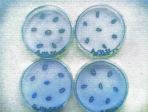
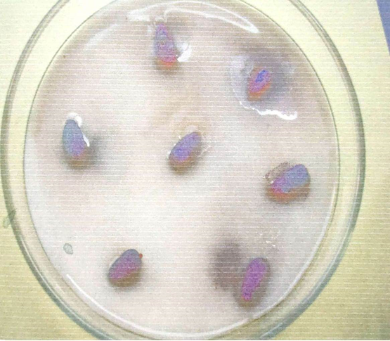
Plating of Sunflower Seeds
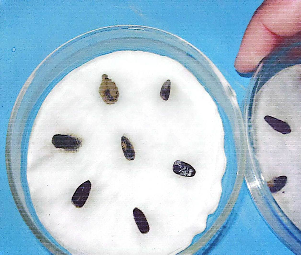
Growth of fungi on seeds
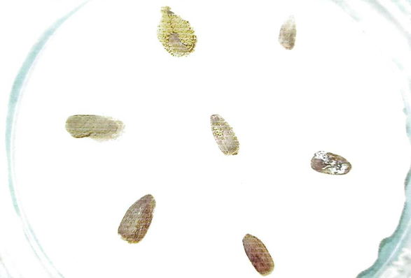
Growth of Aspergillus flavus on seeds
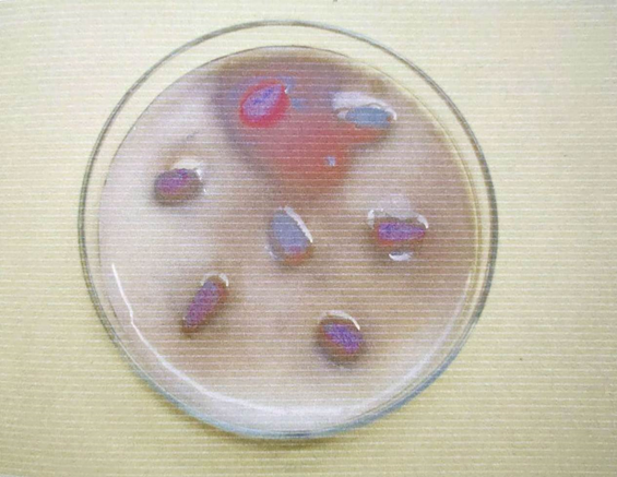
Helminthosporium Pigmention on sunflower Seed
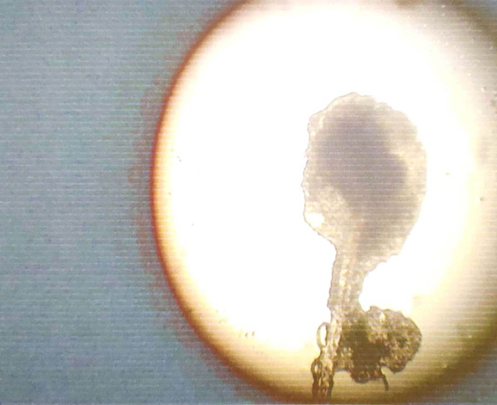
Microscopic view of Aspergillus conidiophores and conidia
Aspergillus niger
| Source of variation | DF | SS | MS | F-value |
| Methods | 1 | 1573.819 | 1573.819 | 24.8650** |
| Fungus | 6 | 21987.488 | 3664.581 | 57.8973** |
| Interaction(M x F) | 6 | 1144.149 | 190.691 | 3.0128* |
| Error | 28 | 1772.247 | 63.295 | |
| Total | 41 | 26477.703 |
**=Highly significa Ns=Non-significant
Table 1: Analysis of variance for isolation of fungi from variety A-88
| Methods | |||
| Fungi | Blotter paper method | Agar plate method | Means |
| F₁=Aspergilus flavus | 80.93 bc | 95.23 a | 88.08 A |
| F₂=Aspergilus niger | 66.63 de | 90.47 ab | 78.55 B |
| F₃=Alternaria alternate | 61.87 e | 76.17 cd | 69.02 C |
| F₄=Macrophomina phaseolina | 57.10 e | 61.87 e | 59.48 D |
| F₅=Rhizopus arrhizus | 33.37 f | 61.87 e | 47.62 E |
| F₆=Fusarium moniliforme | 33.37 f | 33.37 f | 33.37 F |
| F₇=Pencillin spp. | 19.07 f | 19.07 f | 19.07 G |
| 50.33B | 62.58 A | ||
LSD value (M) = 5.029 LSD value (F) = 9.409 LSD value (M x F) =13.31
Table 2: Comparison of mean values for isolation of fungi from variety A-88
| Source of variation | DF | SS | MS | F-value | LSD |
| Methods (M) | 1 | 953.334 | 953.334 | 12.2645** | 5.573 |
| Fungus | 6 | 27271.678 | 4545.280 | 58.4743** | 10.431 |
| Interaction (M x F) | 6 | 272.655 | 45.442 | 0.5846ns | |
| Error | 28 | 2176.473 | 77.721 | ||
| Total | 41 | 30674.140 |
**= Highly significant.
Table 3: Analysis of variance for isolation of fungi from variety A-133
| Methods | |||
| Fungi | Blotter paper method | Ager plate method | Means |
| F₁= Aspergilus flavus | 80.9 | 100.0 | 90.47 A |
| F₂= Aspergilus niger | 76.1 | 90.4 | 83.32 AB |
| F₃= Alternaria alternate | 76.1 | 80.9 | 78.55 B |
| F₄=Macrophemina phaseolina | 61.8 | 71.4 | 66.63 C |
| F₅=Rhizopus arrhizus | 47.6 | 57.1 | 52.38 D |
| F₆=Fusarium arrhizus | 23.8 | 28.6 | 26.22 E |
| F₇=Penicilium spp. | 19.0 | 23.8 | 21.45 E |
| Means | 55.10 B | 64.62 A | |
Table 4: Comparison of mean values for isolation of fungi from variety A-133
| Source of variance | DF | SS | MS | F-value | LSD |
| Methods (M) | 1 | 2796.269 | 2796.269 | 41.0639** | 5.21 |
| Fungus (F) | 6 | 24554.056 | 4092.343 | 60.0971** | 9.759 |
| Interaction (MxF) | 6 | 1825.036 | 304.173 | 4.4669** | 13.80 |
| Error | 28 | 1906.673 | 68.095 | ||
| Total | 41 | 31082.034 |
**= Highly significant Ns= Non-significant
Table 5: Analysis of variance for isolation of fungi from variety G-10
| Methods | |||
| Fungi | Blotter paper method | Ager plate method | Means |
| F1=Aspergilus flavus | 85.70 ab | 100.0 a | 92.85 A |
| F2=Aspergilus niger | 66.63 c | 95.23 ab | 80.93 B |
| F3=Alternaria alternate | 66.63 c | 85.70 ab | 76.17 B |
| F4=Macrophomina phaseolina | 47.63 d | 80.93b | 64.28 C |
| F5=Rhizopus arrhizus | 38.13 de | 61.87 c | 50.00 D |
| F6=Fusarium moniliforme | 33.37 def | 33.37 def | 33.37 E |
| F7=Penicilium spp. | 23.83 ef | 19.07 f | 21.45 F |
| Means | 51.70 B | 68.02 A | |
Table 6: Comparison of mean values for isolation of fungi from variety G-10
| Source of variation | DF | SS | MS | F-value | LSD |
| Methods (M) | 1 | 952.381 | 952.381 | 19.5882** | 4.408 |
| Fungus (F) | 6 | 30872.088 | 5145.348 | 105.8273** | 8.246 |
| Interaction (M x F) | 6 | 340.822 | 56.804 | 1.1683ns | |
| Error | 28 | 1361.367 | 48.620 | ||
| Total | 41 | 33526.658 |
- **= Highly significant Ns= Non-significant
Table 7: Analysis of variance for isolation of fungi from variety G-134
| Methods | |||
| Fungi | Blotter paper method | Ager plate method | Means |
| F₁=Aspergilus flavus | 80.9 | 90.4 | 85.70 A |
| F₂=Aspergilus niger | 71.4 | 90.4 | 80.93 A |
| F₃=Alternarai alternate | 61.8 | 76.1 | 69.02 B |
| F₄=Macrophomina phaseolina | 42.9 | 52.3 | 47.63 C |
| F₅=Rhizopus arrhizus | 23.8 | 33.3 | 28.60 D |
| F₆=Fusarium moniliforme | 19.0 | 23.8 | 21.45DE |
| F₇=Pencilium spp | 14.3 | 14.3 | 14.30 E |
| Means | 44.90 B | 54.42 A | |
Table 8. Comparison of mean values for isolation of fungi from variety G-134
Discussion
Seed is a crucial substrate for seed bone fungus to live on. Some of these are externally linked with the seed. Other fungi, on the other hand, enter the seed and take up residence in various parts of the seed soma usually in the form of mycelium. The influence of this fungus association is felt first in the field and then later throughout the stage. Sunflower seeds, like other seeds, carry fungus. Seeds get increasingly mouldy as a result of storage fungus in a favourable environment, resulting in poor germination.
Microorganisms associated with the seed might affect germination potential and ultimately kill seedlings, or they can induce seedling death before or after emergence. For the interogation, theagar plate and blotter paper procedures were apply and two types of seed were analysed: unsterilized and surface sterilised seed. A total of seven distinct fungus were discovered, each belonging to a separate genus and family. All fungi were identified based on their physical and cultural traits. Aspergillus flavus Link exray, F. moniliforme Sheldon, Macrophomina phaseolina (Tasssi) Goid, and Penicillium spp., were detected among them. It is stated that treated seeds were produced a lower number of seed-borne fungus seeds that haven't been treated, suggesting that certain contaminating fungi were partially eliminated. Nahar et al. (2001) discovered that sterilized sunflower seeds reduced the frequency rate of fungus. Other seeds, such as seed for groundnet (Rashhed et al., 2004) and seed for legumenous by Embaby and Abdel-Galil (2006), have shown are similar in findings. Both the thr ager and blotter paper techniques of fungal isolation were found to be effective, routine, and consistent, and to produce trustworthy findings in the current investigation. Under non-sterile conditions, 7 fungi were isolated using the ager plate method and 7 fungi using the blotting paper method. Aspergillus flavus was recovered from all four seed samples and demonstrated 100 % seed infection in both techniques indicating that it was the most prevalent fungus in sunflower seeds. In the blotter paper method, In four seed samples and three seed samples using the ager plate method, Rhizopu arrhizus was found. Penicilium spp., was found into two seed samples, both sterilized and unsterilized, using the ager plate method. The presence of Alternaria alternate was detected case in 42–71% of patients (Siddiqui and Upadhy (2008). Alternaria helianthi was isolated from sunflower seeds (Wagan et al., 2006). The ager plate approach was found to be more suitable for isolation of Macrophomina phaseolina, however the blotter method also isolated A. helianthi. The blotter approach was demonstrated to be effective in identifying A. helianthi by Saulastiano (2006). A. helianthi was not found in our trials using blotter paper or the ager plate approach. The traditional blotter approach, according to Gowder et al., 2007), proved superior at isolating a several different fungi species. Sunflower seeds with and without fungal inoculation were used in pathogenic experiments, and that results show the non-inoculated (control) were seeds germination of an extend of 85% and had a subsequent seedling viability of 60%. When compared to the non-inoculated seeds. The infected seeds had a germination rate of 55-65 % and a seedling survival rate of 45-70 %. Except for Rhizopus arrhizus, which had a very little negative effect on seed germination and seedling viability for unexplained reasons, all of the fungal isolates decreased seed germination. Macrophomina phaseolina had the most dramatic influence on seed germination. It was also shown that most fungus attack sunflower seeds at temperatures of 20°C and above. At 25°C, Aspergillus flavus attacks the most effectionate. On the temperature 15°C, 20°C, 25°C, 30°C, the highest quantities of Alternaria alternata and Aspergillus niger. This suggests that the high temperature makes sunflower seeds more susceptible to fungal infestation.
Four fungusAspergillus flavus, Aspergillusniger, Macrophomina phaseolina, and Alternaria alternata, were used to test the efficacy of Trimaltex Forte, Acrobat, Tazolen, and Alert plus on seed germination. Initially, fungal isolation was used to the infected seeds, followed by fungicide solutions to determine how they influenced seed germination, and ultimately, they recovered the fungus. At the control of one batch of fungal isolate infected seedlings was grown without any fungicide suspension treatment. Fungicide suspensions from increased the seed germination compared to controlsof fungus. It is two fungicide suspensions that increased seed germination, Tazolen and Alert plus, produced the best results when compared to other fungicide suspensions. The results of this investigation revealed that the impact of fungicides on seed germination varies substantially. Tazolen and Alert plus were more effective and this might be attributable to the molecules swift entrance into the fungal mycelia rapid contact andpossible of destruction of the fungus protoplasm. These two fungicides chemical components may stop fungus propagules from sprouting. This assertion was supported by Bhutta et al., 2001). However, in these tests 4 fungicides, Trimaltex Forte, Acrobat, Tazolen, and Alert plus, had nearly identical fungus control effects. Tazolen, on the other hand, has been shown to have a promising impact against mycoflora linked with watermelon seeds (Bharath et al., 2005). Hussain et al., 2000), found that Tazolen at 500 ppm was effective against Rhizoctonia bataticola. It can be confidently inferred and suggested that fungicidal seed treatment is extremely successful cost efficient and simple to use since it may minimise seed-borne mycoflora promote seed germination and protect seedlings for a considerable amount of time period. However, it shall on followed with extreme caution and attention because it can create major problems such as toxicity, phytotoxicity, environmental and soil contamination and bioaccumulation among other things.
Conclusion:
According to the present research work it is concluded that if seed treatment of sunflower seeds with proper fungicides is done before sowing of seeds, this is not only helpful for good germination but also helpful for controlling different field diseases like Charcoal rot (Macrophomina Phaseolina). Head rot (Aspergillus spp., Rhizopus arrhiza), Seedling wilt (Fusarium spp.), and Sclerotial wilt (Sclerotium rolfsi).
References
- Abdullah, A., A. Peeters., M. de Courten., and J. Stoelwinder., 2010. The magnitude of association between overweight and obesity and the risk of diabetes: a meta-analysis of prospective cohort studies. Diab. res. and clini. prac., 89: 309-319.
View at Publisher | View at Google Scholar - Achbani, E.H., A. Lamrhari., N. Laamaraf., M.H. Bahsine., M.N. Serrhini., A. Douira and D.T. de. Labrouche. 2000. Downy mildew (Plasmopara halstedii): Importance and geographical distribution on sunflower in Morocco. Phytopath. Medit., 39: 283-288.
View at Publisher | View at Google Scholar - Afzal, R., S. Amlani., T. Nadarajah., R. Pal-Sayal., J.W. Eikelboom., and M.K. Natarajan., 2010. Mortality and morbidity following a major bleed in a registry population with acute ST elevation myocardial infarction. J. of throm. and thrombo., 30: 434-440.
View at Publisher | View at Google Scholar - Badouin, H., J. Gouzy., C.J. Grassa., F. Murat., S.E. Staton., L. Cottret., C. Lelandais-Brière., G.L. Owens., S. Carrère., B. Mayjonade., and L. Legrand., 2017. The sunflower genome provides insights into oil metabolism, flowering and Asterid evolution. Nat., 546: 148-152.
View at Publisher | View at Google Scholar - Basra, S.M.A., M.S.M.A. Farooq., and K. Hafeez., 2006. Seed invigoration by osmohardening in coarse and fine rice. Se. Sci. and Tech., 34: 181-187.
View at Publisher | View at Google Scholar - Bharath, B.G., S. Lokesh., and H.S. Shetty., 2005. Effect of fungicides and bio agents on seed mycoflora, growth and yield of watermelon. Integr.Biosci., 9: 75-78.
View at Publisher | View at Google Scholar - Bhattacharya, P., J. Qian., W.A. Henderson., W. Xu., M. Engelhard., O. Borodin., and J.G. Zhang., 2015. High rate and stable cycling of lithium metal anode. Nat. comm., 6: 1-9.
View at Publisher | View at Google Scholar - Bhutta, A.R., M.H. Rahber Bhatti., and I. Ahmad., 2001. Effect of seed diffusates on fungal population and germination of sunflower seeds. Helia., 24 : 77-81.
View at Publisher | View at Google Scholar - Butt, A.M., M. Ali. 2005. Implications of increased oil-seed productions on cropping patterns. Pro. Nat. Conf. Pakistan, March 15-17. : 31.
View at Publisher | View at Google Scholar - Gop. 2020-21. Pakistan Economic Survey finance. Ministry of Finance, Islamabad, Pakistan.
View at Publisher | View at Google Scholar - Gorkovenko, A.N., N.A. Kulesh., P.A. Panchenko., and V.O. Vaskovskiy., 2020. Exchange Bias in Films of Iron Group Metals and Alloys. Inorg. Mater. Appl. Res., 11: 172-176.
View at Publisher | View at Google Scholar - Grisi, C.V.B., P. Veiga-Santos., L.T. Silva., E.C. Cabral-Albuquerque., and J.I. Druzian., 2008. Evaluation of the viability of incorporating natural antioxidants in bio-based packagings. Nov. Sci. Pub. Fo. Chem. Res. Dev.
View at Publisher | View at Google Scholar - Gowdar, S. B., H. N. R. Babu., N. A. Reddy., N. Rajeshwari., and M. Krishnappa., 2007. Seed-borne mycoflora associated with sunflower seeds. Res. on Cr., 8: 469-473.
View at Publisher | View at Google Scholar - Habib, A., S. T. Sahi., M. U. Ghazanfar., and S. Ali., 2007. Location of seed-bome mycoflora of eggplant (Solanum melongena L.) in different seed components and impact on seed germinability. Int. J. Agri. Biol., 9: 514-515.
View at Publisher | View at Google Scholar - Haque, A. H. M. M., M. A. H. Akon, M.A. Islam., K. M. Khalequzzaman, and M. A. Ali., 2007. Study of seed health, germination and seedling vigor of farmers produced rice seeds. Int. J. Sus. Cr. Prod., 2:34-39.
View at Publisher | View at Google Scholar - Hassan, A., M. Al Kuwaiti., M. Szolics., H. El Hasin., N. Soliman., Y. Statsenko., T.M. Al Mansoori., K.N. Von Gorkom., and M. Ljubisavljevic., 2019. Unusual intracranial hemorrhagic complications of sickle cell disease after multiple blood transfusions: One or multiple clinical Radiological entity. J. of the Neu. Sci., 405, : 64.
View at Publisher | View at Google Scholar - ISTA., 1979. International Seed Testing Association. Handbook for Seedling Evaluation. In Bekendam, J., and R. Grob, ISTA Germination Committee. (eds). Zurich Switzerland.
View at Publisher | View at Google Scholar - ISTA., 1993. International rules for seed testing proceedings. Int. Seed Testing Association Zurich, Switzerland., 13: 300-520.
View at Publisher | View at Google Scholar - ISTA., 2001. International rules for seed testing proceedings. Int. Seed Testing Association Zurch, Switzerland., 13: 300-520.
View at Publisher | View at Google Scholar - Jaiswal, P., P. Dharmawardhana., L. Ren., V. Amarasinghe., M. Monaco., J. Thomason., D. Ravenscroft., S. McCouch., and D.Ware., 2013. A genome scale metabolic network for rice and accompanying analysis of tryptophan, auxin and serotonin biosynthesis regulation under biotic stress. Ri., 6: 1-15.
View at Publisher | View at Google Scholar - Kakde, R.B., and A.M. Chavan., 2012. Nutritional changes in soybean and safflower oil due to storage fungi. Cur. Bot., 3: 4.
View at Publisher | View at Google Scholar - Kononenko, G.P., and A.A Burkin., 2008. A survey on the occurrence of citrinin in feeds and their ingredients in Russia. Mycot. Res., 24: 3-6.
View at Publisher | View at Google Scholar - Kononenko, I., and M. Robnik-Šikonjaand., 2009. Explaining classifications for individual instances. IEEE Trans. on Knowl. and Da. Engin., 20: 589-600.
View at Publisher | View at Google Scholar - Kotlyarova, E.G., V.I. Cherniavskih., and E.V. Dumacheva., 2013. Ecologically Safe Architecture of Agrolandscape is basis for sustainable development. Sust. Agric. Res., 2: 526-2016-37959.
View at Publisher | View at Google Scholar - Masirevic, S., S. Jasnic., 2006a. Leaf and stem spot of Sunflower. Bi. Le. Pl. Do., 34: 326-333.
View at Publisher | View at Google Scholar - Morar, M.V., Z. Dancea., C. Bele., D. Salegean., A. Beke., and I. Baonca. 2004. An approach upon the qualities of the raw material and raw oil from sunflower seeds resulting in process of low capacities. Bulletin of the University of Agricultural-Sciences and Veterinary Medicine Cluj
View at Publisher | View at Google Scholar - Nahar, S., M. Mushtaq., and M. H. Hashmi., 2005. Seed-Borne mycoflora os sunflower (Helianthus annus L.) Pak. J. Bot., 37: 451-457.
View at Publisher | View at Google Scholar - Rasheed, S., S. Dawar., A. Ghaffar., and S.S. Shaukat. 2004. Seed borne mycoflora of groundnut. Pak. J. Bot., 36: 199-202.
View at Publisher | View at Google Scholar - Saha, S.K., B. Mondal., D. Dinda., M.E. Ahmed., and S. Mandal., 2016. Amorphous molybdenum sulfide quantum dots an efficient hydrogen evolution electrocatalyst in neutral medium. J. of Mat. Chem. 4: 15486-15493.
View at Publisher | View at Google Scholar - Steel, R.G.D., J.H. Torrie and D.A. Dickey., 1997. Principles and procedures of Statistics, Abiometrical approach. McGraw Hill Co., New York., 178-182.
View at Publisher | View at Google Scholar - Vaidehi, N., S. Schlyer., R.J. Trabanino., W.B. Floriano., R. Abrol., S. Sharma., M. Kochanny., S. Koovakat., L. Dunning., M. Liang., and J.M. Fox., 2006. Predictions of CCR1 chemokine receptor structure and BX 471 antagonist binding followed by experimental validation. J. of Bio. Chem., 281: 27613-27620.
View at Publisher | View at Google Scholar - Zala, D., J.D. Godin., K. Colombo., M. Molina-Calavita., G. Keryer., B.C. Charrin., P. Dietrich., M.L. Volvert., F. Guillemot., I. Dragatsis., and Y. Bellaiche., 2010. Huntingtin is required for mitotic spindle orientation and mammalian neurogenesis. Ne., 67: 392-406.
View at Publisher | View at Google Scholar - Zou, Y., X. Wang., Z. Chen., W. Yao., Y. Ai., Y. Liu., T. Hayat., A. Alsaedi., N.S. Alharbi., and X.
View at Publisher | View at Google Scholar
