

Case Report | DOI: https://doi.org/10.31579/2835-8325/008
Uterine Fibroid Diagnosed During Pregnancy: A Case Report
1 Populations Genetics, Department of Animal Biology, Faculty of Science and Technology, Cheikh Anta Diop University, Dakar, Senegal.
2 Maternity and Gynecology Obstetrics Service, Idrissa Pouye Hospital, Dakar, Senegal.
3 Biology Center for Population Management (GENGESPOP), Institute of Research and Development, Department of Animal Biology, Faculty of Science and Technology, University Cheikh Anta Diop of Dakar, IRD/Bel-Air, Dakar, Senegal.
*Corresponding Author: Codou Diop, Populations Genetics, Department of Animal Biology, Faculty of Science and Technology, Cheikh Anta Diop University, Dakar, Senegal, (+221772916883).
Citation: Codou Diop, Bineta Keneme, Daouda Ciss, Alfousseyni Gaye and Pape Mbacke Sembene (2023), Uterine Fibroid Diagnosed During Pregnancy: A Case Report, J. Clinical Research and Clinical Reports, 2(3); DOI:10.31579/2835-8325/008
Copyright: © 2023, Codou Diop. This is an open-access article distributed under the terms of the Creative Commons Attribution License, which permits unrestricted use, distribution, and reproduction in any medium, provided the original author and source are credited.
Received: 17 February 2023 | Accepted: 24 March 2023 | Published: 02 May 2023
Keywords: fibroid; pregnancy; benign; COMT; Senegal
Abstract
Uterine fibroids arise from the smooth muscle cells of the myometrium and contain large amounts of extracellular matrix. It is not uncommon for these tumours to be diagnosed during gestation and their volumetric changes remain a matter of debate. The fibroid can be a source of obstetric complications in pregnant women and pregnancy can influence the condition of the fibroid. The objective is to evaluate the behaviour of fibroids coexisting with pregnancy according to their genetic profile in order to determine the penetrance of the COMT gene in Senegalese patients. We analysed the variability of the COMT gene in 6 pregnancy patients with fibroids by PCR-sequencing. Molecular analyses of the COMT gene show an involvement of exon 4 of the said gene in this pathology.
Introduction
The terminology used to name the benign uterine pathology we are describing has not yet been given a single, definitive and consensual name, since the terms myoma, leiomyoma, fibroleiomyoma and, more frequently, fibroma are also used, which reflets the histopathological diversity of the condition [1]. Uterine fibroids are benign monoclonal neoplasms of the smooth muscle layer of the uterus [2,3]. Moreover, two essential features of leiomyomas are increased smooth muscle cell proliferation and excessive extracellular matrix deposition [4]. Common causes of fibroids are factors variables, such as genetics, endocrine factors and lifestyle factors [5,6]. It is now accepted that uterine fibroids are dependent on steroid hormones, including oestrogen [7]. However, myomatous uterine pathology is common, with approximately 25% of genitally active women being carriers. This means that there is a high probability of its interaction with pregnancy. The increase in the age of the first pregnancy, which increased from 24 years in 1978 to 30 in 2012, saw an increase in the occurrence of uterine fibroids during pregnancy [8,9]. Nevertheless, fibroids can have consequences probably on all stages of fetal development. In addition to affecting fertility by mechanically preventing conception and implantation, they can also complicate the course of pregnancy, delivery and postpartum. Pregnancy, on the other hand, can facilitate the progression of myomas to complications [10].
Presentation of cases and methodology
The study was conducted in 6 pregnant Senegalese patients diagnosed with uterine fibroids. The age range was [28-41 years]. These patients are managed at the Maternity and Obstetrics Gynecology Departement of the General Hospital Idrissa Pouye after obtaining ethical approval (Reference: Protocol 0267/2017/CER/UCAD). As fibroids are benign tumours, the surgeries were performed on the tumour tissue only. These samples were directly sent to the Genomics laboratory of the Population Genetics and Management Team at UCAD where they were stored in tubes containing 96% alcohol for molecular analysis. The gene related to the metabolism of the estrogen Catechol-O-Methyltransferase (COMT) encoded by a single gene in mammals, which in the human species is located in band q11.21 of chromosome 22 [11,12], was studied. The extraction of total DNA from each sample was done using the Zymo protocol (Zymo research kit). For electrophoretic migration, 5 μl of DNA extracts and 3μl of bromophenol blue (loading blue), were deposited on a 2% agarose gel (with Safeview 10μl) and migrated at 100 volts for 30 minutes. Exon 4 of the COMT gene is amplified from the DNA extracts made. Thus, PCR conditions were optimised for each primer pair (sense primer: 5’-TACTGTGGCTACTCAGCTGTGC-3’ and the antisense primer 5’-GTGAACGTGGTGTGAACACC-3’) and applied uniformly to all 6 samples in a 25μl reaction volume. Amplification conditions were 94°C/3 min; 35cycles at 93°C/45s, 55°C/1min, 72°C/4 min; 72°C/4min and hold at 10°C [12]. After sequencing, the result is presented in the form of a chromatogram representing the succession of bases making up the DNA fragment. The sequences obtained after senquencing were carefully checked, corrected and aligned with BioEdit version 8.0.5 software [13] to determine site homologies among others. The mutation search was done with the Mutation Surveyor version 5.0.1 software (www.softgenetics.com) which compares the submitted chromatograms with the reference sequence Accession (NG_011526) of the said gene. To see the pathogenecity of substitutions in exon 4 of the COMT gene, the nucleotide sequences are translated using MEGA7 [14] into protein sequences and these are submitted to SIFT (http://www.sift.jcvi.org), Mutation Taster (http://www.mutationtaster.org), Polyphen-2 (http://www.genetics.bwh.harvard.edu/pph2), and UMDPredictor (UMD (umd-predictor.eu). Genetic characterisation is the determination of the population identity card in a global analysis that shows the number of sites, haplotypic and nucleotide diversity, mutation rate, percentage of transitions and transversions, total number of mutations, number of haplotypes, nucleotide frequencies as well as substitution types. The determination of these parameters was done with the Dnasp software version 5.10 [15]. However, the nucleotide frequencies, the nature of the mutations and the mutation rate are performed with the program MEGA version 7.0.14. The codon selection test, which allow us to see if the mutations are heterogeneously distributed, was performed with MEGA version 7.0.15 [14].
Results
We analysed the variability of the COMT gene in 6 pregnancy patients. The alignment is done with the reference sequence LRG_1010, Accession (NG_011526), Version (NG_011526.1).
Mutation research
Chromatogram analysis shows the presence of mutations (c.408C>G, c.438C>T et c.472G>A) in the COMT gene (Figure 1) in 3 patients. The other 3 patients do not show any variability in the COMT gene.
All mutations found are listed in the dbSNP database.
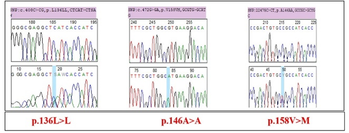
Most of the mutations in exon 4 of the COMT gene are synonymous mutations that do not induce an amino acid change and have no effect on coding, unlike the non-synonymous c.472G>A mutation that induces a substitution of valine for methionine (Table 1).
Fibroids and pregnancy | |||
Variants | dbSNP | Effect on coding | Amino acid assigned |
c.408C>G | rs4818 | Synonymous mutation | p.136L>L |
c.438C>T | rs8192488 | Synonymous mutation | p.146A>A |
c.472G>A | rs4680 | Missence mutation | p.158V>M |
Table 1: Nature and position of the mutations.
Pathogenicity of mutations
For exon 4 of the COMT gene c.472G>A missence variant is considered benign polymorphic in contrast to the synonymous variants which are considered probably harmful (Table 2).
| Variants | UMD Predictor | SIFT (Score) | Mutation Taster (score) | Polyphen2 (score) |
c.472G>A p.158V>M | Polymorph | Tolerated (0.12) | Missence (0.49) | Benign (0.005) |
Table 2: Pathogenicity of mutations.
Genetic characterisation
The amplified region of the COMT gene is 182pb. Three polymorphic sites were found, one of which is informative. The total number of mutations is 3. The number of haplotypes is 4. Concerning the diversity indices we note a high haplotypic diversity (hd) and a low nucleotide differences k is 1.2. The mutation rate R is 2.621. The mutations in exon 4 of the COMT gene are much more transitional in nature (Table 3).
| Parameters | Pregnancy patients with fibroma |
| Number of sequences | 6 |
| Number of sites | 182 |
| Monomorphic sites | 179 |
| Polymorphic sites | 3 |
| Singleton sites | 2 |
| Parcimony informative site | 1 |
| Total number of mutations (Eta) | 3 |
| Number of haplotypes | 4 |
| Haplotypic diversity (hd) | 0.8 ± 0.02963 |
| Nucleotide diversity (Pi) | 0.0065 ± 0.0000040 |
| Average number of nucleotide difference (k) | 1.2 |
| % transition | 73.22 |
| % transversion | 26.8 |
| Mutation rate (R) | 2.621 |
| A+T | 39.83 |
| C+G | 60.17 |
Table 3: Parameters of diversity.
Codon selection test
The codon selection test, which allows us to see if the mutations are heterogeneously distributed, shows that the difference between dN and dS is zero for all codons except codons 1 and 31, where it is negative and codon 67, where it is greater than zero (Méthionine) with p-values >0,05. No codon is under selection, so the mutations are heterogeneously distributed (Table 4).
| Codon | Triplet | dN-dS | p-value |
| 1 | CTC | -0,923 | 1 |
| 31 | GCC | -1 | 1 |
| 67 | ATG | 0.399 | 0.83 |
Table 4: Codon selection test of exon 4 of the COMT gene.
Discussion
In this report, the nuclear catechol-O-methyltransferase (COMT) gene involved in oestrogen metabolism was investigated in 6 cases of fibroids and pregnancy. The presence of mutations shows that the COMT gene is involved in the occurrence of uterine fibroids in Senegalese women. This is in agreement with the study by Cong et al. (2012) [16] which suggests that a large number of genes are associated with the risk of uterine leiomyoma, and the COMT gene is one of them. The c.472G>A (p.158V>M) polymorphism (rs 4680) (score 0.49) is found in 2 of the 3 patients with mutations. Thus the c.472G>A variant appears to be associated with the incidence of uterine fibroids in the Senegalese population. This differs from the study (PCR- pyrosequencing in genotyped women) by Denschlag et al. (2006) [17] which shows no statiscally significant difference in allele frequency and distribution of the COMT G158A genotype in Caucasian women. COMT converts 2-hydroxyestradiol to 2-methoxyestradiol. 2-Hydroxyestradiol has been shown to act as an anti-estrogen in many tissue systems [18,19]. On the other hand, 2-methoxyestradiol has been shown to have a mitogenic effect on different cell types [20,21,22]. Therefore, the high activity COMT genotype (Val/Val) would result in a rapid and efficient conversion of the anti-estrogenic metabolite (2-hydroxyestradiol) into its more mitogenic counterpart (2-methoxyestradiol), creating a high estrogenic cellular environment. Conversely, the low activity COMT genotype (Met/Met) would lead to the accumulation of 2-hydroxyestradiol, creating a low estrogenic environment [23]. WHO has demonstrated lower levels of 2-hydroxyestradiol in leiomyomas compared to adjacent normal myometrium and implicated it in tumour genesis. According to Rutherford et al. (2008) and Bilder et al. (2004) [24,25] the Val158Met polymorphism (rs4680) is considered the main cause of variation in COMT activity. This single nucleotide (SNP), involving a G to A transition at codon 158, results in an amino acid change and makes the enzyme prone to active site distortion and protein aggregation at physiological temperature. We found 3 variable sites which stipulates that mutations in exon 4 of the said gene are not the only genetic alterations in uterine fibroids. The 4 haplotypes identified show that there is a similarity between individuals which could be explained either by tumour size or by the same tumour location. The percentage of transition is 26.8% so the COMT gene would be more likely to have mutations in individuals whose fibroids do not coexist with pregnancy. We note a high haplotypic diversity (hd) and a low nucleotide diversity (Pi) which shows a signal of population growth and therefore an increase in mutations over time. This could be explained by the fact that epidemiological factors such as age, obesity, parity, gestational age, etc. are important in the growth mechanism of fibroids. According to the codon selection test no codon is under selection. This shows that there are more synonymous mutations than non-synonymous mutations.
Conclusion
The results of the molecular analyses obtained show an involvement of exon 4 of the COMT gene in uterine fibroids coexisting with pregnancy in Senegalese women. The Val158Met polymprphism (rs4680) is considered to be the main cause of COMT activity dysfunction.
References
- Racinet, C. (2009). Epidemiology, risk factors and symptomatology of uterine myomas. Médecine de la Reproduction, 11(2) : 118-122.
View at Publisher | View at Google Scholar - Whiteman, M. K., Kuklina, E., Jamieson, D. J, Hillis, S. D. et Marchbanks, P. A. (2010). Hospitalisation pour troubles gynécologiques aux États-Unis. American journal of obstetrics and gynecology, 202(6) :541-541.
View at Publisher | View at Google Scholar - Stewart, E. A, Cookson, C. L., Gandolfo, R. A. et Schulze‐Rath, R. (2017). Epidémiologie des fibromes utérins une revue systématique. BJOG : An International Journal of Obstetrics & Gynaecology, 124(10), 1501-1512.
View at Publisher | View at Google Scholar - Grudzien, M. M., Low, P. S., Manning, P. C., Arredondo, M., Belton Jr, R. J. et Nowak, R. A. (2010). Le médicament antifibrotique halofuginone inhibe la prolifération et la production de collagène par le léiomyome humain et les cellules musculaires lisses du myomètre. Fertilité et stérilité, 93(4) :1290-1298.
View at Publisher | View at Google Scholar - He, Y, Zeng, Q, Dong, S, Qin, L., Li, G., & Wang, P. (2013). Associations between uterine fibroids and lifestyles including diet, physical activity and stress : a case-control study in China. Asia Pacific journal of clinical nutrition, 22(1) :109-117.
View at Publisher | View at Google Scholar - Vlahos, N. F, Theodoridis, T. D. et Partsinevelos, G. A. (2017). Myomes et adénomyose : impact sur les résultats de la reproduction. BioMed Research International.
View at Publisher | View at Google Scholar - Bineta, K., Ciss, D., Ka, S., Mbaye, F., Dem, A., & Sembene, M. (2018). Fibromes utérins au Sénégal : polymorphisme du gène MED12 et corrélation avec des facteurs épidémiologiques. American Journal of cancer research and reviews, 2 :4.
View at Publisher | View at Google Scholar - Cramer SF, Patel A. The frequency of uterine leiomyomas. Am J Clin Pathol.1990. 94 :435-438.
View at Publisher | View at Google Scholar - Poovathi, M, & Ramalingam, R. (2016). Maternal and Fetal Outcome in Pregnancy with Fibroids : A Prospective Study. International Journal of scientific study, 3(11) :169-172.
View at Publisher | View at Google Scholar - Chauveaud-Lambling, A., & Fernandez, H. (2004). Fibrome et grossesse. EMC-Gynécologie-Obstétrique, 1(3) :127-135.
View at Publisher | View at Google Scholar - Zhai, J, Jiang, L, Wen, A., Jia, J, Zhu, L., & Fan, B. (2019). Analysis of the relationship between COMT polymorphisms and endometriosis susceptibility. Medicine, 98(1).
View at Publisher | View at Google Scholar - De Oliveira, E., de Aquino Castro, R., Gomes, M. T. V., da Silva, I. D. C. G., Baracat, E. C., de Lima, G. R., ... & Girão, M. J. B. C. (2008). The catechol-O-methyltransferase (COMT) gene polymorphism and prevalence of uterine fibroids. Maturitas, 60(3-4) :235-238.
View at Publisher | View at Google Scholar - Hall TA (1999). BioEdit : à user-friendly biological sequence alignment editor and analysis program for Windows 95/98/NT.Nucl. Acids. Symp. Ser, 41 :95-98 :14.
View at Publisher | View at Google Scholar - Librado, P. and Rozas, J. (2009). DnaSP v5 : à software for comprehensive analysis of DNA polymorphism data. Bioinformatics, 25(11) : 1451-1452.
View at Publisher | View at Google Scholar - Tamura, K., Stecher, G., Peterson, D, Filipski, A. and Kumar, S. (2013). MEGA6 : molecular evolutionary genetic analysis version 6.0. Molecular biology and evolution, 30(12) : 27252729.
View at Publisher | View at Google Scholar - Cong, R. J., Huang, Z. Y., Cong, L., Ye, Y., Wang, Z., Zha, L, Cao, L. P., Su, X. W., Yan, J., & Li, Y. B. (2012). Polymorphisms in HSD17B1 and HSD17B2 genes and risk of uterine leiomyoma in Chinese women. Archives of Gynecology and obstetrics, 286(3) :701-705.
View at Publisher | View at Google Scholar - Denschlag, D, Bentz, E. K., Hefler, L, Pietrowski, D, Zeillinger, R., Tempfer, C, & Tong, D. (2006). Genotype distribution of estrogen receptor alpha, catechol-O-methyltransferase, and cytochrome P450 17 gene polymorphisms in Caucasian women with uterine leiomyomas. Fertility and sterility, 85(2) :462-467.
View at Publisher | View at Google Scholar - Bradlow HL, Telang NT, Sepkovic DW, Osborne MP. (1996). 2-hydroxyestrone : the 'good' estrogen. J Endocrinol. 259-265.
View at Publisher | View at Google Scholar - Vandewalle B. (1989). Opposite effects of estrogen and catecholestrogen on hormone-sensitive breast cancer cell growth and differentiation. Mol Cell Endocrinol, 61 :239-246.
View at Publisher | View at Google Scholar - Liu Z-J, Zhu BT. (2004). Concentration-dependent mitogenic and antiproliferative actions of 2-methoxyestradiol in estrogen receptor positive human breast cancer cells. J Steroid Biochem Mol Biol, 88 :265-275.
View at Publisher | View at Google Scholar - Banerjee SN, Sengupta K, Banerjee S, Saxena NK, Banerjee SK. (2003). 2-Methoxyestradiol exhibits a biphasic effect on VEGF-A in tumor cells and upregulation is mediated through ER-alpha : a possible signaling pathway associated with the impact of 2-ME2 on proliferative cells. Neoplasia, 5 :417-426.
View at Publisher | View at Google Scholar - Lippert C, Seeger H, Mueck AO. (2003). The effect of endogenous estradiol metabolites on the proliferation of human breast cancer cells. Life Sci, 72 :877-883.
View at Publisher | View at Google Scholar - Othman EE, Al-Hendy A. (2008). Molecular genetics and racial disparities of uterine leiomyomas. Best Pract Res Clin Obstet Gynaecol. 22(4) :589-60 :25.
View at Publisher | View at Google Scholar - Rutherford, K., Le Trong, I., Stenkamp, R. E. and Parson, W. W. (2008). Crystal structures of human catechol-O-methyltransferase 108V and 108M. Journal of molecular biology, 380(1) : 120-130.
View at Publisher | View at Google Scholar - Bilder, R. M., Volavka, J., Lachman, H. M. and Grace, A. A. (2004). Catechol-O-methyltransferase polymorphism : relationships with the tonic-phase dopamine hypothesis and neuropsychiatric phenotypes. Neuropsychopharmacology, 29 (11) :1943-1961.
View at Publisher | View at Google Scholar
