

Review Article | DOI: https://doi.org/10.31579/2834-8532/010
Retrosplenial and Cingulate Cortex of The Rat Brain – Cyto- And Chemoarchitectonics
- Bon E.I *
- Maksimovich N.Ye
- Zimatkin S.M
- Misyuk V.A
Grodno State Medical University, 80, Gorkogo St., 230009, Grodno, Republic of Belarus.
*Corresponding Author: Lizaveta I. Bon, Candidate of biological science, assistant professor of pathophysiology department named D.A. Maslakov, Grodno State Medical University, Belarus.
Citation: Bon E.I, Maksimovich N.Ye, Zimatkin S.M, Misyuk V.A, (2023). Retrosplenial and Cingulate Cortex of The Rat Brain – Cyto- And Chemoarchitectonics. Clinical Genetic Research, 2(1); Doi:10.31579/2834-8532/010
Copyright: © 2023 Bon E.I, This is an open-access article distributed under the terms of the Creative Commons Attribution License, which permits unrestricted use, distribution, and reproduction in any medium, provided the original author and source are credited.
Received: 09 February 2023 | Accepted: 15 February 2023 | Published: 21 February 2023
Keywords: retrosplenial cortex, cingulate cortex, rat, cytoarchitectonics, chemoarchitectonics
Abstract
The retrosplenial cortex of rats is divided into granular and agranular regions. The difference in the granular region of the retrosplenial cortex is that layers II-III consist of large neurons. The cingulate cortex, which lies above the corpus callosum, on the medial wall of the hemisphere, is an intermediate formation between the paleocortex and neocortex; in its complex cellular composition, it is similar to the neocortical formation. It contains five layers of neurons: molecular, small cell, mediopyramidal, large cell and multiform. The data presented in the article can serve as a fundamental basis for further study of the parts of the rat brain in normal and pathological conditions with further extrapolation of the obtained data to humans.
Retrosplenial cortex
The retrosplenial cortex is located on the medial surface of the cerebral hemispheres of the rat brain [13] (Figure 1).
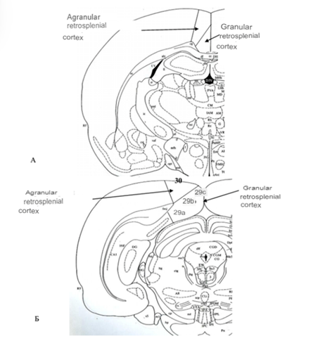
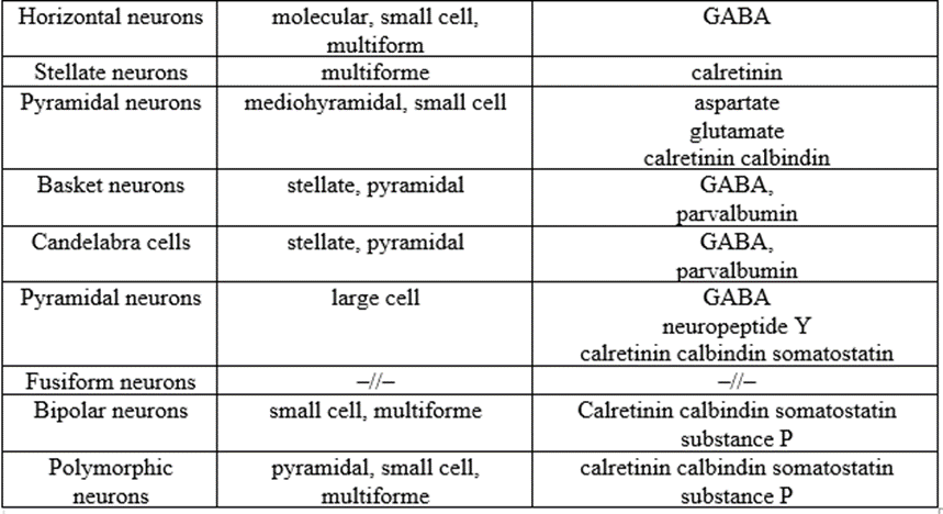
It includes seven layers: molecular, stellate, granular, reticular, mediopyramidal, large pyramidal, and multiform layer [13]. The fourth layer in rats is usually weakly expressed (table 1).

The retrosplenial cortex of rats is divided into granular and agranular regions [3,9]. The difference in the granular region of the retrosplenial cortex is that layers II-III consist of large neurons.
The granular retrosplenial cortex includes the following fields: 29a, 29b and 29c. The classification is based on the features of cytoarchitectonics, mainly of the mediopyramidal and large-pyramidal layers. In field 29a, layers II, III, and VII are thin, and the mediopyramidal layer is barely visible. Field 29b, on the contrary, has a well-defined layer II, formed by densely located bodies of stellate neurons [19], the granular and mediopyramidal layers are less pronounced, the neuron perikarya are more dispersed in them. Layer VI contains the bodies of large typical pyramidal neurons. The multiform layer, although thin, is clearly defined. In field 29c, the granular layer is most pronounced and, in general, the neurons of other layers are smaller in size compared to fields 29a and 29b. The predominant types of neurons are fusiform neurons and the transitional type of neurons - stellate pyramids. The apical dendrites of these neurons form bundles reaching the molecular layer [15, 19]. The stellate pyramidal neurons of old rats form noticeably fewer branches than in young animals [11]. The mediopyramidal and macropyramidal layers are well expressed, their organization is similar to the organization of the pyramidal layer of the frontal (frontal) isocortex cortex. The agranular area of the retrospenny cortex is represented by field 30. Microscopic examination of this area reveals a narrow granular layer. For this reason, field 30 cannot be called agranular in the full sense of the term. The retrosplenial cortex forms connections with the thalamic nuclei [12,17], the raphe nuclei, the nuclei of the medial geniculate body, and the motor cortex [2,3,5]. It is involved in the processes of visual memory [2,6,9,14] and regulation of behavior to predict and prevent situations that lead to painful sensations [3,5,6].
Сingulate cortex
The cingulate cortex, which lies above the corpus callosum, on the medial wall of the hemisphere, is an intermediate formation between the paleocortex and neocortex; in its complex cellular composition, it is similar to the neocortical formation [12]. It contains five layers of neurons: molecular, small cell, mediopyramidal, large cell and multiform [7] (Table 2).
Predominantly small cells are densely located in the upper layers [7]. In the second layer, two types of cells are distinguished: bipolar neurons and multipolar neurons with spherical branching of dendrites. The cingulate cortex receives afferent connections from the neocortex [17], mainly from the association zones of the posterior hemispheres and the frontal cortex [12], sending projections to the hippocampus through the entorhinal region [1,4,5].
The cingulate cortex is involved in the regulation of autonomic and endocrine functions, in the processes of emotional learning [20], vocalization, assessment of the motivational content and emotional valence of internal and external stimuli [5,6], and in interactions between mother and offspring.
The data presented in the article can serve as a fundamental basis for further study of the parts of the rat brain in normal and pathological conditions with further extrapolation of the obtained data to humans.
References
- Neurochemical development of the hippocampal region in the fetal rhesus monkey, III: calbindin-D28K, calretinin and parvalbumin with special mention of cajal-retzius cells and the retrosplenial cortex/ J Comp Neurol. 1996 Mar 18;366(4):674-99 /B Berger, C Alvarez.
View at Publisher | View at Google Scholar - Structural and functional brain network of human retrosplenial cortex/ Neurosci Lett.2018 May 1;674:24-29. /Panlong Li, Han Shan, Shengxiang Liang, Binbin Nie, Shaofeng Duan, Qi Huang, Tianhao Zhang, Xi Sun, Ting Feng, Lin Ma, Baoci Shan, Demin Li, Hua Liu .
View at Publisher | View at Google Scholar - Distinct Contribution of Granular and Agranular Subdivisions of the Retrosplenial Cortex to Remote Contextual Fear Memory Retrieval/ J Neurosci.2022 Feb 2;42(5):877-893. /Tsung-Chih Tsai, Ting-Hsuan Yu, Yu-Chieh Hung, Lok-Ieng Fong, Kuei-Sen Hsu
View at Publisher | View at Google Scholar - Cingulate cortex in the three limbic subsystems/ Handb Clin Neurol. 2019;166:39-51. /Brent A Vogt.
View at Publisher | View at Google Scholar - Atypical functional connectivity between the anterior cingulate cortex and other brain regions in a rat model of recurrent headache/ Mol Pain.2019 Jan-Dec;15:1744806919842483. /Zhihua Jia, Xiaoyan Chen, Wenjing Tang, Dengfa Zhao, Shengyuan Yu.
View at Publisher | View at Google Scholar - Contributions of the rodent cingulate-retrosplenial cortical axis to associative learning and memory: A proposed circuit for persistent memory maintenance/ Neurosci Biobehav Rev.2021 Nov;130:178-184. /Sydney Trask, Nicole C Ferrara, Aaron M Jasnow, Janine L Kwapis.
View at Publisher | View at Google Scholar - Cytoarchitecture of mouse and rat cingulate cortex with human homologies/ Brain Struct Funct.2014 Jan;219(1):185-92. /Brent A Vogt, George Paxinos.
View at Publisher | View at Google Scholar - Immunohistochemical studies of localization and co-localization of glutamate, aspartate and GABA in the anterior thalamic nuclei, retrosplenial granular cortex, thalamic reticular nucleus and mammillary nuclei of the rat/ J Chem Neuroanat.1996 Dec;12(2):77-84. /A Gonzalo-Ruiz, J M Sanz, A R Lieberman.
View at Publisher | View at Google Scholar - The separate and combined properties of the granular (area 29) and dysgranular (area 30) retrosplenial cortex/ Neurobiol Learn Mem.2021 Nov;185:107516. /John P Aggleton, Steliana Yanakieva, Frank Sengpiel, Andrew J Nelson.
View at Publisher | View at Google Scholar - Localization of amino acids, neuropeptides and cholinergic markers in neurons of the septum-diagonal band complex projecting to the retrosplenial granular cortex of the rat/ Brain Res Bull. 2000 Aug;52(6):499-510. /A Gonzalo-Ruiz, L Morte.
View at Publisher | View at Google Scholar - Functional connectivity with the retrosplenial cortex predicts cognitive aging in rats/ Proc Natl Acad Sci U S A.2016 Oct 25;113(43):12286-12291. /Jessica A Ash, Hanbing Lu, Lisa R Taxier, Jeffrey M Long, Yihong Yang, Elliot A Stein, Peter R Rapp
View at Publisher | View at Google Scholar - Cingulate gyrus: cortical architecture and connections/ Brain Nerve.2011 May;63(5):473-82. /Yasushi Kobayashi
View at Publisher | View at Google Scholar - Human retrosplenial cortex: where is it and is it involved in emotion?/ Trends Neurosci.2000 May;23(5):195-7. /B A Vogt, J R Absher, G Bush
View at Publisher | View at Google Scholar - NMDA receptor-dependent oscillatory signal outputs from the retrosplenial cortex triggered by a non-NMDA receptor-dependent signal input from the visual cortex/ Brain Res.2005 May 31;1045(1-2):12-21. /Hiroshi Yoshimura, Tokio Sugai, Makoto Honjo, Natsuki Segami, Norihiko Onoda
View at Publisher | View at Google Scholar - Parvalbumin positive dendrites co-localize with apical dendritic bundles in rat retrosplenial cortex/ Neuroreport.2002 May 7;13(6):757-61. /Noritaka Ichinohe, Kathleen S Rockland
View at Publisher | View at Google Scholar - NMDA receptor antagonist neurotoxicity and psychotomimetic activity/ Masui.2003 Jun;52(6):594-602. /Shinichi Nakao, Atsushi Nagata, Munehiro Masuzawa, Etsuko Miyamoto, Makiko Yamada, Nobuyasu Nishizawa, Koh Shingu
View at Publisher | View at Google Scholar - Organization of subcortical pathways for sensory projections to the limbic cortex. I. Subcortical projections to the medial limbic cortex in the rat/ J Comp Neurol.1987 Nov 8;265(2):175-88. /S M Thompson, R T Robertson
View at Publisher | View at Google Scholar - Comparison of substance P and enkephalin distribution in rat brain: an overview using radioimmunocytochemistry/ Neuroscience.1985 Mar;14(3):837-52. /S McLean, L R Skirboll, C B Pert
View at Publisher | View at Google Scholar - Dendritic bundling in layer I of granular retrosplenial cortex: intracellular labeling and selectivity of innervation/ J Comp Neurol. 1990 May 1;295(1):33-42. /J M Wyss, T Van Groen, K Sripanidkulchai
View at Publisher | View at Google Scholar - The cingulate cortex and limbic systems for emotion, action, and memory/ Brain Struct Funct.2019 Dec;224(9):3001-3018.
View at Publisher | View at Google Scholar
