

Research Article | DOI: https://doi.org/10.31579/2835-8325/111
Post-Natal Effects of Aqueous Extract of Soybeans (Glycine max) on The Cerebrum, Cerebellum and Hippocampus of Juvenile Wistar Rats
- Akporobo Ejeguo 1*
- Kate Oneshorona Sunday 1
- Maliki Samuel Ozavogu 1
- Fredrick Ohiomokhai Tobalu 1
- Emekwue Chukwudi Alex 2
- Fabian Ewaensetin Omosigho 3
1Department of Anatomy, School of Basic Medical Sciences, College of Medical Sciences, University of Benin, Edo State, Nigeria.
2University of Nigeria Teaching Hospital, Ituku-ozalla, Enugu.
3Department of Family Medicine, Federal Medical Center, Owo, Ondo State, Nigeria.
*Corresponding Author: Akporobo Ejeguo, Department of Anatomy, School of Basic Medical Sciences, College of Medical Sciences, University of Benin, Edo State, Nigeria.
Citation: Akporobo Ejeguo, Kate O. Sunday, Maliki S. Ozavogu, Fredrick O. Tobalu, Alex Emekwe, et al, (2024), Post-Natal Effects of Aqueous Extract of Soybeans (Glycine max) on The Cerebrum, Cerebellum and Hippocampus of Juvenile Wistar Rats, Clinical Research and Clinical Reports, 6(2); DOI: 10.31579/2835-8325/111
Copyright: © 2024, Akporobo Ejeguo. This is an open-access article distributed under the terms of the Creative Commons Attribution License, which permits unrestricted use, distribution, and reproduction in any medium, provided the original author and source are credited.
Received: 26 November 2024 | Accepted: 03 December 2024 | Published: 19 December 2024
Keywords: glycine max; postnatal effects; cerebrum; cerebellum; hippocamp
Abstract
Increased interest on the effects of maternal consumption (of food, herbs, drugs) during pregnancy on the offspring's development, cognition, motor functions, and overall well-being has been witnessed in recent years. Plants rich in antioxidants have been reported to enhance both cognitive and motor functions of the brain, as well as promote general health, and Glycine max is one of such antioxidant-rich plants. Accordingly, this study was aimed at investigating the postnatal effect of aqueous extract of Glycine max (GM) on the development of cerebrum, cerebellum and hippocampus of juvenile Wistar rats. Pregnant rats were randomly assigned into groups A (control), B (250mg/kg body weight [BW] of GM) and C (500 mg/kg BW of GM) and administered GM from GD 0 (gestational day 0) until term. Administration continued until the offspring were weaned. Weaned offspring were selected on postnatal day twenty-one and orally administered with GM until day forty-two (42). Neurobehavioral activities were evaluated and rats were sacrificed to harvest the cerebra, cerebella, and hippocampi for biochemical and histological assessments. Findings from the study showed that GM is a rich source of nutrients as was evident in the presence of phytochemicals such as saponins, and flavonoids. There was a significant increase (p<0.05) in post-natal weight, rearing, ambulation, spontaneous alternations, open arm and total arm entries, as well as CAT activity (250 mg/kg GM-treated rats) and a significant decrease (p<0.05) in immobility and closed arm entry in rats treated with GM when compared to control. There was also a significant decrease (p<0.05) in SOD and CAT activity in 500 mg/kg GM-treated rats compared to control. There was a decrease in MDA concentration in the GM-treated groups when compared to control. Histological findings revealed normal and intact histoarchitecture of the cerebrum, cerebellum and hippocampus of 250 mg/kg GM-treated rats when compared to control. However, there were mild histological alterations in the cerebellum and hippocampus of rats treated with 500 mg/kg GM. Conclusively, GM exhibits optimum neuroprotective benefits at a precisely determined dose, highlighting the importance of dosage specificity in harnessing its therapeutic potential for protecting the brain and promoting neuronal health.
Introduction
The use of herbal medicinesand phytonutrients (nutraceuticals) continues to increase as many people now opt for the use of these products for treatment of various health problems across the world (W.H.O, 2004) In the past decade there has been a notable increase in acceptance and public interest in natural therapies as approximately four billion people (80% of the world population) use medicinal products as alternative source of healthcare (Mukherjee, 2002; Bodeker and Ong, 2005; Bandaranayake, 2006).
Glycine max commonly known as soybean is a specie of Leguminosae family that originated from China, East Asia (Singh et al., 2020). It is commonly consumed around the world as a source of protein-rich foods and beverages. It is also used in major culinary arts in some parts of the world like Japan, China, Korea and many others (Li et al., 2018). The seed is majorly composed of oil and protein. The protein consists of a balanced proportion of all nine essential amino acids that is needed by the human body (Shah et al., 2022). G. max and its composites possess antioxidant, anti-obesity, anti-proliferative and anti-inflammatory properties (Huang et al., 2016). Consumption of G. max has been associated with numerous potential health benefits and decrement of various chronic illnesses like immune disorders, cardiovascular diseases, diabetes, obesity and certaintypes of cancer(Huang et al., 2016).
Methods and materials
Plant Material
Glycine max seeds were obtained from New Benin market, Benin City, Edo state, Nigeria. It was authenticated in the Herbarium unit of the Department of Plant Biology and Biotechnology, University of Benin, Benin City, and assigned an herbarium number UBH- G470. The G. max seeds were first dried in the oven at a temperature of 100℃ for about 4hours. The dried seeds were then pulverized to powder form. The powder weighing 1.5kg was extracted thrice in distilled water (41.25 L) at room temperature on shaker for 48 hours. The extract was filtered using a Buchner funnel and Whatman No.1 filter paper. The filtrate of aqueous extract obtained was quickly frozen at -40°Cand dried for 48 h using a freeze dryer. The resultingextract was reconstituted with distilled water to give desired concentrations used in this study.
Phytochemical Screening
The qualitative assessment of the chemical composition of G. max was done using standard methods. Compounds such as alkaloids, phenols, flavonoids, saponins and proteins were tested. Briefly, for alkaloids, 3 ml of aqueous extract was stirred with 3 ml of 1% HCl on a steam bath. Thereafter, Mayer’s and Wagner’s reagents were then added to the mixture, and the turbidity of the resulting precipitate was taken as evidence for the presence of alkaloids. For saponin, 0.2 g of plant extract was shaken with 4 ml of distilled water and then heated to boil on a water bath; the appearance of small bubbles showed the presence of saponin. For reducing sugar, 2 ml of distilled water and 0.2 g of plant extract were mixed together and thoroughly shaken in a test tube. One milliliter each of Fehling’s solutions A and B was added to the mixture, and a brick red precipitate at the bottom of the test tube confirmed the presence of reducing sugar. For phenol, 0.5 g of plant extract was added to 1 ml of 10?Cl3 solution; a deep bluish-green coloration showed the presence of phenol. For flavonoid, 5 ml of distilled water and 0.2 g of plant extract were mixed thoroughly. Thereafter, 1 mL of 1% AlCl3 solution was added and a light-yellow precipitate showed the presence of flavonoids. For terpenoid, the plant extract was mixed with 2 ml of chloroform in a test tube and 3 ml of conc. H2SO4 was added along the sides of the test tube, and a reddish brown indicate the presence of terpenoids. For protein, using Biuret test, the extract was added 5% NaOH solution and copper sulphate, deposition of blue coloration indicates the presence of protein.
Animals
Eighteen (18) juvenile Wistar rats weighing an average of 60g were bred and housed in the Animal House of the Department of Anatomy, University of Benin, Benin City. They were kept in clean cages and allowed access to feed (Grower's mash, a product of Premier Feed Mills Co Ltd) and water ad libitum throughout the experimental period.
Experimental Design
The eighteen (18) juvenile rats were randomized into three groups of six (6) rats each. Group A served as control and groups B and C served as treatment groups. Rats in groups B and C were treated with 250 mg/kg and 500 mg/kg body weight of G. max respectively, for twenty-one (21) postnatal days via an orogastric tube.
Evaluation of Neurobehavioral Activity
On the 22nd day, neurobehavioral activities were evaluated using novel object recognition (NOR), open field test (OFT), elevated plus maze (EPM), and Y-maze tests. NOR test was used to evaluate cognition in experimental rats. The test was performed as described by Pitsikas, (2015) with minor modifications. The test was performed in a wooden open box apparatus (80 × 60 × 40 cm). The objects to be discriminated was of two different colours (yellow and pink), made up of painted wood, around 10 cm in height and heavy (so it will not be displaced by the animals during test). The day before testing, rats were allowed to explore the apparatus for a 2 min session of habituation. Twenty-four hours later, the first 3 min sample trial test (T1), with two similar objects (termed as sample objects FO1 and FO2) presented at the corners of the box, commenced. Following T1, all the rats were placed back in their home cage and a delay of 60 min was given as inter-trial interval for T2. In the second 5-min choice trial (T2), one of the objects (FO2) presented in T1 was replaced by a new object (NO). To evaluate the effect on long term memory, animals were again exposed to the apparatus and time spent by rats in exploring FO1 and NO was recorded. Exploratory behavior was defined as when the rat approached an object with its nose within a 2 cm distance, either by sniffing or making physical contact with the object using its nose. To avoid the presence of olfactory trails, the apparatus and the objects were cleaned thoroughly with 70 % ethanol after each trial. The discrimination between familiar and novel objects during T2 was evaluated by comparing exploration times. To control for individual variability in exploration, a discrimination index (D) was calculated as: D = (Time spent exploring novel object - Time spent exploring familiar object) / (Total time spent exploring both objects) (Pitsikas, 2015).
Open field test was used to evaluate locomotion, exploration, and anxiety in the experimental rats (Millan, 2003). A wooden arena measuring 72 × 72 × 36 cm whose floor was divided into 16 equal squares, via 4 × 4 cm grid lines, represented the open field used in this study. A day before the test (day 21), rats were given free access to explore the box for 3 min. On day 22, experimental rats were tested for a period of 5 min each. The open-field maze was sanitized with 70% ethanol after each test to eliminate any olfactory cues that might affect the behavior of the subsequent rat. The activities evaluated include frequency of rearing episodes (the number of times in which the rat stood on its hind limbs with its forearm against the wall of the testing cage), self-grooming activity (the number of times paws or tongue is used to clean/scratch body), ambulation (the rate of movement), and immobility (complete absence of body movements) (Cauli and Morelli, 2002).
Sun and colleagues highlighted that while the EPM is often used to measure anxiety-like behavior in rodents, it can also be used to assess cognitive function, making it a valuable tool for investigating various aspects of behavior (Sun et al., 2010). The device is made up of two open arms (50 × 10 cm) crossed by two closed arms with walls that are 30 cm high. The device resembles a plus sign since the arms are joined by a central square (10 × 10 cm). The maze is 60 cm above ground level. Each rat was given five minutes to freely explore the elevated plus maze after being placed in the middle, facing an open arm. The parameter measured include; open arm entries, closed arm entries, open/closed arm quotient (ratio between open and closed arm entries), and total arm entries. Arm entry is defined as when the hind paws of the rats are completely within the arm. The testing apparatus was thoroughly disinfected with 70% ethanol between trials to prevent any potential bias caused by residual odors from previous animals (Casteller et al., 2006; Naqvi et al., 2012).
The Y-maze is used to assess short-term memory in rodents, leveraging their natural inclination to explore novel environments (Kraeuter et al., 2019). Animals were individually placed in the Y-maze, comprising three identical arms (33×11×12cm each) spaced at 120° (Dall'Igna et al., 2007). The test was used to assess spontaneous alternations. Each rat was placed in arm A and allowed to explore all three arms freely for 5 minutes. The number and sequence of arm visits were recorded, with alternations defined as consecutive visits to all three arms (Monte et al., 2013). The percentage of alternation was calculated as described previously (Dall'Igna et al., 2007). After each session, the apparatus was disinfected with 70% ethanol to remove any residual odors.
Determination of Brain Weight
After the neurobehavioral tests were completed, the rats were euthanized with low level anesthesia, followed by cervical dislocation. The rats’ brains were excised from their skulls, blotted clean of blood and weighed and recorded in the nearest decimal.
Assessment of Biochemical Parameters
The brains were homogenized in ice-cold 20 mM Tris–HCl buffer (pH 7.4), and the homogenates were then centrifuged at 10,000 g for 10 min at 4 °C (Montilla et al., 2005). The supernatants were collected and evaluated for superoxide dismutase (SOD) (Misra and Fridovich, 1972), catalase (CAT) (Cohen, 1983), gluthathione peroxidase (GPx) (Nyman, 1959) and malondialdehyde (MDA) (Buege and Aust, 1978).
Histological Evaluation
The cerebrum, cerebellum and hippocampus were fixed in 10% buffered formal saline for 72 hours. Thereafter, samples were processed through the paraffin wax embedding method as previously reported. The hematoxylin and eosin staining (Drury and Wallington, 1980) was also performed.
Statistical Analysis
The data were analyzed using the IBM statistical package for social science (SPSS) version20. Values were presented as mean ± standard error of mean (SEM), and one-way analysis of variance (ANOVA) followed by Tukey’s multiple comparisons post hoc test was used to determine statistical significance (p<0>
Results
Phytochemical Screening
The qualitative phytochemical analysis revealed that G. max contains alkaloids, saponins, reducing sugars, phenols, flavonoids, terpenoids and proteins. This indicates that G. max is rich in phytoconstituents.
S/N | Phytochemicals | Results |
1 | Alkaloids | + |
2 | Saponins | ++ |
3 | Reducing sugars | + |
4 | Tannins | - |
5 | Phenols | + |
6 | Flavonoids | + |
7 | Terpenoids | + |
8 | Proteins | ++ |
9 | Steroids | - |
Table 1: Qualitative phytochemical screening of aqueous extract of Glycine max
Present (+), highly present (++), absent (-)
Effect of Treatment on Body and Brain Weights
Figures 1 and 2 respectively show the body and organ weights of animals across the experimental groups. There was a significant increase (p<0>G. max when compared to control. Also, there was an insignificant increase (p>0.05) in the brain weights of rats treated with 250 mg/kg and 500 mg/kg of G. max extract when compared to control.
Effect of Treatment on Neurobehavioral Activity
Figure 3 shows the effect of treatment on discrimination index across experimental groups. There was an increase in the discrimination index of rats treated with 250 mg/kg and 500 mg/kg B.W of G. max extract when compared to control, however, this increase was not significant.
Figure 4 shows the effect of treatment open field parameters across experimental groups. For rearing frequency, there was a significant increase (p<0>G. max extract when compared to control. For grooming, there was a decrease in rats treated with 250 mg/kg and 500 mg/kg of the extract, however, this decrease was not significant. For ambulation, there was a significant increase (p<0>G. max extract when compared to control. For immobility, there was a significant decrease (p<0>G. max extract when compared to control.
Figure 5 shows the effect of treatment on EPM parameters across experimental groups. For open arm entries, there a significant increase (p<0>G. max extract when compared to control. For closed arm entries, there was a significant decrease (p<0>G. max extract when compared to control. there was an increase in the open/closed arm quotient of rats treated with G. max extract, however, this increase was not significant. There was a significant increase (p<0>G. max extract when compared to control.
Figure 6 shows the effect of treatment on spontaneous alternation across experimental groups. There was a significant increase (p<0>G. max extract when compared to control.
Effect of Treatment on Antioxidant Enzymes Activity and Lipid Peroxidation
Figure 7 shows the effect of treatment on antioxidant enzymes (SOD and CAT) activity across experimental groups. For SOD, there was a significant decrease (p<0>G. max extract when compared to control. There was also an increase in SOD activity in rats treated with 250 mg/kg B.W of G. max extract, however, this increase was not significant. There was a significant increase (p<0>G. max extract, however, there was a significant decrease (p<0>G. max extract when compared to control.
Figure 8 shows the effect of treatment on MDA concentration across experiment groups. There was a significant decrease in MDA concentration in rats treated with 250 mg/kg and 500 mg/kg B.W of G. max extract when compared to control.
Effects of Treatment on Histology of the Cerebrum, Cerebellum and Hippocampus
Figure 9 shows histological sections of the cerebral cortex stained with H&E, revealing normal morphology in both the control and G. max-treated groups, with a characteristic arrangement of cells, indicating healthy cerebral cortex.
Figure 10 shows histological sections of the cerebellum stained with H&E. Both the control group and the groups treated with G. max exhibited normal morphology, with distinct visualization of the molecular, Purkinje, and granular layers, indicating healthy cerebellum.
Figure 11 displays histological sections of the hippocampal CA1 region stained with H&E, revealing normal morphological features in both the control group and the G. max-treated groups, with a characteristic arrangement of cells, indicative of a healthy hippocampus, although there was a reduced population of pyramidal cells in stratum pyramidale.
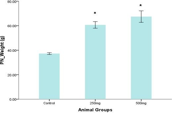
Figure 1: Body weight of rats across experimental groups. * p<0>
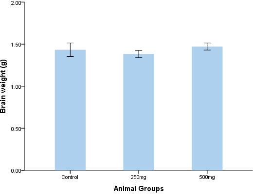
Figure 2: Brain weight of rats across experimental groups.
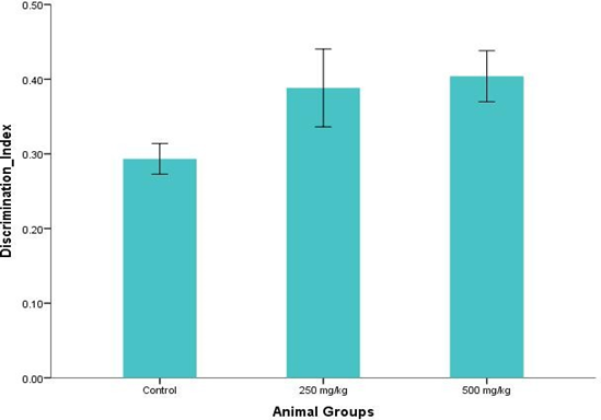
Figure 3: Discrimination index across experimental groups.
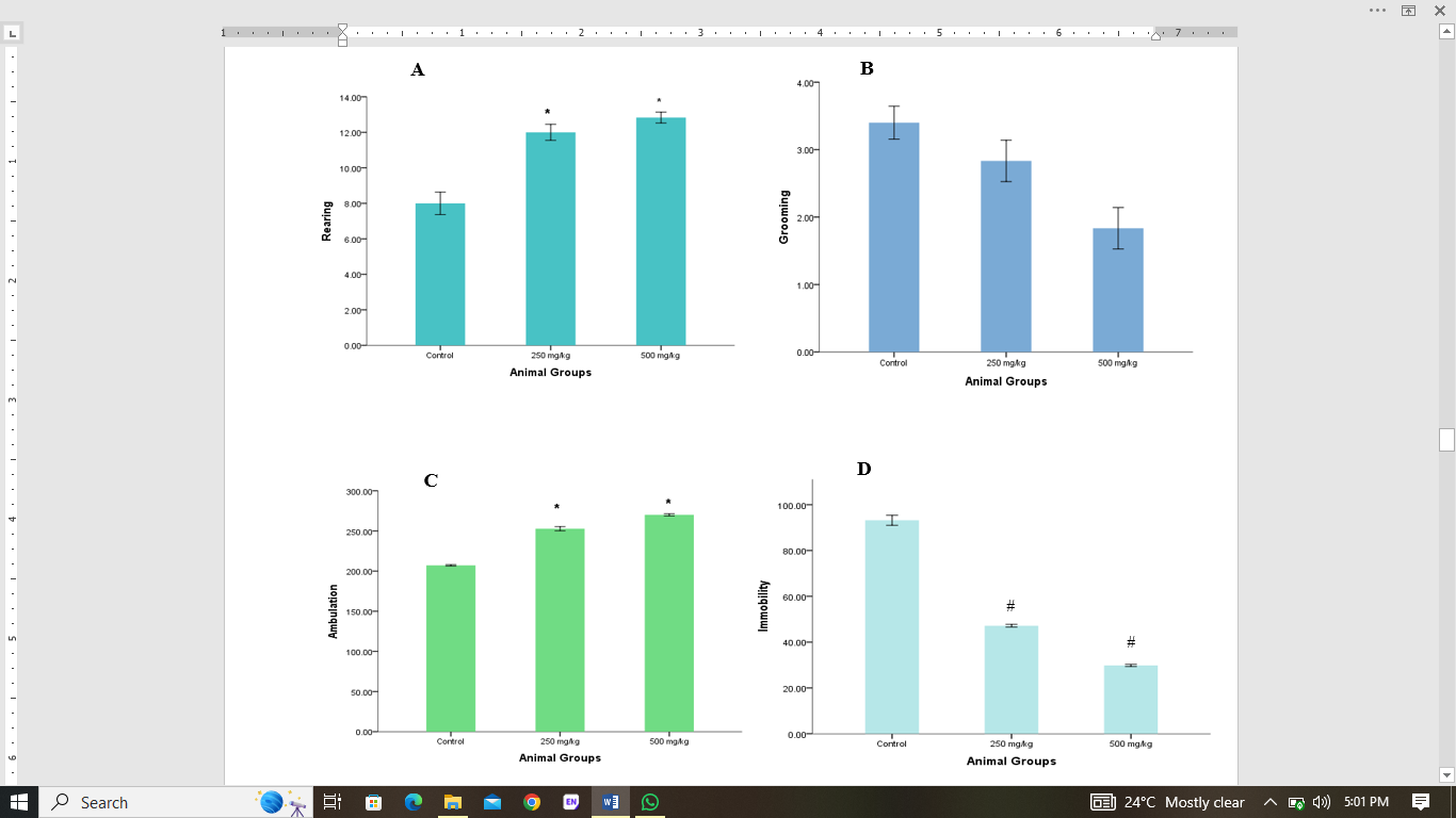
* p<0>
# p<0>
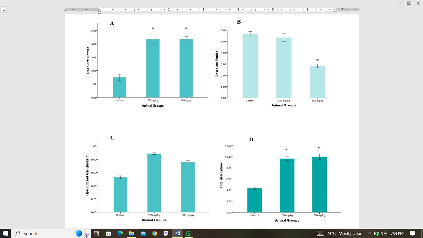
Figure 5: The elevated plus maze activities include: A open arm entries, B closed arm entries, C open/closed arm quotient, and D total arm entries.
Values are given as mean ± SEM.
* p<0>
# p<0>
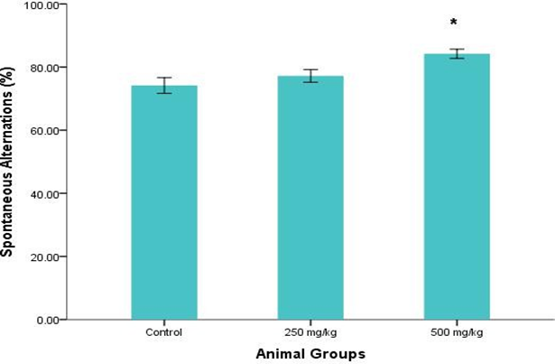
Figure 6: Spontaneous alternation across experimental groups.
Values are given as mean ± SEM.
* p<0>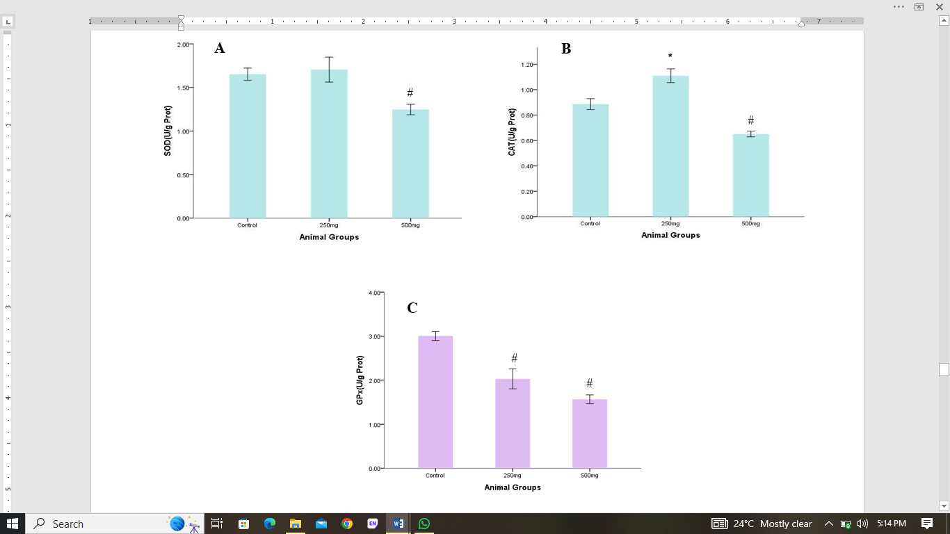
Figure 7: The antioxidant enzymes activities include: A superoxide dismutase (SOD), B catalase (CAT). Values are given as mean ± SEM. * p<0>p<0>
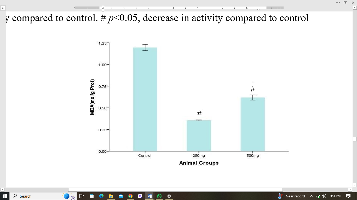
Figure 8: MDA concentration across experimental groups. Values are given as mean ± SEM. # p<0>
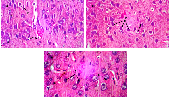
Figure 9: Photomicrograph of the cerebral cortex in control and G. max-treated groups. A control, B 250 mg/kg G. max-treated group and C 500 mg/kg G. max-treated group. Both control and treatment groups show normal histological features: pyramidal cells (p) and granular cells (g). H&E 400×
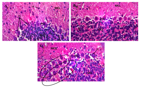
Figure 10: Photomicrograph of the cerebellum in control and G. max-treated groups. Both A control, and B 250 mg/kg G. max-treated group showed normal morphology with distinct identification of the molecular layer (MCL), Purkinje cell layer (PCL), and granular layer (GCL). C 500 mg/kg G. max-treated group showed disruption of the PCL. Stellate cells (sc) and Purkinje cells (pc) were clearly visible. H&E 400×
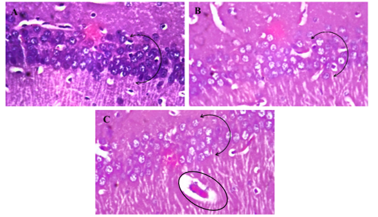
Figure 11: Photomicrograph of the hippocampus (CA1) in control and G. max-treated groups. A control, with populated, intenselystained basophilic large round pyramidal cells. G. max treated groups, B (250 mg/kg G. max) and C (500 mg/kg G. max), with less populated, less intensely stained pyramidal cells, and vacuolations. H&E 400×
Discussion
Glycine max is an exceptionally rich and affordable source of protein for both human consumption and animal feed (Riaz, 2006). Notably, it is one of the rare plant-based sources that provide all the essential amino acids necessary for the body to build and repair tissues (Michelfelder, 2009). It is described as a treasure trove of bioactive compounds. Result from the preliminary qualitative phytochemical screening showed the presence ofalkaloids, saponins, reducing sugar, phenols, flavonoids, terpenoids, and proteins. This corresponds with findings from previous studies (Lisanti and Arwin, 2019; Vishnupriya and Kowsalya, 2022). Studies have shown that these compounds possess several potential health benefits, such as antioxidant, anti-diabetic, anti-obesity, anti-proliferative, and anti-inflammatory activities, which can contribute to the prevention and management of various health conditions (Huang et al., 2016). Phenolic acids and flavonoids in G. max have been reported to be potent antioxidants (Alghamdi et al., 2018) that shield the body from oxidative stress and related diseases (Valko et al., 2006). Also, G. max contains saponins, which have immunomodulatory effects (Tezuka and Imai, 2024), terpenoids and alkaloids, which exhibit antimicrobial activity (Huang et al., 2022). The proteins in G. max are also vital for tissue growth and repair (Hughes et al., 2011), highlighting the importance of this plant in maintaining overall health and well-being.
Research has demonstrated that a increase in body weight can serve as a vital indicator of overall health and well-being in rodents and experimental animals, providing valuable insights into their general health status (Al-Shabanah et al., 2002). Findings from this study showed a significant increase in body weights in rats in G. max-treated groups compared to control. This increase in body weights could be attributed to high-quality proteins, essential amino acids, and unsaturated fats contents of G. max (Dorward et al., 2007). Messina and colleagues reported a significant increase inbody weight (Messina and Gleason, 2016) which is consistent with our findings. The relative brain weight of rats in the G. max-treated groups showed no significant difference compared to the control group, and this is consistent with the findings of Cristane et al. (2013) who observed no significant change in relative brain weight in rats fed with G. max extract. This may be due to the fact that G. max has a minimal effect on the brain's lipid and cholesterol levels (Kim et al., 2018).
The Novel Object Recognition (NOR) test is a behavioral test used to assess cognitive function in animals, particularly rodents (Lueptow, 2017). It evaluates an animal's ability to recognize and distinguish between novel and familiar objects, providing insights into their working memory, recognition memory, cognitive flexibility. Notably, the cerebrum is crucial for this process, playing a central role in facilitating these cognitive functions (Abhang et al., 2016; Stewart, 2023). A decrease in discrimination index implies impaired memory while an increase in discrimination index suggest an improved memory and cognitive function (Hem et al., 2016). Result from this study showed an increase in discrimination index in rats treated with G. max extract when compared to control, however, this increase was not significant. Studies have shown that G. max isoflavones may have a positive impact on brain function, improving overall cognitive abilities and memory (Cui et al., 2020).
According to Kraeuter and colleagues, the OFT is a tool used to evaluate anxiety, general locomotor activity, and exploratory behavior in rats, providing insights into their emotional state and willingness to explore new environments (Kraeuter et al., 2019). In this study, rearing, grooming, ambulation and immobility were assessed. A decrease in rearing behavior in rodent models is a sign of increased stress and anxiety levels (Borta and Schwarting, 2005). On the other hand, ambulation, typically measured by the number of lines crossed by the animal, is a direct indicator of motor activity, with higher ambulation rates indicating enhanced movement and motor function (Ewalds-Kvist et al., 1999). An increase in grooming behavior is a telltale sign of stress and anxiety in animals (Kalueff and Tuohimaa, 2005). Conversely, immobility or freezing, characterized by a lack of movement, is a direct indicator of reduced motor activity and impaired motor function (Roelofs, 2017). Findings from the present study showed a significant increase in the rearing ability and ambulation in rats treated with G. max extract when compared to control indicative of the fact that G. max decreased the stress and anxiety levels in the rats. Rats treated with G. max extract showed a significant decrease in immobility time, compared to the control group elucidating an improved activity of the cerebellum. Additionally, there was a decrease in grooming activity in rats treated with G. max extract, however, this decrease was not statistically significant when compared to the control group. These findings suggest that G. max may have neuro-stimulatory effects and the ability to enhance motor function, potentially leading to improved brain health and mobility.
The elevated plus maze is a well-established behavioral test, widely used to assess anxiety levels in rodents (Walf and Frye, 2007). In this study, open arm entries, closed arm entries, time spent in open arms, and open/closed arm quotient were used to evaluate anxiety-like behaviours. Results from this study showed that there was a significant increase in the open arm and total arm entries in rats treated with G. max extract when compared to control. However, there was a significant decrease in closed arm entries in rats treated with G. max extract when compared to control. This highlights the potential antidepressant properties of G. max isoflavones, which is consistent with previous studies (Messina and Gleason, 2016).
The Y-maze test evaluates short-term spatial memory, a crucial aspect of navigation and episodic memory formation (Kim et al., 2008). The hippocampus plays a key role in spatial learning and memory. Spontaneous alternation, a measure of spatial working memory, is assessed by allowing rodents to explore the maze, driven by their innate curiosity to visit new arms (Kraeuter et al., 2019). Findings from this study showed a significant increase in the spontaneous alternation in rats treated with a higher dose of G. max extract when compared to control. This increase in alternation indicates improved learning ability of rats that has been exposed to G. max prenatally and may be attributed to the neuro-stimulatory effects of G. max (Sharma et al., 2020), and the beneficial effects of G. max isoflavones on cognition (Cui et al., 2020).
Antioxidants mitigate the damaging effects of oxidative stress by protecting cells from free radical damage (Engwa et al., 2022). Results from this study showed an increase in SOD activity in rats treated with 250mg/kg of G. max extract, however, this increase was not significant. Conversely, there was a significant decrease in SOD activity in rats treated with 500 mg/kg of the extract when compared to control. Similarly, there was a significant increase in CAT activity in rats treated with 250 mg/kg G. max extract, while there was a significant decrease in CAT activity in rats treated with 500 mg/kg G. max extract when compared to control. The decreased observed in group C might be due to a reversal effect as a result of possible overdose of the plant. This indicates that G. max extract upregulated antioxidant enzymes in a dose-dependent manner. Our findings corroborate the findings of Ekor and colleagues who reported an increase in SOD and CAT activities in rats treated with only 250 mg/kg G. max extract in comparison to the control group (Ekor et al., 2010). Oxidative stress and lipid peroxidation are reflected in MDA levels, which are produced through the breakdown of polyunsaturated fatty acids in cell membranes (Aoyama, 2021). Consequently, a reduction in MDA levels indicates a reduction in oxidative stress and an overall enhancement of cellular well-being. Findings from this study showed a significant decrease in MDA concentration in rats treated with G. max extract when compared to control. This is consistent with the previous study by Risfianty and co-workers, who reported a decrease in MDA levels in rats fed with dietary fibers of G. max (Risfianty et al., 2016).
The histological appearance of the cerebrum, cerebellum, and hippocampus is crucial in understanding the structure and function of a healthy brain. These three regions are essential for controlling various physiological processes, including movement, cognition, emotion, and memory (Maldonado and Alsayouri, 2019). Results from the hematoxylin and eosin staining of the cerebrum of rats treated with G. max extract maintained histoarchitectural integrity, displaying normal pyramidal and granular cells when compared to control. Our findings align with the study by Abbasabadi and colleague, which found that a soy diet had no significant impact on the number of neurons in the molecular, pyramidal, and multiform layers of the cerebral cortex (Abbasabadi and Tadjalli, 2016). The cerebellum of rats treated with 250 mg/kg of the extract showed normal histology with distinct molecular, Purkinje and granular cell layers. However, rats treated with 500 mg/kg of the extract showed a disruption of the Purkinje cell layer (PCL) when compared to the control. In the 250 mg/kg G. max-treated rats, mild changes were observed in the CA1 region of the hippocampus, including pyramidal neuron differentiation and spine intensity in basal dendrites. The pyramidal cells were relatively normal when compared to control; however, in the rats administered 500 mg/kg of the extract, the CA1 region exhibited fewer pyramidal cells with reduced staining intensity, and additionally, displayed vacuolar changes, indicating a deleterious effect on neuronal population and structure. These histological alterations observed in the PCL of the cerebellum CA1 region of the hippocampus could be attributed to the inability of the prenatal rats to effectively metabolize the increased dosage of the extract, thereby resulting in mild toxicity. Conversely, a previous study found that adult rats administered 1000 mg/kg of G. max extract showed no abnormalities in the histology of the cerebellum and cerebrum layers, with both regions appearing normal and intact (Orheruata and Enogieru, 2024).
In conclusion, G. max extract demonstrated dose-dependent neuro-stimulatory effects on the cerebrum, cerebellum, and hippocampus of juvenile Wistar rats. The extract's antioxidant and anti-inflammatory properties have been shown to protect the brain from damage, promoting neuronal survival and maintaining cellular homeostasis.
Conflict of Interest: None declared
References
- Abbasabadi, B. M., and Tadjalli, M. (2016). Effect of soy milk on circulating 17-β estradiol, number of neurons in cerebral cortex and hippocampus and determination of their ratio in neonatal ovariectomized rats. Veterinary Research Forum, 7(4):347.
View at Publisher | View at Google Scholar - Abhang, P. A., Gawali, B. W., and Mehrotra, S. C. (2016). Introduction to Emotion, Electroencephalography, and Speech Processing. Introduction to EEG- and Speech-Based Emotion Recognition, 1-17.
View at Publisher | View at Google Scholar - Al-Shabanah, A. O., El-Hadiyah, T. M., and Al-Majed, A. A. (2002). Effect of prolonged vigabatrin treatment on hematological and biochemical parameters in plasma, liver and kidney of Swiss albino mice. Scientia Pharmaceutica, 70(2):135-145.
View at Publisher | View at Google Scholar - Alghamdi, S. S., Khan, M. A., El-Harty, E. H., Ammar, M. H., Farooq, M., and Migdadi, H. M. (2018). Comparative phytochemical profiling of different soybean (Glycine max (L.) Merr) genotypes using GC–MS. Saudi Journal of Biological Sciences, 25(1):15-21.
View at Publisher | View at Google Scholar - Aoyama, K. (2021). Glutathione in the Brain. International Journal of Molecular Sciences, 22(9):5010.
View at Publisher | View at Google Scholar - Bandaranayake, W. M. (2006). Quality control, screening, toxicity, and regulation of herbal drugs. Modern phytomedicine: turning medicinal plants into drugs, 25-57.
View at Publisher | View at Google Scholar - Bodeker, G., and Ong, C.-K. (2005). WHO global atlas of traditional, complementary and alternative medicine (Vol. 1): World Health Organization.
View at Publisher | View at Google Scholar - Borta, A., and Schwarting, R. K. (2005). Inhibitory avoidance, pain reactivity, and plus-maze behavior in Wistar rats with high versus low rearing activity. Physiology and Behavior, 84(3):387-396.
View at Publisher | View at Google Scholar - Buege, J. A., and Aust, S. D. (1978). Microsomal lipid peroxidation. Methods in Enzymology, 52:302-310.
View at Publisher | View at Google Scholar - Casteller, G., Fraile, M., Laconi, M., Landa, A. I., Cabrera, R., and Gargiulo, P. A. (2006). Desinhibitory effect of allopregnanolone within the medial prefrontal cortex of male rats on the plus maze test. International Journal of Neurodegenerative Disorders, 2:120-126.
View at Publisher | View at Google Scholar - Cauli, O., and Morelli, M. (2002). Subchronic caffeine administration sensitizes rats to the motor-activating effects of dopamine D 1 and D 2 receptor agonists. Psychopharmacology, 162:246-254.
View at Publisher | View at Google Scholar - Cohen, G. (1983). Catalase, glutathione peroxidase, superoxide dismutase, and cytochrome P-450. In Handbook of Neurochemistry: Volume 4 Enzymes in the Nervous System, 315-330.
View at Publisher | View at Google Scholar - Cui, C., Birru, R. L., Snitz, B. E., Ihara, M., Kakuta, C., Lopresti, B. J., Aizenstein, H. J., Lopez, O. L., Mathis, C. A., and Miyamoto, Y. (2020). Effects of soy isoflavones on cognitive function: a systematic review and meta-analysis of randomized controlled trials. Nutrition Reviews, 78(2):134-144.
View at Publisher | View at Google Scholar - Dall'Igna, O. P., Fett, P., Gomes, M. W., Souza, D. O., Cunha, R. A., and Lara, D. R. (2007). Caffeine and adenosine A2a receptor antagonists prevent β-amyloid (25–35)-induced cognitive deficits in mice. Experimental Neurology, 203(1):241-245.
View at Publisher | View at Google Scholar - Dorward, D. W., Sharma, A., and Preuss, H. G. (2007). A soy-based dietary supplement attenuates indices of reproductive toxicity in mature male rats. Food and Chemical Toxicology, 45(11):2155-2163.
View at Publisher | View at Google Scholar - Drury, R., and Wallington, E. (1980). Carleton’s histological technique 5th ed New York: Churchill Livingstone.
View at Publisher | View at Google Scholar - Ekor, M., Emerole, G. O., and Farombi, E. O. (2010). Phenolic extract of soybean (Glycine max) attenuates cisplatin-induced nephrotoxicity in rats. Food and Chemical Toxicology, 48(4):1005-1012.
View at Publisher | View at Google Scholar - Engwa, G. A., Nweke, F. N., and Nkeh-Chungag, B. N. (2022). Free radicals, oxidative stress-related diseases and antioxidant supplementation. Alternative Therapies in Health and Medicine, 28(1).
View at Publisher | View at Google Scholar - Ewalds-Kvist, S. B. M., Selander, R.-K., and Kvist, M. O. (1999). Open-field Δ-ambulation as a selection tool for bidirectional responses in maze learning in Mus musculus L. Psychobiology, 27:123-132.
View at Publisher | View at Google Scholar - Hem, S., Albite, R., Loresi, M., Rasmussen, J., Ajler, P., Yampolsky, C., Chabot, J. D., Gerszten, P. C., and Goldschmidt, E. (2016). Pathological changes of the hippocampus and cognitive dysfunction following frontal lobe surgery in a rat model. Acta Neurochirurgica, 158:2163-2171.
View at Publisher | View at Google Scholar - Huang, S.-S., Su, S.-Y., Chang, J.-S., Lin, H.-J., Wu, W.-T., Deng, J.-S., and Huang, G.-J. (2016). Antioxidants, anti-inflammatory, and antidiabetic effects of the aqueous extracts from Glycine species and its bioactive compounds. Botanical studies, 57:1-11.
View at Publisher | View at Google Scholar - Huang, W., Wang, Y., Tian, W., Cui, X., Tu, P., Li, J., Shi, S., and Liu, X. (2022). Biosynthesis investigations of terpenoid, alkaloid, and flavonoid antimicrobial agents derived from medicinal plants. Antibiotics, 11(10):1380.
View at Publisher | View at Google Scholar - Hughes, G. J., Ryan, D. J., Mukherjea, R., and Schasteen, C. S. (2011). Protein digestibility-corrected amino acid scores (PDCAAS) for soy protein isolates and concentrate: Criteria for evaluation. Journal of Agricultural and Food Chemistry, 59(23):12707-12712.
View at Publisher | View at Google Scholar - Kalueff, A. V., and Tuohimaa, P. (2005). The role of hair in swimming of laboratory mice: implications for behavioural studies in animals with abnormal hair. Laboratory Animals, 39(4):370-376.
View at Publisher | View at Google Scholar - Kim, Y., Seo, C.-W., Khan, A. L., Mun, B.-G., Shahzad, R., Ko, J.-W., Yun, B.-W., Park, S.-K., and Lee, I.-J. (2018). Exo-ethylene application mitigates waterlogging stress in soybean (Glycine max L.). BMC Plant Biology, 18:1-16.
View at Publisher | View at Google Scholar - Kim, Y. T., Yi, Y.-J., Kim, M.-Y., Bu, Y., Jin, Z. H., Choi, H., Doré, S., and Kim, H. (2008). Neuroprotection and enhancement of spatial memory by herbal mixture HT008-1 in rat global brain ischemia model. The American Journal of Chinese Medicine, 36(02):287-299.
View at Publisher | View at Google Scholar - Kraeuter, A.-K., Guest, P. C., and Sarnyai, Z. (2019). The Y-maze for assessment of spatial working and reference memory in mice. Pre-clinical models: Techniques and Protocols, 105-111.
View at Publisher | View at Google Scholar - Li, Y., Chang, R., and Qiu, L. (2018). Utilizing the Diversity of Wild Soybeans in China for Accelerating Soybean Breeding in the Genome Era. In Applied Mathematics and Omics to Assess Crop Genetic Resources for Climate Change Adaptive Traits 71-80, CRC Press.
View at Publisher | View at Google Scholar - Lisanti, E., and Arwin, A. (2019). Phytochemical screening and proximate analysis of soybeans (Glycine max) variety Gamasugen 1 and Gamasugen 2 derived from gamma rays irradiation. Paper presented at the Journal of Physics: Conference Series.
View at Publisher | View at Google Scholar - Lueptow, L. M. (2017). Novel object recognition test for the investigation of learning and memory in mice. Journal of Visualized Experiments, 126:55718.
View at Publisher | View at Google Scholar - Maldonado, K. A., and Alsayouri, K. (2019). Physiology, brain. https://www.ncbi.nlm.nih.gov/books/NBK551718/.
View at Publisher | View at Google Scholar - Messina, M., and Gleason, C. (2016). Evaluation of the potential antidepressant effects of soybean isoflavones. Menopause, 23(12), 1348-1360.
View at Publisher | View at Google Scholar - Michelfelder, A. J. (2009). Soy: a complete source of protein. American family physician. 79(1):43-47.
View at Publisher | View at Google Scholar - Millan, M. J. (2003). The neurobiology and control of anxious states. Progress in Neurobiology, 70(2):83-244.
View at Publisher | View at Google Scholar - Misra, H. P., and Fridovich, I. (1972). The role of superoxide anion in the autoxidation of epinephrine and a simple assay for superoxide dismutase. Journal of Biological Chemistry, 247(10):3170-3175.
View at Publisher | View at Google Scholar - Monte, A. S., de Souza, G. C., McIntyre, R. S., Soczynska, J. K., dos Santos, J. V., Cordeiro, R. C., Ribeiro, B. M. M., de Lucena, D. F., Vasconcelos, S. M. M., and de Sousa, F. C. F. (2013). Prevention and reversal of ketamine-induced schizophrenia related behavior by minocycline in mice: possible involvement of antioxidant and nitrergic pathways. Journal of Psychopharmacology, 27(11):1032-1043.
View at Publisher | View at Google Scholar - Montilla, P., Barcos, M., Munoz, M. C., Bujalance, I., Munoz-Castaneda, Juan, R., and Tunez, I. (2005). Red wine prevents brain oxidative stress and nephropathy in streptozotocin-induced diabetic rats. BMB Reports, 38(5):539-544.
View at Publisher | View at Google Scholar - Mukherjee, P. K. (2002). Quality control of herbal drugs: an approach to evaluation of botanicals. New Delhi, India: Business Horizons Publishers.
View at Publisher | View at Google Scholar - Naqvi, F., Haider, S., Batool, Z., Perveen, T., and Haleem, D. (2012). Sub-chronic exposure to noise affects locomotor activity and produces anxiogenic and depressive like behavior in rats. Pharmacological Reports, 64(1):64-69.
View at Publisher | View at Google Scholar - Nyman, N. (1959). Determination of glutathione peroxidase in tissue. Analytical Biochemistry, 28:481.
View at Publisher | View at Google Scholar - Orheruata, A. R., and Enogieru, A. B. (2024). Is aqueous extract of soybeans neurotoxic? Preliminary evidence from histological evaluation of the cerebellum and cerebrum of Wistar rats. Journal of Experimental and Clinical Anatomy, 21(1):8-13.
View at Publisher | View at Google Scholar - Pitsikas, N. (2015). The role of nitric oxide in the object recognition memory. Behavioural Brain Research, 285:200-207.
View at Publisher | View at Google Scholar - Riaz, M. N. (2006). Processing of soybeans into ingredients. Soy applications in food, 40-62.
View at Publisher | View at Google Scholar - Risfianty, D., Mahdi, C., Wuragil, D., and Am, A. A. (2016). Dietary fiber of Glycine max (L.) Merr. compound as antihypercholesterolemia to MDA levels and hepar histopatology on hyper cholesterolemic rats. International Journal of Pharmaceutical and Clinical Research, 8(7):695-698.
View at Publisher | View at Google Scholar - Roelofs, K. (2017). Freeze for action: neurobiological mechanisms in animal and human freezing. Philosophical Transactions of the Royal Society B: Biological Sciences, 372(1718):20160206.
View at Publisher | View at Google Scholar - Shah, F.-u.-H., Sharif, M. K., Ahmad, Z., Amjad, A., Javed, M. S., Suleman, R., Sattar, D.-e.-S., Amir, M., and Anwar, M. J. (2022). Nutritional characterization of the extrusion-processed micronutrient-fortified corn snacks enriched with protein and dietary fiber. Frontiers in Nutrition, 9:1062616.
View at Publisher | View at Google Scholar - Sharma, A., Weber, D., Raupbach, J., Dakal, T. C., Fließbach, K., Ramirez, A., Grune, T., and Wüllner, U. (2020). Advanced glycation end products and protein carbonyl levels in plasma reveal sex-specific differences in Parkinson's and Alzheimer's disease. Redox Biology, 34:101546.
View at Publisher | View at Google Scholar - Singh, G., Dukariya, G., and Kumar, A. (2020). Distribution, importance and diseases of soybean and common bean: A review. Biotechnology Journal International, 24(6):86-98.
View at Publisher | View at Google Scholar - Stewart, S. (2023). Lobes of the brain. Https://www.kenhub.com/en/library/anatomy/lobes-of-the-brain.
View at Publisher | View at Google Scholar - Sun, T., He, W., Hu, G., and Li, M. (2010). Anxiolytic-like property of risperidone and olanzapine as examined in multiple measures of fear in rats. Pharmacology Biochemistry Behavior, 95(3):298-307.
View at Publisher | View at Google Scholar - Tezuka, H., and Imai, S. (2024). Fine-tuning of mononuclear phagocytes for improved inflammatory responses: role of soybean-derived immunomodulatory compounds. Frontiers in Nutrition, 11:1399687.
View at Publisher | View at Google Scholar - Valko, M., Rhodes, C., Moncol, J., Izakovic, M. M., and Mazur, M. (2006). Free radicals, metals and antioxidants in oxidative stress-induced cancer. Chemico-Biological Interactions, 160(1):1-40.
View at Publisher | View at Google Scholar - Vishnupriya, S., and Kowsalya, S. (2022). Phytochemical profiling of various extracts of Glycine max (L.) seeds and in-silico approach for hepato-protective activity, 607-616.
View at Publisher | View at Google Scholar - W.H.O. (2004). WHO Guidelines on Safety Monitoring of Herbal Medicines in Pharmacovigilance Systems. Geneva, Switzerland: World Health Organization.
View at Publisher | View at Google Scholar - Walf, A. A., and Frye, C. A. (2007). The use of the elevated plus maze as an assay of anxiety-related behavior in rodents. Nature Protocols, 2(2):322-328.
View at Publisher | View at Google Scholar
