

Research Article | DOI: https://doi.org/10.31579/2834-8788/32
Impact of Nanographene Oxide on Cisplatin Induced Acute Kidney Injury Managed by Stem Cells Therapy
1 Department of Animal Biology, Faculty of Biological Sciences, Kharazmi University, Tehran, Iran.
2 Department of Nephrology, Shahid Labbafinejad Medical Center and Urology and Nephrology Research Center, Shahid Beheshti University of Medical Sciences, Tehran, Iran.
*Corresponding Author: Tahereh Foroutan, Department of Animal Biology, Faculty of Biological Sciences, Kharazmi University, Tehran, Iran.
Citation: Parvin Karimzadeh, Tahereh Foroutan, Mohsen Nafar, Sahar Kalavati, (2025), Impact of Nanographene Oxide on Cisplatin Induced Acute Kidney Injury Managed by Stem Cells Therapy, Journal of Heart and Vasculature, 4(4); DOI:10.31579/2834-8788/32
Copyright: © 2025, Tahereh Foroutan. This is an open access article distributed under the Creative Commons Attribution License, which permits unrestricted use, distribution, and reproduction in any medium, provided the original work is properly cited.
Received: 19 June 2025 | Accepted: 02 July 2025 | Published: 12 July 2025
Keywords: graphene oxide; mesenchymal stem cells; kidney injury
Abstract
Introduction. Graphene-based nanomaterials have shown some degrees of stem cell protection against cell death. Due to their distinctive function, the kidneys are exposed to many toxic substances. On the other hand, minor and trivial effects of stem cells have been reported for the treatment of acute kidney injury (AKI). Here, we explain the use of Graphene oxide (GO) for improving the efficacy of mesenchymal stem cells (MSCs) in the treatment of Cisplatin-induced AKI.
Methods. In this study, GO particles were synthesized in our lab. Cisplatin-induced AKI was modeled on rats. Thirty adults male Wistar Albino rats were divided into five groups: control group (did not receive any treatment), Cisplatin group (received 5 mg/ kg cisplatin intraperitoneally), sham group (received 500 µL saline intraperitoneally 5th days after Cisplatin injection), [Cisplatin + MSCs] group (received 5×10⁶ /kg MSCs after Cisplatin injection), and [Cisplatin+ MSCs + GO] group (received 1.5 mg/kg GO + MSCs after Cisplatin injection. Biochemical analysis of serum creatinine (Cr) and blood urea nitrogen (BUN) levels, as well as histological study of the kidneys in diverse groups were compared. The one- way analysis of variance (ANOVA) and Dunnett’s test were used for comparisons between the study groups.
Results. GO improved the effects of MSCs transplantation on serum Cr and BUN in AKI rat models. It also reduced cell death, hyaline casts, and cell debris in the animal models compared to the MSCs group.
Conclusion. It could be concluded that GO can enhance the efficacy of MSCs transplantation in the treatment of damaged kidneys.
Introduction
Due to the unique role of the kidneys, they are exposed to various toxic compounds which may result from the metabolism of food or environmental materials. [1] This can lead to acute kidney injury (AKI) that affects up to 7% of hospitalized patients. Acute kidney injury (AKI) is reversible potentially but can be a determining element of multiple organ failure. The mortality rate of hospital-acquired acute kidney injury ranges 30 to 80%.2 Common methods for the treatment of kidney damages have shown limited therapeutic effects by stem cell therapy.[2] Today, many strategies are used to increase the efficiency of cells in regenerative medicine, such as laser therapy, nanomaterials, and 3D printing.3-6 Nano graphene is considered as a substrate to stimulate the adult stem cells differentiation.[7] GO, as the first two-dimensional material in the world, has unique characteristics such as remarkable electrical and thermal conductivities, high surface area, strong mechanical strength, and great biological effects on stem cells, which could make it as a promising material for tissue engineering and regenerative medicine application. [8, 9] Numerous carboxyl- and hydroxyl-functional groups are present on the edges of GO that makes the molecule quite hydrophilic. [10] Moreover, its specific topography makes GO capable of absorbing and binding to small molecules with high affinity, and further to bind to extracellular matrix proteins. [11,12] Several reports have shown that GO has the ability to enhance the potential differentiation of mesenchymal stem cells (MSCs).9, [13] These properties make GO extremely attractive for various applications including drug delivery. [14, 15] Previously, we showed a partially protective effect of intraperitoneal injections of human bone marrow MSCs at various times, on the improvement of gentamicin-induced AKI. [16] In the present project we were looking for a method to increase the protective effects of MSCs on the treatment of AKI. After stem cell transplantation, the loss of MSCs adhesion due to apoptosis will limit their regenerative ability and reduce their therapeutic effects. [17] Sufficient number of MSCs in addition to tissue microenvironment are required for promoting tissue repair. MSCs secrete cytokines and growth factors with anti- inflammatory, immunosuppressive, anti-apoptotic and proliferative properties. [18,19] Growth factors and cytokines secreted by MSCs ameliorate renal tubular injury.13
The aim of the present study was to apply GO
to increase the recovery effect of MSCs in the treatment of AKI. It seems that the injection of MSCs mixed with GO would increase the effects of recovery of MSCs in AKI models. Herein, we developed the fabrication form of GO according to our previous studies. 2, [20-23] GO was mixed with MSCs in order to possibly improve the effect of stem cells in the treatment of cisplatin- induced AKI in the adult rats. The biochemical markers of serum of animals such as blood urea nitrogen (BUN), creatinine (Cr), and some histological changes were evaluated in experimental groups.
Materials and Methods
In the present study, the used GO made on the basis of Hummer’s method and GO nano particles were developed according to our previous studies.2, [24] Natural flake graphite to prepare GO was obtained from Qingdao Dingding Graphite Products. We prepared GO out of graphite using H2SO4 (98%), H2O2 (30%), and potassium permanganate (KMnO4), all from Aldrich Co. Styrene, sodium dodecyl sulfate, benzoyl peroxide, and octanol were bought from Sigma–Aldrich. All purchased compounds were used as received, with no further purification except for styrene.
Characterization
Prepared GO nanosheets were characterized using X-ray diffraction (XRD, Philips Xpert MPD, Co K irradiation, 1.78897 A), scanning electron microscopy (SEM, Philips XL30 microscope with an accelerating voltage of [25] kV), transmission electron microscopy (TEM, Philips, EM208S, Netherlands, at 100 kV of acceleration voltage), atomic force microscopy (AFM, VEECO, CP-Research), and micro-Raman spectroscopy (Almega Thermo Nicolet Dispersive Raman Spectrometer, excitation wavelength of 532 nm).
Statistical Analysis
One-way ANOVA and P < .05 were considered as significantly different data.Design Model of Acute Kidney Injury Thirty adults male Wistar Albino rats weighing 180 to 220 g were housed at 12 hours of light-dark cycles with controlled temperature (24 ± 3 °C). All animal care, experimental and surgical processes, and postoperative euthanasia were performed in strict accordance with the ethical principles of the National Institute of Health Guide for the Care and Use of Animals and the approval of the Ethics Committee at Kharazmi University (IR. KHU.REC.1401.040).
MSCs Culture
Human bone marrow MSCs cultured in Dulbecco’s Modified Eagle’s containing 10?tal bovine serum and 1% penicillin-streptomycin were obtained from Royan institute. The cell supernatant was used for the treatment of rats after the incubation time. Each rat received 5 × 10⁶ MSCs per kilogram of body weight intraperitoneally at 3 equal volumes for 3 consecutive days.
Study Groups
The study groups, each consisting of six rats, included control group (did not receive any treatment), Cisplatin group (received intraperitoneal Cisplatin at a dosage of 5 mg/kg) sham group (received 500 µl saline intra peritoneally on the 5th day after Cisplatin injection), Cisplatin + MSCs group (received 5×10⁶ /kg MSCs after Cisplatin injection), and Cisplatin + MSCs + GO group (received 1.5 mg/kg GO + MSCs after Cisplatin injection). On the 9th day after Cisplatin injection, kidney and blood samples of all rats were collected for histological and biochemical analysis.
Kidney Functional Indices
On the 5thday after GO injection, all groups were anesthetized and blood samples taken from the heart, and the kidneys were removed and fixed in formalin. The collected blood samples were centrifuged and serum Cr and BUN were measured. The kidney tissue sections were prepared and stained with hematoxylin and eosin (H&E). Acute cell swelling, necrosis, aggregation inflammatory cells, glomerular injury, and hyaline cast were all studied by light microscopy.
Statistical Analysis
The statistical analyses were conducted using SPSS software (Statistical Package for the Social Sciences, version 16.0, SPSS Inc, Chicago, IL, USA). The one-way analysis of variance and Dunnett’s test were used for comparisons between the study groups.
Results
Graphene oxide was synthesized to improve the effects of MSCs therapy on acute kidney injury in a rat model. Figure 1 shows the schematic

Figure 1: (a) Synthesis of the GO nanoparticles from graphite, (b) SEM, (c) AFM, (d) TEM image of the synthesized GO nano-sheets, (e) Raman spectrum, (f) XRD pattern of GO nano-sheets representation of synthesis of GO (Figure 1). This hypothesis was confirmed using SEM image (Figure 2).
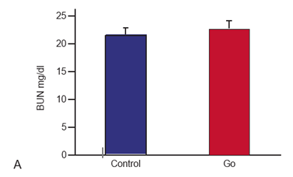
Our results showed significantly higher levels of serum Cr and BUN after Cisplatin administration (Figure 3). Histological sections showed that Cisplatin increased necrosis, cysts formation, and intra tubular debris (Figure 4). In the current study a dose of 1.5 mg/kg GO was used for the treatment of AKI. However, the results showed that 1.5 mg/kg of GO did not change Cr and BUN A levels (Figure 3, 5).
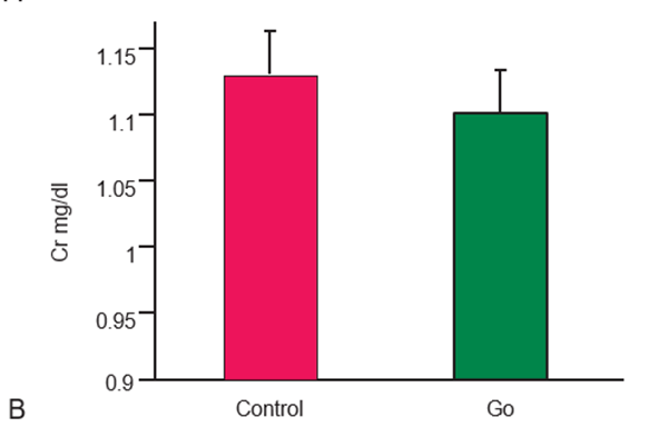
Figure 3: Intraperitoneal injection of 1.5 mg/kg dose of GO reduced the level of serum Cr and BUN on the ninth day after Cisplatin injection insignificantly compared to the control group (n = 6, GO: Graphene oxide)
Furthermore, the effects of MSCs and MSCs + GO in the treatment of AKI were compared with each other separately. Our findings showed that treatment of the animals in all these four groups with MSCs led to the improvement of the AKI. MSCs transplantation in the third passage had a regenerative effect on AKI. This effect was confirmed by serum biochemical tests and histological study of the kidney tissue (Figure 5). MSCs injection significantly repaired tissue damage, including cell Control Go necrosis, cyst formation, and intra-tubular debris. The results of this study showed that AKI caused apoptosis and necrosis of epithelial cells in kidney tubules (Figure 4).

Figure 2 A: SEM images of the interaction between GO (blue arrows), MSCs (green arrows), and macromolecules (yellow arrows) derived from stem cells (Magnification: 2000×), B: green arrows indicate the GO and yellow arrows show macromolecules secreted from stem cells (Magnification: 2500×)

Figure 3: Intraperitoneal injection of 1.5 mg/kg dose of GO reduced the level of serum Cr and BUN on the ninth day after Cisplatin injection insignificantly compared to the control group (n = 6, GO: Graphene oxide)
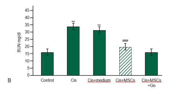
Figure 4: A) Intraperitoneal injection of MSCs and MSCs+ GO reduced the level of serum Cr (mg/dL) in AKI rats significantly. Values with asterisks (***) were significantly different from control group (***P < .001). Values with squares (###) were significantly different from Cisplatin group (###P < .001). B) Intraperitoneal injection of MSCs and MSCs+ GO on the ninth day after Cisplatin injection reduced the level of serum BUN in AKI rats significantly. Values with asterisks (***) were significantly different from control group (***P < .001. Values with squares (###) were significantly different from Cisplatin group (###P < .001, n = 6).
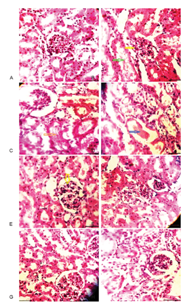
Figure 5: Histopathological study of the AKI treated with different medium. The figure shows less pathological damages necrosis, debries, and hyaline casts in the group treated with GO compared to the other groups. (A): Normal kidney (control group). B, C, and D: AKI induced by cisplatin. Intra-tubular debris (green and brown arrows), necrosis (yellow arrow), hyaline cast (dark blue arrow). E: AKI treated with GO transplantation (yellow arrow indicates necrosis). F: AKI treated by MSCs transplantation. G and H: AKI treated with GO mixed with MSCs. G and H show normal histology of treated AKI.
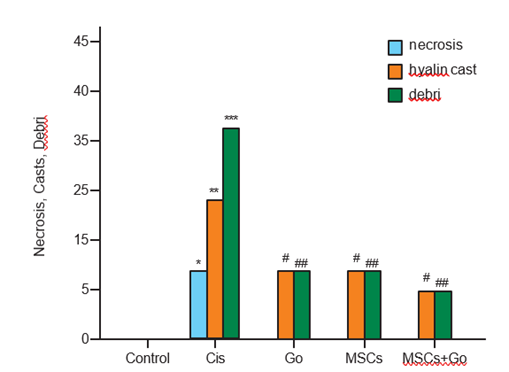
Figure 6: Histopathological analysis of the animal model kidney treated with different conditions. Significant differences between Cis and control groups is P < 0 n=6>
Biochemical and histological markers showed that the selected dose of 1.5 mg/kg GO made no significant change on biochemical markers of the blood, and histological features of the kidneys (Figure 4, 5). MSCs + GO caused a higher level of improvement in AKI than the others. In addition, the improvement induced by GO was greater than that of MSCs alone (Figure 4, 5). The serum levels of BUN and Cr in GO group was lower than those of MSCs group. Histological studies also showed that cellular necrosis, formation of cysts, and intratubular debris were significantly less in MSCs + GO compared to those who received only MSCs (Figure 4). Histological findings of the present study also revealed a reduction in necrosis and cyst formation in GO + MSCs group compared to the MSCs group (Figure 6). The SEM images showed that edges of GO hybrid show good connections with MSCs and biomolecules such as growth factors (Figure 2). Comparison of MSCs cultured with and without GO showed that they have been attached to the edges of GO and have formed a single unit (Figure 2).
Discussion
Our SEM images showed that the edges of GO show good connections with transplanted stem cells. Researchers showed that GO is not toxic for MSCs. [20-22] Comparison of MSCs with and without GO showed that the cells have been attached to the edges of GO and have formed a single unit. It seems that the enhanced therapeutic effect of MSCs + GO compared to MSCs is closely related to the role of GO in the stimulation of stem cells activities. Also, GO might be a good, engineered platform to enhance the role of MSCs transplantation. Drug loading and in vitro release properties of the GO showed much higher drug loading capacity as a result of increased drug surface interactions in comparison with the Graphene sample. [25] The reports on the therapeutic benefits of stem cells implantation are limited due to the poor survival of implanted cells.12,13 Here, we showed that surface attachment of GO on diatom silica structures increased therapeutic effects of MSCs transplantation in the treatment of AKI. We also found greater therapeutic effects for MSCs+ GO compared to GO and MSCs alone in the treatment of AKI. Presumably, the larger adsorption capacity of MSCs on GO is the main reason for its higher effect on cell therapy in AKI compared to MSCs. In this study, we proposed the use of GO attached to the MSCs within the blood flow, to protect implanted MSCs that supply micro-environments suitable for long-term in vivo survival. The GO’s specific topography (such as a large number of hydroxyl groups on its surface) leads to its high ability to absorb small molecules and extra cellular matrix (ECM) proteins. [26] These properties make GO very attractive for various applications including drug delivery. [27] Our SEM images showed that edges of GO exhibited a good connection with MSCs and molecules within grow medium. MSCs attached to the edges of GO formed a single unit. Based on the results of this study, GO acts as a cellular adhesion that provides surfaces for cell adhesion. One of the main causes for the poor survival of MSCs transplantation is reactive oxygen species (ROS) regenerated in damaged region, which hinder the adhesion of MSCs to the ECM by disrupting focal contacts.13 On the other hand, the loss of MSC adhesion induces the MSC apoptosis, thereby limits the therapeutic effects of MSCs implanted for tissue repair.13 Graphene based materials are found to be useful as a cellular adhesion in preventing the implanted cells from cell death.[28] GO has a great ability to attach to MSCs and their ECM proteins such as integrin β1 (a major receptor for ECM adhesion) and protects MSCs against apoptosis that can be followed by increasing the secretion of growth factors from MSCs.13 Reactive oxygen species prevent cell adhesion in addition to induction of cell apoptosis.[29] We believe that GO not only induces adhesion of transplanted MSCs and growth factors within blood flow to each other, by preventing MSC apoptosis, but also repairs kidney tissue by increasing paracrine factor secretion from MSCs. It appears that the ability of GO in the delivery of important biological factors is a good strategy for tissue repair and differentiation of stem cells. It seems that the adhesion of GO to MSCs is the most important reason for prevention of death of implanted cells, thereby enhancing the therapeutic efficiency of MSCs through maintaining MSCs integrity.
Our results showed that GO acts as a cellular adhesive substance that provides a proper surface for cell adhesion. In other words, we found a higher ability to connect to biomolecules and stem cells for GO. We also found that GO and GO + MSCs injection reduced the rate of cell death in kidney tissue compared with the cisplatin and sham groups. A possible mechanism for the attenuation of stem cell death by GO could be reduction of ROS by scavenging hydroxyl radicals. [30] Comparison of MSCs with and without GO showed that MScs attached to the edges of GO and formed a single unit. The presence of large numbers of hydroxyl groups on the surface of GO increases their hydrophilicity and biocompatibility, large surface-to-volume ratio, and its specific topography. These inherent features lead GO to be able to strongly absorb small molecules and ECM proteins. [26] GO forms micro- environments around cells and plays a significant role in controlling their behavior. Flexibility in size, high thermal and electrical conductivity, and low cytotoxicity of GO enhance cellular signals transduction.26 GO showed the best absorption of materials and growth factors because of a strong electrostatic interaction between components of the protein and oxygenated groups present on its surface.[26] Hence, in addition to the effects that are individually applied on the damaged tissue (through absorption and loading of molecules on its surface), GO collects the factors secreted from injected cells and expose them to the kidney cells in order to intensify the protective and regenerative effects of stem cells.
Reduced stem cell adhesion and survival rate after transplantation is the main cause of decreased success rate of cell therapy.13 It seems that the increased adhesion of GO to MSCs is the most important reason for prevention of death of implanted cells, thereby enhancing the therapeutic efficiency of MSCs through maintaining MSCs integrity. Indeed, the implantation of MSC + GO allows a significantly larger number of molecules to engraft at the lesion site. 17 On the other hand, previous studies proved that the use of graphene- based materials facilitates and accelerates stem cells differentiation. [31,32] It seems that the major mechanism of tissue repair by stem cell implantation is paracrine factor secretion from the implanted cells rather than the differentiation of the engrafted stem cells into the lesion site. [24]
In addition, ECM molecules adsorbed on GO play a significant role in mediating MSC adhesion to GO and preventing cell death. GO has a great ability to attach to ECM proteins, and binding to MSCs, increasing the adhesion of cells to the ECM such as integrin. This can be followed by increasing the secretion of growth factors from MSCs. [33] It has been shown that ECM protein-adsorbed GO increased integrin β1 expression, which is one of the key receptors by which cells adhere to ECM molecules, and inhibited p38 activation in MSCs cultured under toxic conditions. [34] It seems that GO inhibits MSCs death and facilitates their movement in blood flow. Previous studies showed that the increased therapeutic angiogenesis and the inhibition of apoptosis of cardiac myocytes contribute to the improvement in the function of the infarcted heart. 33 GO can also facilitate the regenerative effects of MSCs in the injured heart tissue through expression of angiogenic growth factor and gap junction protein. 34,30 Enhanced functional improvements in AKI may be due to the strong adhesion of implanted cells to each other and to GO, which prevents cell death. Enhanced cell survival rate in MSCs-GO treatment increases secretion of vascular endothelial growth factors, which enhance angiogenesis of damaged tissues.
Conclusion
The results of the present study showed that GO increases the efficiency of MSCs transplantation through binding to the MSCs. Moreover, GO enhanced the interaction of cells with each other and with the ECM. It could be proposed that GO improves regenerative and protective effects of stem cells for cell therapy.
Conflict of Interest
The authors declare no conflict of interest.
Acknowledgment
The authors would like to express their appreciation to all participants and staff of Kharazmi University for their kind cooperation. This project was supported by Kharazmi University.
Ethical Considerations
This study was reviewed and approved by the ethics committee of Kharazmi University (license code: IR.KHU.REC.1401.040).
References
- Moghadasali R, M.Mutsaers HA, Azarnia M, et al. Mesenchymal stem cell-conditioned medium accelerates egeneration of human renal proximal tubule epithelial cells after gentamicin toxicity. Cytoterapy 2013; 65: 5, 595-600.
View at Publisher | View at Google Scholar - Foroutan T, Nafar M, Motamedi E. Intraperitoneal injection of graphene oxide nanoparticle accelerates stem cell therapy effects on acute kidney injury. Stem Cells and Cloning 2020; 13 21–32
View at Publisher | View at Google Scholar - Foroutan T, Amid R, Karimi MR. Comparison of manual tools, ultrasonic and erbium-doped yttrium aluminum garnet (Er: YAG) laser on the debridement effect of the surface of the root of teeth suffering from Periodontitis. 2013; Journal of lasers in medical sciences 4 (4), 199.
View at Publisher | View at Google Scholar - Salehi PM, Foroutan T, Javeri A, et al. Extract of mouse embryonic stem cells induces the expression of pluripotency genes in human adipose tissue-derived stem cells. Iranian journal of basic medical sciences 2017; 20 (11), 1200.
View at Publisher | View at Google Scholar - Eini F, Foroutan T, Bidadkosh A, et al. The effects of freeze/thawing process on cryopreserved equine umbilical cord blood-derived mesenchymal stem cells. Comparative Clinical Pathology 2012; 21, 1713-1718
View at Publisher | View at Google Scholar - Ayobian-Markazi N, Fourootan T, Kharazifar MJ. Comparison of cell viability and morphology of a human osteoblast-like cell line (SaOS-2) seeded on various bone substitute materials: An in vitro study. 2012; Dental research journal 9 (1), 86.
View at Publisher | View at Google Scholar - Bressan E, Ferroni L, Gardin C, et al. Graphene based scaffolds effects on stem cells commitment. J. Transl. Med. 2014; 12, 296.
View at Publisher | View at Google Scholar - Park S.Y, Park J, Sim S.H, et al. Enhanced differentiation of human neural stem cells into neurons on graphene. Adv Mater. 2011; 23, 263–267.
View at Publisher | View at Google Scholar - Halim A, Luo Q, Ju Y, et al. A mini review focused on the recent applications of graphene oxide in stem cell growth and differentiation. Nanomaterials 2018; 8, 736.
View at Publisher | View at Google Scholar - Kumeria T, Bariana M, Altalhi T, et al. Graphene oxide decorated diatom silica particles as new nano-hybrids: towards smart natural drug microcarriers. J Mater ChemB. 2013;1, 6320.
View at Publisher | View at Google Scholar - Ryu S, Kim B.S. Culture of neural cells and stem cells on graphene. Tissue Engin Regen Med. 2013; 10 (2), 39.
View at Publisher | View at Google Scholar - Yoon H.H, Bhang S.H, Kim T, et al. Dual roles of graphene oxide in chondrogenic differentiation of adult stem cells: Cell-adhesion substrate and growth factor-delivery carrier. Adv.Func. Mater. 2014; 24, 6455.
View at Publisher | View at Google Scholar - Feng Z.Q, Yan K, Shi C, et al. Neurogenic differentiation of adipose derived stem cells on graphene-based mat. Mater. Sci. Eng. C 2018; 90, 685–692.
View at Publisher | View at Google Scholar - Sun X, Liu Z, Welsher K, et al. Nano-graphene oxide for cellular Iiaging and drug delivery. Nano Res. 2008; 1:203.
View at Publisher | View at Google Scholar - Bai H, Li C, Wang X, Shi G. A pH-sensitive graphene oxide composite hydrogel. Chem. Commun 2010; 46: 2376.
View at Publisher | View at Google Scholar - Abedi A, Azarnia M, Jamali M, et al. Effect of different times of intraperitoneal injections of human bone marrow mesenchymal stem cell conditioned medium on gentamicin-induced acute kidney injury. Urology journal 2016; 13: 2708.
View at Publisher | View at Google Scholar - Park J, Kim B, Han J, et al. Graphene oxide flakes as a cellular Adhesive: Prevention of reactive oxygen species mediated death of implanted cells for cardiac repair. ACS Nano 2015; 9: 4987–4999.
View at Publisher | View at Google Scholar - Humphreys BD, Bonventre JV. Mesenchymal stem cells in acute kidney injury. Annu Rev Med. 2008; 59: 311-325.
View at Publisher | View at Google Scholar - Abedini F, Foroutan T, Jahangiri L. Alkaline phosphatase and CD34 reaction of deciduous teeth pulp stem cells. Pakistan Journal of Biological Sciences: PJBS 2007; 10 (18), 3146-3149.
View at Publisher | View at Google Scholar - Foroutan T, Nazemi N, Tavana M, et al. Suspended graphene oxide nanoparticle for accelerated multilayer osteoblast attachment. J Biomed Mater Res Part A 2018; 106: 293–303.
View at Publisher | View at Google Scholar - Foroutan T, Kassaee MZ, Salari M, et al. Magnetic Fe3O4@graphene oxide improves the therapeutic effects of embryonic stem cells on acute liver damage. Cell Proliferation 2021; 54 (11).
View at Publisher | View at Google Scholar - Foroutan T, Kabiri F, Motamedi E. Silica magnetic graphene oxide improves the effects of stem cell- conditioned medium on acute liver failure. ACS Omega 2021; 6 (33), 21194-21206.
View at Publisher | View at Google Scholar - Foroutan T, Ahmady F, Moayer F, et al. Effects of intraperitoneal injection of magnetic graphene oxide on the improvement of acute liver injury induced by CCl4. Biomat. Res. 2020; 24 (14).
View at Publisher | View at Google Scholar - Hummers Jr WS, & Offeman RE. Preparation of graphitic oxide. J. Am. Chem. Soc. 1958; 80(6): 1339-1339.
View at Publisher | View at Google Scholar - Solanki A, Chueng S.T, Yin P.T, et al. Axonal alignment and enhanced neuronal differentiation of neural stem cells on graphene-nanoparticle hybrid structures. Adv. Mater. 2013,25, 5477.
View at Publisher | View at Google Scholar - Kim J, Choi K.S, Kim Y, et al. Bioactive effects of graphene oxide cell culture substratum on structure and function of human adipose derived stem cells. J. Biomed. Mater. Res. Part A 2013; 101 (12): 3520-3530.
View at Publisher | View at Google Scholar - Sun X, Liu Z, Welsher K, et al. Nano-graphene oxide for cellular imaging and drug delivery. Nano Res.2008; 1:203.
View at Publisher | View at Google Scholar - Wagner A.S, Glenske K, Henß A, et al. Cell behavior of human mesenchymal stromal cells in response to silica/ collagen based xerogels and calcium deficient culture conditions. Biomed Mater. 2017; (12), 045003.
View at Publisher | View at Google Scholar - Mirotsou M, Jayawardena M, Schmeckpeper J, et al. Paracrine mechanisms of stem cell reparative and regenerative actions in the heart. J. Mol. Cell. Cardiol. 2011;50 (2), 280-289.
View at Publisher | View at Google Scholar - Qiu Y, Wang ZY, Owens ACE et al. Antioxidant chemistry of graphene-based materials and its role in oxidation protection. Technology Nanoscale 2014; 6: 11744–11755.
View at Publisher | View at Google Scholar - Palipoch S. Review of oxidative stress in acute kidney injury: Protective role of medicinal plants-derived antioxidants. Afr J Tradit Complement Altern Med. 2013; 10(4): 88–93.
View at Publisher | View at Google Scholar - Park J, Kim IY, Patel M, et al. 2D and 3D hybrid systems for enhancement of chondrogenic differentiation of tonsil- derived mesenchymal stem cells. Adv. Funct. Mater. 2015; 25: 2573–2582.
View at Publisher | View at Google Scholar - Simon HU, Haj-Yehia A, Levi-Schaffer F. Role of reactive oxygen species (ROS) in apoptosis induction. Apoptosis 2000; 5: 415–41.
View at Publisher | View at Google Scholar - Lee TJ, Parkm S, Bhang SH, et al. Graphene enhances the cardiomyogenic differentiation of human embryonic stem cells. Biochem Biophys Res Commun 2014; 12:174–180.
View at Publisher | View at Google Scholar
