

Review Article | DOI: https://doi.org/10.31579/2835-2882/093
Hydrogels: Properties, Classifications, Characterizations, and Biomedical Applications
- Fatemeh Sadat Shariati 1
- Tahereh Ebrahimi 2
- Alireza Shariati 3
- Maryam Maghsood 4
- Salehi Pegah 5
- Zeinab Zakeria 6
- Arefe Sadat Khavari 7
- Masoud Delashoub 8*
1Department of Influenza and Respiratory Viruses, Pasteur Institute of Iran, Tehran, Iran.
2Department of Nanobiotechnology, New Technologies Research Group, Pasteur Institute of Iran, Tehran, Iran.
3Department of Materials Engineering, Tarbiat Modares University, Tehran, Iran.
4Department of Biological Sciences, Friedrich Schiller University, Jena, Germany.
5Department of Biological Sciences, Islamic Azad University, North Tehran Branch, Tehran, Iran.
6Department of Stem cell and tissue regeneration, Pasteur Institute of Iran, Tehran, Iran.
7Department of Biology, College of Basic Sciences, Shahed University, Tehran, Iran.
8Department of Basic Science, Faculty of Veterinary Medicine, Tabriz Medical Sciences, Islamic Azad University, Tabriz Iran.
† These authors contributed equally to this work.
*Corresponding Author: Masoud Delashoub, Department of Basic Science, Faculty of Veterinary Medicine, Tabriz Medical Sciences, Islamic Azad University, Tabriz Iran.
Citation: Fatemeh S. Shariati, Tahereh Ebrahimi, Alireza Shariati, Maryam Maghsood, Masoud Delashoub et al., (2025), Hydrogels: Properties, Classifications, Characterizations, and Biomedical Applications, Clinical Research and Studies, 4(5); DOI:10.31579/2835-2882/093
Copyright: ©2025, Masoud Delashoub. this is an open-access article distributed under the terms of the Creative Commons Attribution License, which permits unrestricted use, distribution, and reproduction in any medium, provided the original author and source are credited.
Received: 04 August 2025 | Accepted: 28 August 2025 | Published: 05 September 2025
Keywords: hydrogels; biocompatibility; biomedical applications; drug delivery; tissue engineering
Abstract
Hydrogels are three-dimensional, cross-linked networks of polymers capable of absorbing significant amounts of water, making them highly relevant for various applications in the biological and biomedical fields. Their hydrophilic nature, biocompatibility, and non-toxic characteristics have established hydrogels as critical materials in medical science. These materials are employed extensively in wound care, controlled drug delivery systems, tissue engineering, biosensors, and artificial organs, among other uses. The ability of hydrogels to closely mimic the properties of natural tissues has propelled their use in advanced therapeutic applications. This review explores properties, classifications, characterizations, and biomedical applications of hydrogels. A detailed analysis of their physical properties, such as swelling behavior, mechanical strength, and biodegradability, is presented alongside the latest characterization techniques. Biomedical applications are highlighted, particularly focusing on the role of hydrogels in tissue regeneration and drug delivery, where their responsiveness to stimuli like temperature and pH allows for precise and controlled therapeutic interventions.
Introduction
Hydrogels are defined as hydrophilic polymer chains forming a network capable of absorbing large quantities of water without dissolution [1]. Their introduction in 1960 marked a significant breakthrough, as they have high-water absorption capacity and similarity to natural tissue, making them ideal for various biomedical applications [2, 3]. Their broad range of applications, from biomedicine to environmental science, underscores their versatility. Figure 1 demonstrates the wide-ranging applications of hydrogels across both biomedical and environmental fields. It highlights their use in drug delivery systems, wound healing, tissue engineering, and biosensors in medicine, as well as their roles in waste management, soil moisture retention, and water purification in environmental applications. The figure emphasizes the adaptability of hydrogels in addressing diverse challenges. In biomedicine, hydrogels are shown to be crucial in drug delivery systems, where their ability to respond to physiological stimuli is leveraged for controlled release. The figure also depicts their role in tissue engineering, where they provide scaffolds that support cell growth and regeneration. Additionally, the use of hydrogels in wound dressings is highlighted, showcasing their capacity to maintain a moist environment that promotes healing. The figure also includes examples from the environmental sector, where hydrogels are employed in water purification processes, capturing pollutants and improving water quality. This visual summary underscores the broad applicability of hydrogels, demonstrating their impact across multiple fields.
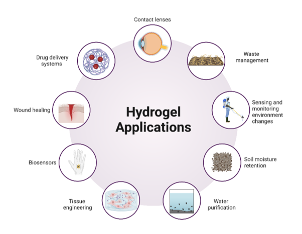
Figure 1: Diverse applications of hydrogels in biomedical and environmental Fields.
Properties of hydrogels
Structure and swelling
Hydrogels are formed by linking polymer chains through strong covalent bonds, electrostatic interactions, and hydrogen bonds. This crosslinked structure enables hydrogels to absorb significant amounts of fluid, causing them to swell and closely mimic the characteristics of living tissues. When dried out, these polymer networks are typically dehydrated using vacuum and heat, resulting in a rubbery or glass-like solid [3].
Hydrogels, as hydrophilic materials, naturally tend to swell when exposed to water or other suitable solvents. The swelling process involves three key steps: (1) water diffuses into the hydrogel network, (2) the polymer chains loosen, and (3) the network expands as it absorbs more fluid. The high absorption capacity of hydrogels is due to the presence of hydrophilic functional groups within the polymer chains, such as hydroxyl (OH), amine (NH2), sulfate (SO42-), and amide (CONH, CONH2) groups. These groups facilitate the migration of water molecules into the polymer network, leading to the hydrogel's expansion and increased volume. Typically, the polymer can absorb water more than 10% of its weight. The extent of water absorption varies depending on the chemical structure of the synthetic or biopolymer, environmental conditions, and the density of crosslinks [4]. The equilibrium swelling of the hydrogel is obtained from the following formula:

Where, Ws represents the weight of the swollen hydrogels and Wd is the weight of the hydrogels after freeze-drying. In hydrogels, drug release primarily occurs through the swelling or contraction of the hydrogel and the subsequent drug penetration through the polymer network. Previous studies have indicated that swelling kinetics and equilibrium are affected by various factors such as crosslinking ratio, synthesis state, the chemical nature of dried polymers, pH, and ionic media. Moreover, the extent of crosslinking significantly influences the swelling ratio of hydrogels. Highly crosslinked structures exhibit a lower swelling ratio, while less crosslinked structures show a higher swelling ratio. The swelling behavior of polymers is crucial for drug release, as it dictates the release rate and the overall efficiency of drug delivery systems. Stimuli-responsive hydrogels, also known as smart hydrogels, can undergo significant changes in swelling behavior in response to slight variations in environmental factors such as pressure, temperature, electric field, pH, light, carbohydrates, and antigens [5-8]. For example, anionic hydrogels exhibit increased deprotonation and swelling when the pH exceeds that of the linked ionizable groups. Conversely, cationic hydrogels swell when the pH is lower than the pKa of the linked ionizable groups [9]. Among these stimuli, pH-sensitive hydrogels have been extensively studied for the development of innovative drug delivery systems. A well-known example of temperature-sensitive hydrogels is poly (N-isopropyl acrylamide) (PNIPAM), which responds to temperature changes by altering its swelling behavior [5]. Temperature-sensitive hydrogels can be categorized as follows: (i) positively temperature-sensitive, which swell at temperatures higher than the upper critical solution temperature (UCST); (ii) negatively temperature-sensitive, which swell at temperatures lower than the lower critical solution temperature (LCST); and (iii) thermally reversible hydrogels that exhibit a sol-gel transition above and below a defined temperature [10-13]. A desired pH-responsive behavior and swelling capacity can be achieved by using different monomers. Common ionic polymers used in pH-sensitive hydrogels include polyacrylamide (PAAm), polymethacrylic acid (PMAA), polyacrylic acid (PAA), poly diethylamino ethyl methacrylate (PDEAEMA), poly dimethylaminoethyl methacrylate (PDMAEMA), and derivatives of phosphoric acid [14]. Previous studies have shown the significant influence of polymer type, size, ionic charge, flexibility of polymer chains, crosslink type, and crosslink density on the structure and water retention capabilities of hydrogels [15].
Biocompatibility and biodegradability
Naturally-based hydrogels are typically more biocompatible than synthetic ones, though synthetic hydrogels often possess greater chemical strength and stability. The selection of polymer type for hydrogel construction largely depends on the intended application. For biomedical applications, especially where the hydrogel comes into contact with blood, the polymers should be biodegradable and biocompatible. Common natural polymers used for these applications are chitosan, agar, and fibrin [16-18]. Polysaccharide-based hydrogels are widely considered to be biologically compatible and non-toxic, and as a result, they have been approved for use in dietary applications. On the other hand, several synthetic polymers, such as polyvinyl alcohol, polyacrylamide, polyethylene oxide, and polyethylene glycol, are commonly used in the preparation of hydrogels for various medical applications due to their tunable properties and robustness.
The biodegradability of hydrogels refers to their ability to break down into harmless end products within a living organism. This process is influenced by several factors, including the method of preparation, molecular weight, the components present within the system, polymer-water interactions, and the hydrophilicity of the hydrogel. Additionally, environmental factors like temperature and pH can affect the degradation rate through solubilization. Hydrogels composed of polysaccharides, proteins, and synthetic polypeptides can undergo degradation via enzyme hydrolysis. This hydrolysis is facilitated by different enzymes, which catalyze the breakdown of C-C, C-O, and C-N bonds. In polysaccharide-based hydrogels, biodegradation primarily occurs due to the action of glycosidases [18]. For instance, chitosan can be broken down through enzyme hydrolysis, targeting bonds such as glucosamine-glucosamine, glucosamine-N-acetyl-glucosamine, and N-acetyl-glucosamine-N-acetyl-glucosamine. In addition to enzyme hydrolysis, the degradation of chitosan can also occur through redox reactions and the action of free radicals. Controlled degradation is a crucial aspect of hydrogels, especially in biomedical applications like drug delivery systems. This controlled degradation is mainly determined by the chemical composition and the structure of hydrogels. These hydrogels are typically designed to ensure that they function as intended during degradation [19, 20].
Mechanical and physical properties
The desired mechanical properties of hydrogels can be achieved by incorporating specific polymers and cross-linkers, as well as adjusting the degree of crosslinking. Increasing the crosslinking degree can create a stronger gel network, though this often results in reduced elongation and elasticity, making the gel more brittle. However, enhanced elasticity can improve the flexibility of the crosslinked network, which is beneficial for the movement of incorporated therapeutic agents. Assessing the mechanical properties and strength of hydrogels is critical for ensuring their physiological functionality in biomedical applications, including tissue engineering, wound dressings, tendon and ligament repair, cartilage replacement, and as matrices for drug delivery. The rheological properties of hydrogel, which describe the material's flow and deformation under external influences such as stress, strain, and temperature over time, can be characterized using oscillation modes on a rheometer [21]. To enhance the mechanical strength and thermal stability of hydrogels, several methods have been developed, including double network hydrogels [22], topological hydrogels [23], nanocomposite hydrogels (NCHs) [24], hydrogen bonding-reinforced hydrogels [25], and microgel-reinforced hydrogels [26]. Higher stiffness in hydrogels can be achieved by increasing the crosslinking degree. However, the mechanical properties of hydrogels depend on various factors and require specific analyses tailored to the material in question. The mechanical strength of hydrogels is crucial because it is essential for the hydrogel to maintain its physical integrity during the controlled release of therapeutic agents over a specific period. Frequency-based sinusoidal testing, commonly conducted using a rheometer, involves loading the hydrogel sample onto a specified geometry instrument and performing various types of sweep measurements to evaluate its properties [13].
Traditional hydrogels often faced challenges with mechanical stability, being prone to brittleness and fragility under stress due to uneven distribution of their network and lack of energy dissipation mechanisms. Recent advancements have focused on improving these properties by designing network structures that include non-covalent interactions like electrostatics, hydrogen bonding, and supramolecular recognition. These interactions enable hydrogels to show reversible damage and recovery, leading to significant improvements in their strength, toughness, and self-healing abilities [27]. Furthermore, hydrogels have been engineered to respond to environmental triggers (e.g., pH, temperature, light), which, combined with improved mechanical properties, opens up new applications in areas like soft robotics and artificial muscles. The development of strong, tough, and responsive hydrogels marks a significant advancement in overcoming the material's historical limitations, enabling their use in a broader range of applications and highlighting their potential in cutting-edge technological and biomedical fields [28]. Figure 2 outlines the key properties of hydrogels, such as water absorption, swelling behavior, mechanical properties, biocompatibility, degradability, stimuli-responsiveness, and others. Each property plays a critical role in determining the suitability of hydrogels for specific applications. The figure also highlights factors like permeability, surface properties, and electrical conductivity, which are essential for their performance in drug delivery, tissue engineering, and biosensing.
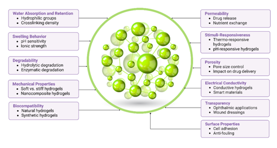
Figure 2: Key properties of hydrogels and their functional characteristics
Classification of hydrogels
Hydrogels can be categorized based on various criteria including their origin, environmental responsiveness, polymeric composition, cross-linking method, configuration, and ionic charge [27, 29, 30]. This classification system helps to understand the wide range of hydrogel structures and their potential applications in different fields [3]. Figure 3 provides a comprehensive visual summary of the different ways hydrogels can be classified. It illustrates the classification based on source, composition, physical structure, crosslinking method, charge, stimuli-responsiveness, degradability, and functionality. Each of these classifications highlights the versatility of hydrogels, allowing them to be tailored for various applications, from biomedical to environmental fields.
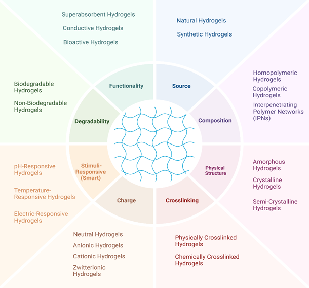
Figure 3: Comprehensive classification of hydrogels based on source, structure, functionality, and other key properties.
Classification based on the source
Natural hydrogels
These hydrogels are derived from biodegradable polymers such as starch, cellulose, chitosan, collagen, and hyaluronic acid. While natural hydrogels are attractive for their biocompatibility, bioactivity, and biodegradability, they often face limitations in mechanical strength and stability [31].
Synthetic hydrogels
Synthetic hydrogels are formed through the polymerization of monomers such as polyvinyl alcohol (PVA), poly acrylic acid (PAA), and polyethylene glycol (PEG). Synthetic hydrogels are widely utilized due to their high water absorption capacity and versatility across various applications. However, concerns about the environmental impact, non-renewability, and degradation challenges restrict their application. The properties of synthetic hydrogels, including their sensitivity to external stimuli like solvent, temperature, electric and magnetic fields, pressure, and light intensity, can be precisely tailored by adjusting factors such as the degree of cross-linking, charge density, and the type of monomer used. Due to the stronger chemical structure of these polymers, various cross-linking strategies can be employed (physical, chemical, and enzymatic), each uniquely influencing the final properties of the developed hydrogel [3].
Semisynthetic hydrogels
These hydrogels are composed of chemically modified natural polymers (e.g. methacryloyl-modified gelatin or acrylate-modified hyaluronic acid) or a combination of natural and synthetic polymers. The goal of semisynthetic hydrogels is to combine the bioactivity of natural hydrogels with the mechanical stability of synthetic ones. Types of hydrogels within this category include physically crosslinked, chemically crosslinked, and interpenetrating network (IPN) hydrogels, which encompass both semi-IPN and Full-IPN structures [27].
Classification based on the synthetic strategy
Chemically-synthesized hydrogels
These hydrogels can be made by covalent bonds created during the chemical cross-linking process [32]. Cross-linking agents such as glutaraldehyde and formaldehyde are commonly used to create a stable polymer network capable of significant water absorption. Chemical cross-linking alters the hydrogel's swelling properties and is often favored for its durability and environmental safety [30].
Physically-synthesized hydrogels
These hydrogels are formed through non-covalent interactions among polymer chains, such as hydrogen bonding, electrostatic interactions, or physical entanglements [32]. For instance, physical cross-linking can occur in polyvinyl alcohol (PVA) hydrogels combined with chitosan or starch through repeated freeze-thaw cycles. This process generates microcrystalline structures, resulting in hydrogels with enhanced elasticity and porosity, making them suitable for applications like molecular and whole-cell immobilization in biotechnology [30].
Enzymatic-synthesized hydrogels
In this approach, enzymes facilitate the formation of covalent bonds within the hydrogel network. This method allows for more controlled and specific cross-linking, which can be particularly advantageous in biomedical applications where mild reaction conditions are necessary. Hydrogels produced by tyrosinases, transferases, and lysyl oxidases show interesting characteristics and are used as dynamic scaffolds and controlled release matrices [33, 34].
Classification based on cross-linking method
This type of chemical cross-linking involves the interaction of ionic polymers with divalent or trivalent counter-ions. For example, sodium alginate can form gels when exposed to calcium ions (Ca²⁺), creating a cross-linked structure through the ionic interaction of oppositely charged species [2]. Thermal method involves using temperature changes to induce the formation or modification of hydrogel structures. Polymers like poly(N-isopropylacrylamide) (PNIPAM) exhibit thermos-responsive behavior, undergoing phase transitions in response to temperature fluctuations. At temperatures around its lower critical solution temperature (LCST) of 32 °C, PNIPAM becomes hydrophobic and collapses, making it useful in applications such as drug delivery, tissue engineering, and soft robotics [35, 36].
Smart hydrogels and types of stimuli
Researchers have been working diligently to create a category of hydrogels, often referred to as smart or intelligent hydrogels, by modifying their physicochemical characteristics. These advanced hydrogels are capable of reacting to a wide range of stimuli, including physical (such as temperature, light, electromagnetic fields, pressure, and ultrasound radiation), chemical (like pH, glucose, and ionic strength), and biological factors (including enzymes and antigen/antibody interactions). Non-contact stimuli include light, temperature, and magnetic/electric fields. These stimuli can non-invasively trigger phase transitions, stiffness changes, or other properties in hydrogels. Contact stimuli involve direct interaction with substances like pH changes, ion concentration variations, and biochemical reactions. These stimuli can activate hydrogels in a bio-environment, making them suitable for targeted biomedical applications [37]. Physical responsive hydrogels are a type of smart materials that undergo a change in their physical properties in response to external physical stimuli such as temperature, light, and mechanical stress. These stimuli-responsive hydrogels are particularly interesting for a wide range of applications due to their ability to mimic natural processes and respond to environmental changes [38]. Stimuli-responsive hydrogels hold great promise in biotechnology, targeted drug delivery, tissue engineering, smart biosensors, and 3D cell culture. They offer dynamic responses to environmental cues, enabling smart biomedical applications [39].
Thermo-sensitive hydrogels
Temperature-responsive hydrogels or thermos-gels are known for their sensitivity to temperature changes in the surrounding environment [40]. They comprise both hydrophobic and hydrophilic groups in their matrix. One of the particular characteristics of these polymers is the presence of a critical solution temperature at which the phase of polymer and solution is discontinuously changed based on their composition [40]. These hydrogels undergo phase transition in response to temperature changes. For example, poly(N-isopropylacrylamide) (PNIPAM) is a well-known temperature-sensitive hydrogel that exhibits a lower critical solution temperature (LCST) around 32 °C, above which it becomes hydrophobic and collapses. Temperature-sensitive hydrogels are used in drug delivery systems where the release of drugs can be controlled by body temperature or externally applied heat, tissue engineering scaffolds that can morph with temperature changes, and valves or actuators in microfluidic devices [41].
Photosensitive hydrogels
Light-sensitive hydrogels contain chromophores that absorb specific wavelengths such as UV, visible, or infrared, leading to a physical or chemical change in the hydrogel structure [42]. This change can be a result of photo-induced crosslinking, cleavage, or isomerization processes. Light-sensitive hydrogels are promising in the development of optical switches, display units, ophthalmic drug delivery devices, photo-responsive artificial muscles, and cartilage tissue engineering [43]. These hydrogels can be categorized as UV-sensitive and visible light-sensitive hydrogels. Visible light is simply available, inexpensive, clean, safe, and easily manipulated compared to UV lights [43]. Visible light-sensitive hydrogels are generally prepared by the introduction of chromophores to the temperature-sensitive hydrogel network. UV-sensitive hydrogels can be synthesized by incorporating of leucocyanide molecule to the matrix. These hydrogels are useful in creating controlled drug delivery systems, where light triggers drug release, in developing photo-patternable scaffolds for tissue engineering, and in creating soft robots that can move or change shape upon light exposure [44]. Huang et al. generated smart near-infrared (NIR) light-responsive poly N-isopropylacrylamide (PNIPAM) nanocomposite hydrogels through the incorporation of sparse chemical crosslinking of small molecules [45]. This highlights the versatility of light-responsive hydrogels, which can be tailored for different wavelengths, such as UV or NIR, depending on the desired application.
Enzyme sensitive hydrogels
These hydrogels contain polymers or linkers that are sensitive to specific enzymes present in the body or overexpressed in certain conditions like a disease. The interaction with the enzyme leads to the cleavage of the polymer backbone or crosslinking agents, causing a change in the hydrogel's properties, such as its porosity, degradation rate, or mechanical strength. Enzyme-responsive polymers can be a potential candidate for tumor tissue-targeted delivery, and site-specific triggered release from drug-nano carriers or polymer-drug conjugates in comparison with conventional stimuli (i.e., pH, temperature, light) [46, 47]. These hydrogels can be used for vary of application including tissue engineering, health monitoring, targeted therapeutic delivery, and regulation of biochemical processes [48]. The frequently targeted enzymes in enzyme responsive materials (ERMs) are proteases, kinases, phosphatases, and endonucleases. Proteases and endonucleases are respectively appropriate for peptides cleaving and oligonucleotides, which are applicable in the degradation or disassembly of ERMs. Furthermore, they can able to cleave functional groups from ERMs to be suitable for a range of sensing applications. It is noteworthy that Najafi et al. developed an injectable enzyme-responsive hydrogel capable of contacting a host of enzymes. It contains a core crosslinked (CCL) flower-like micelle and an enzyme-responsive linker P(NIPAM-co-HPMA-Cys)-PEG-peptide-PEG-P(NIPAM-co-HPMA-Cys) (Pep-NC). The results demonstrated that CCL micelles can be delivered intracellularly, for example, in cancer tissue, despite the increased levels of matrix metalloproteinases (MMPs) [49].
Reactive oxygen species sensitive hydrogels
Reactive Oxygen Species (ROS)-sensitive hydrogels are innovative materials designed to react to ROS, important molecules in biological systems that can become harmful at elevated levels associated with diseases like cancer and inflammation. These hydrogels are particularly notable in biomedical applications such as drug delivery and tissue engineering. They contain ROS-responsive elements that alter the hydrogel's structure upon interacting with ROS, allowing for the targeted release of therapeutics within diseased tissues, where ROS levels are high. The design involves incorporating substances such as thioketal linkages, which break down in the presence of ROS, or phenylboronic ester groups, which convert from hydrophobic to hydrophilic upon oxidation by ROS. Other examples include sulfur-containing compounds like thioether or disulfide bonds, which are cleaved in oxidative environments, facilitating controlled therapeutic release with minimal side effects on healthy tissues. Overall, ROS-sensitive hydrogels offer a sophisticated approach to precision medicine, providing tailored treatments by leveraging the body's own biochemical signals [50, 51].
Shape memory hydrogels
Shape memory hydrogels (SMHs) are three-dimensional polymeric networks that can deform and recover their original shape in response to external stimuli, such as heat, pH, light, electricity, or chemical changes. The change from a temporary shape and recovering to the original shape can be achieved by reversible crosslinks. Due to the need for diverse functionalities in practical applications, many SMHs are now developed with additional properties such as self-healing, thermos-plasticity, self-adhesion, antibacterial, and anti-inflammatory features. For example, a study reported an SMH with thermos-plasticity using a hydrophobic polyampholyte network that could alter its permanent shape upon heating, thereby enhancing its versatility. This unique property opens up a wide array of applications across various fields including biomedical devices and tissue engineering, and smart materials [52, 53].
Nanocomposite hydrogels
The creation of nanocomposite hydrogels involves chemically or physically cross-linking polymers with nanomaterials. This innovative approach was first reported in 2002, showcasing hydrogels with significantly improved properties over traditional hydrogels. This integration enhances the mechanical strength, introduces electrical conductivity, and imbues other unique properties such as antibacterial, antioxidation, and magnetic responsiveness. These nanocomposite hydrogels are particularly useful for bone and cartilage tissue engineering. For example, incorporating nanomaterials like hydroxyapatite (HAP) and tricalcium phosphate (TCP) enhances the mechanical properties of hydrogels and supports the proliferation, differentiation, and mineral deposition essential for bone and cartilage regeneration [54].
Chemical responsive hydrogels
Chemically responsive hydrogels are designed to directly convert the chemical potential of their environment into mechanical motion, making them invaluable in a variety of applications. These hydrogels can be categorized into several types based on their response to different chemical stimuli, including solvent-responsive, pH-responsive, and biomolecule-responsive actuators [55].
pH responsive hydrogels
pH responsive hydrogels are designed to undergo significant changes in their properties (e.g., solubility, volume, porosity) in response to pH variations. This behavior is due to the presence of ionizable groups in the polymer chain, which can accept or donate protons depending on the surrounding pH, thus changing the hydrogel's affinity for water [56]. pH-responsive hydrogels can be used to create smart drug delivery systems that release therapeutic agents in a controlled manner at the site of action, where the pH might differ from that of healthy tissue. The ability of these hydrogels to change their physical properties in response to pH makes them suitable as scaffolds for tissue engineering. Injectable pH-responsive hydrogels can be used in minimally invasive surgical procedures for bone repair. For conditions like osteoporosis or bone tumors, where the local pH may be altered, pH-responsive hydrogels can be engineered to respond to these specific conditions, providing targeted therapeutic actions [56-58]. pH-responsive hydrogels, particularly those based on chitosan, offer promising avenues for advancing bone tissue engineering and regenerative medicine. Their ability to respond to pH changes enables the development of smart, adaptive materials that can address the dynamic requirements of tissue healing and regeneration [59].
Biochemically responsive hydrogels
Biochemically responsive hydrogels are designed to respond to biological molecules. These hydrogels can undergo structural changes when exposed to specific biochemical stimuli, leading to the formation or degradation of the hydrogel network. This responsiveness is achieved through the incorporation of biochemically reactive groups or molecules into the hydrogel structure, which interact with the target biomolecule [60]. One notable example of biochemically responsive hydrogels is glucose-responsive hydrogels, which are a specialized type of chemically responsive hydrogel designed to respond to variations in glucose levels. These hydrogels typically incorporate a glucose recognition element, such as glucose oxidase (GOx), which is an enzyme that catalyzes the oxidation of glucose to gluconic acid and hydrogen peroxide. This enzymatic reaction is key to the hydrogel's responsiveness to glucose [61]. The primary application of glucose-responsive hydrogels is in the development of smart insulin delivery systems. Beyond insulin delivery, glucose-responsive hydrogels can be utilized in the fabrication of glucose sensors. Research is also exploring the use of glucose-responsive hydrogels in tissue engineering, particularly in creating scaffolds that can modulate their properties in response to the metabolic activity of the surrounding tissue. Another their potential application is in wound healing, where glucose-responsive hydrogels could release therapeutic agents in response to the metabolic needs of the healing tissue, thereby accelerating the healing process [62]. Glucose-responsive hydrogels represent a significant advancement in biomaterials, offering innovative solutions for diabetes management and beyond. Their ability to respond dynamically to glucose levels makes them a promising tool in personalized medicine, with the potential to greatly improve the quality of life for individuals with diabetes [63].
Classification based on composition
Hydrogels are composed of a wide range of polymers, each contributing to the material's overall properties. Natural polymers such as alginate, chitosan, and cellulose are frequently used for their biocompatibility and biodegradability. On the other hand, synthetic polymers like polyethylene glycol (PEG) and poly acrylic acid (PAA) are valued for their tunable properties and structural stability [64]. The choice of polymer significantly impacts the hydrogel's characteristics, including its ability to retain water, its compatibility with biological tissues, and its potential for use in drug delivery and tissue engineering applications [65].
Homo-polymer
These hydrogels are composed of a single type of polymer, which simplifies their structure and often leads to predictable and consistent properties. Examples of homopolymer hydrogels include polyacrylic acid (PAA), polyvinyl alcohol (PVA), and polyethylene glycol (PEG) which exhibit predictable swelling behaviors, excellent biocompatibility, and versatile applications in drug delivery and tissue engineering [66].
Composite
Formed from a combination of different polymers, composite hydrogels integrate the advantageous properties of each component. This blending allows for enhanced functionality, such as improved mechanical strength or responsiveness to environmental stimuli. For example, Chen et al. developed an injectable composite hydrogel comprising gelatin microspheres embedded in chitosan-alginate hydrogels for the delivery of the anti-cancer drug 5-fluorouracil. This hydrogel was crosslinked through a Schiff base reaction involving the amino groups of carboxyethyl chitosan and the aldehyde groups of oxidized alginate. The composite hydrogel not only formed in situ but also exhibited self-healing behavior, attributed to the dynamic and reversible nature of Schiff base formation under specific conditions. Over a period of five weeks, it demonstrated a controlled and sustained release of 5-fluorouracil, outperforming the release rates from either the hydrogel or the microspheres alone. Additionally, the incorporation of magnetic Fe3O4 nanoparticles within the gelatin microspheres enabled an accelerated drug release when subjected to a magnetic field [67, 68].
Interpenetrating network (IPN)
IPN hydrogels consist of two or more polymer networks that are interlaced but not covalently bonded to each other. This structure provides superior mechanical strength and resilience, making them useful in applications where durability and robustness are required [69]. Suo et al. employed a novel mechanism to form an interpenetrating network (IPN) of gelatin methacryloyl (GeIMA) and chitosan (CS) hydrogels. They found that the semi-IPN and IPN structures significantly enhance the mechanical properties of GeIMA-CS hydrogels compared to single-network CS or GeIMA hydrogels [70]. Cui et al. prepared and characterized interpenetrating network (IPN) hydrogels based on chitosan and gelatin, utilizing genipin as the cross-linker. The results of the swelling tests indicated that the IPN hydrogels are pH-sensitive, exhibiting reversibility and a rapid response to changes in pH [71].
Classification based on network electrical charge
Cationic hydrogels
The electrical charge within a hydrogel network is crucial as it affects its interaction with other molecules and its suitability for various applications [72]. Cationic hydrogels possess positively charged polymer chains, which enable them to interact effectively with negatively charged molecules. This property makes cationic hydrogels particularly useful in gene delivery (DNA or RNA) and drug delivery applications, where their pH responsiveness allows for the controlled release of therapeutic agents in response to environmental changes [73]. Oligochitosan, a cationic natural polymer, was combined with Ca2+ to cross-link with pectin, resulting in the formation of hydrogel microcarriers. These microcarriers are engineered for slow drug delivery in the upper gastrointestinal tract, while also facilitating a rapid drug release in conditions that mimic the physiological environment of the colon [74]. Cationic polymers have contributed significantly to the development of sustained-release matrix hydrogel. These tablets were fabricated using a blend of hydrophobic ethyl cellulose and hydrophilic sodium carboxymethyl cellulose. The in vitro release of losartan potassium, a medication for hypertension, was investigated. The findings revealed that this formulation achieved a prolonged drug release over 12 hours. Additionally, cationic polymers have been utilized in biodegradable micelles that serve as effective drug carriers [75].
Anionic hydrogels
Anionic hydrogels, which composed of polymers with negatively charged chains, are employed in controlled drug delivery and tissue engineering. Their ability to interact with positively charged species and their tunable swelling behavior based on pH make them ideal for these applications [76]. Hyaluronic acid (HA) is an anionic polysaccharide that is naturally found in connective tissues, skin, and joint tissues. Due to its unique properties, including water retention and the ability to form hydrogels, it is recognized as a highly useful material in the production of hydrogels. Hyaluronic acid-based hydrogels are widely used in medical applications, particularly in wound healing, tissue engineering, and drug delivery [77]. Alginate and pectin are other anionic polysaccharide that makes anionic hydrogels and are used a lot in biomedical application [78].
Non-ionic hydrogels
These hydrogels feature neutral polymer chains, meaning they do not carry a net electrical charge. As a result, non-ionic hydrogels remain swelling properties across different pH levels and ionic strengths. They are particularly suitable for applications like wound dressings and tissue engineering, where consistent performance is required [79]. Non-ionic polymers including hydroxyethyl cellulose (HEC), hydroxypropyl methylcellulose (HPMC), polyethylene oxide (PEO), polyvinyl alcohol (PVA), hydroxypropyl cellulose (HPC), and polyvinylpyrrolidone (PVP) can be employed to fabricate hydrogels suitable for a wide range of biomedical applications [80]. Guar gum (GG) is a biocompatible and biodegradable polymeric compound derived from plants, readily available in nature. This non-ionic hydrophilic carbohydrate is a cost-effective hydrocolloid polysaccharide, representing a new generation of plant gums. The easy availability, non-toxicity, eco-friendliness, and biodegradability of GG make it an excellent candidate for drug delivery applications. Additionally, guar gum hydrogels can be utilized as a transdermal drug delivery system, providing a promising method for effective medication administration [81].
Ampholytic hydrogels
Ampholytic hydrogels contain both positively and negatively charged groups within a polymer chain. This dual charge allows them to adjust their swelling behavior in response to pH or ionic strength, providing versatility for use in drug delivery and tissue engineering [66]. Acrylamide, N-[3-(Dimethylamino)propyl] acrylamide, 2-(Dimethylamino)ethyl methacrylate, 2-(Diethylamino)ethyl methacrylate, [2-(Methacryloyloxy)ethyl] trimethylammonium chloride, 2-(Acryloyloxy ethyl)trimethyl ammonium chloride, [3-(Methacryloylamino)propyl] trimethylammonium chloride, 2-Carboxyethyl acrylate, Methacrylic acid, Acrylic acid, Carboxylated poly-L-lysine, 3-Sulfopropyl methacrylate potassium salt, 2-Sulfoethyl methacrylate are the common monomers used in polyampholyte hydrogels [82]. Polyampholyte hydrogels offer a promising choice for tissue engineering because of their overarching characteristics. In addition to being tunable, responsive, and non-fouling, they also feature a considerable moisture-holding capacity which is commonly linked to biocompatibility [82, 83]. Because of the inherent responsive characteristics of polyampholyte polymers mentioned earlier, they have garnered growing interest for use in drug delivery applications.[82, 84, 85].
Complex coacervation
This process involves the formation of hydrogels through the interaction of poly-anions and poly-cations, where opposite charges on the polymers attract and bind together, forming soluble or insoluble complexes. Complex coacervation is highly dependent on the pH and concentration of the solutions. An example of this is the coacervation of poly-cationic chitosan with poly-anionic xanthan, which results in the formation of polyion complex hydrogels. These hydrogels demonstrate the ability of positively charged proteins (below their isoelectric point) to bind with anionic hydrocolloids, leading to gel formation [2].
Characterization methods
The characterization of hydrogels involves examining their swelling behavior, microstructure, mechanical properties, biocompatibility, and biodegradability. These parameters are crucial for determining the suitability of hydrogels for specific applications. Swelling tests evaluate a hydrogel's capacity to absorb and retain water, providing insights into its network structure and potential applications. These tests typically involve immersing the hydrogel in a fluid and measuring weight changes over time. Factors such as pH, temperature, and ionic strength are manipulated to mimic real-world conditions, revealing insights into the material’s responsiveness [86]. Understanding the internal structure of hydrogels is vital. Techniques such as Scanning Electron Microscopy (SEM), Transmission Electron Microscopy (TEM), and Atomic Force Microscopy (AFM) are employed to visualize surface morphology, porosity, and network architecture. These methods highlight pore size, distribution, and the overall architecture critical for applications like cell scaffolding [87]. Additionally, Hydrogels need to exhibit appropriate mechanical strength for their intended use. Mechanical testing includes compression and tensile tests to measure elasticity and durability, Dynamic Mechanical Analysis (DMA) to evaluate viscoelastic properties, and rheological studies to assess the material's flow and deformation behavior under stress [88]. Furthermore, techniques such as Fourier Transform Infrared Spectroscopy (FTIR) and Nuclear Magnetic Resonance (NMR) spectroscopy are employed to identify chemical bonds and functional groups within the hydrogel. These insights are crucial for confirming the chemical composition and monitoring potential degradative changes [86]. For biomedical uses, compatibility with living cells is paramount. In vitro assays, such as MTT and Live/Dead staining, evaluate cell viability and adhesion, while in vivo studies observe tissue response and integration [86]. Biodegradation tests determine how hydrogels break down in physiological conditions. Enzymatic degradation assays and mass loss studies over time are typical approaches, providing data about longevity and bioresorbability [86]. Moreover, rheology evaluates the mechanical properties of hydrogels by measuring their viscosity, elasticity, and viscoelastic behavior. These parameters help to understand hydrogel's response to stress and strain, which is important for applications like injectable hydrogels and tissue scaffolds [89]. Finally, additional methods include X-Ray Diffraction (XRD) to determine the crystalline or amorphous nature of hydrogels, Thermal Analysis (TGA and DSC) to assess thermal stability and transitions, and Mass Spectrometry (e.g., MALDI-TOF) for precise molecular weight and composition analysis [86].
Small-angle neutron scattering (SANS)
SANS helps in visualizing the internal structure and dynamic behavior of hydrogels, especially when exposed to different environmental conditions like pH or ionic strength. It provides a deeper understanding of the nanoscale organization of polymer chains and the role of water channels [90].
These techniques collectively help evaluate crucial parameters such as swelling behavior, mechanical properties, biocompatibility, and biodegradability, all of which are necessary to ensure the hydrogel’s suitability for specific applications like drug delivery, wound healing, and tissue engineering [91].
Applications in biomedicine
The biomedical field has extensively embraced hydrogels due to their diverse applications, ranging from drug delivery systems and tissue engineering to bone regeneration and biosensors. These materials support critical medical processes, such as tissue regeneration, spinal cord recovery, and bone repair, by providing scaffolds that mimic the extracellular matrix. In the 1960s, Wichterle and Lim made significant progress by developing soft contact lenses using poly-2-hydroxyethylmethacrylate (PHEMA)-based hydrogels [92]. This marked a major milestone in the biomedical application of hydrogels, showcasing their biocompatibility and paving the way for their extensive use in modern medical devices and therapeutic innovations [2]. Contact lenses are usually categorized as "hard" or "soft" based on their elasticity. Hard contact lenses are usually made from hydrophobic materials such as poly(methyl methacrylate) (PMMA) or poly(hexafluoroisopropyl methacrylate) (pHFIM), while soft lenses are made of hydrogels [93]. Hydrogels can be customized for specific purposes, such as reacting to environmental changes and working well with living tissues. This makes them very promising for improving medical treatments, regenerative medicine, and drug delivery systems. However, their use is restricted by their weak mechanical strength and inability to withstand high temperatures [26, 32].
Three-dimensional cell cultures
Hydrogels provide a three-dimensional scaffold that closely mimics the extracellular matrix (ECM), enabling cells to grow in all directions. Since hydrogels consist of a hydrophilic polymer network that can absorb significant amounts of water and biological fluids, creating a soft and wet environment similar to the natural ECM is essential for encapsulating cells [94]. This feature is critical for studying cell behavior and developing tissue models [95-97]. Recent advancements in 3D printing further expand their use as biological tissues [98]. Among natural polymers, collagen stands out as the most abundant fibrous protein in the human body. Studies have shown that collagen-based hydrogels significantly enhance cell growth, adhesion, the differentiation of neural cells, and forming tissue-like structures when co-cultured with chondrocytes [99, 100]. For example, Jin G.Z. and Kim H.W. demonstrated that type I collagen maintains the chondrogenic phenotype when used as a scaffold for 3D culturing of rat chondrocytes[101]. Type II collagen also plays a critical role in the differentiation of human mesenchymal stem cells (hMSCs) to chondrocytes. Although collagens I and II support chondrogenesis through different mechanisms, their use in hydrogel scaffolds shows great potential for cartilage tissue engineering [102].
Hyaluronic acid (HA) is another important component of natural hydrogels because it serves as a major structural element within the extracellular matrix (ECM). It is ubiquitously present in various tissues, especially in skin and cartilage, and has a vital role in facilitating cellular processes such as survival, migration, angiogenesis, differentiation, and neural regeneration [103, 104]. The biological activity of hyaluronic acid (HA) depends greatly on its molecular weight. Low-molecular-weight HA (LMW-HA) is particularly known for its ability to promote cell proliferation and migration, which are essential for tissue regeneration and wound healing. Recent studies have highlighted that these effects are mediated through specific interactions with cell surface receptors [105]. Research by Wu et al. indicated that HA-based hydrogels can promote the neural differentiation of human-induced pluripotent stem cell-derived neural progenitor cells (hiPSC-NPCs) [106]. In another study, synthesized adjustable stiffness HA-hydrogels could maintain stem cell characteristics and directly induce cartilage differentiation [107]. Suo et al. developed an HA-hydrogel scaffold through a combination of hydrazone and photo-dual crosslinking processes, resulting in a structure with similar topography and mechanical properties to the ECM found in breast cancer tumors. Their study showed that breast cancer MCF-7 cells cultured in this 3D scaffold exhibited greater migration, invasion abilities, and tumorigenicity compared to those in a 2D culture [108]. Lou et al. further explored HA-collagen mixed hydrogels, which mimic the viscoelasticity and fibrillar structure of the ECM, promoting cell spreading, fiber remodeling, and focal adhesion of hMSCs in 3D cell culture [109]. Other studies have utilized different hydrogel compositions for 3D cell culture. For example, Gorczyca et al. developed an alginate/fibrin interpenetrating network (IPN) hydrogel that can form gas and nutrient exchange spaces similar to native ECM [110]. Hunt et al. utilized an RGD-modified alginate hydrogel to induce pluripotency in human embryonic stem cells (hESCs) and human-induced pluripotent stem cells (hiPSCs) [111]. Moxon et al. reported that alginate-collagen hydrogels enhance cell adhesion of hiPSC-derived neurons and support the formation of complex neuron networks in a 3D culture system [112]. Additionally, Wilkinson et al. demonstrated that polyvinyl alcohol (PVA) hydrogels could replace albumin to enhance the expansion of murine hematopoietic stem cells (mHSCs) [113].
Drug Delivery
Hydrogels are widely used as effective carriers for controlled and targeted drug release, mainly due to their ability to respond to various environmental stimuli such as pH, temperature, and light. This high level of responsiveness enables precise control over drug release profiles, allowing for the exact timing of drug delivery. Recent advances in hydrogel technology have led to the development of innovative designs, such as 3D-printed hydrogels, nanocomposite hydrogels, injectable hydrogels, and self-healing hydrogels, expanding their potential applications in biomedical engineering and drug delivery systems [2]. The suitability of hydrogels as drug carriers is mainly attributed to their similarity to the extracellular matrix (ECM) found in the human body. Their affinity for water and the ability to manipulate the cross-link density within the gel matrix creates an optimal environment for drug loading. The release of the drug is regulated by the diffusion properties of the hydrogel, which depend on factors such as the molecular size of the drug and the properties of the hydrogel matrix [114, 115]. Hydrogels are beneficial for controlling drug release, reducing dosing frequency, and targeting delivery to specific sites, thereby enhancing drug efficacy. This targeted delivery is especially relevant in cancer treatment, where the distinct pH differences between the intracellular and extracellular environments of cancer cells can be exploited. Hydrogels can be designed to release drugs in response to these pH changes, making them excellent candidates for delivering chemotherapy drugs directly to tumor sites [116, 117]. In regenerative medicine and drug delivery, collagen-based hydrogels are commonly used due to their biocompatibility and versatility. The characteristics of collagen-based hydrogels are influenced by various factors, including the preparation methods, collagen source, and processes of purification, fibril formation, and cross-linking. For instance, colon-specific hydrogels have been created using polysaccharides, which are highly effective due to the abundant presence of polysaccharide-degrading enzymes in the gastrointestinal tract. Dextran-based hydrogels have been specifically formulated for drug delivery to the colon, while bio-adhesive hydrogels are being explored for rectal drug delivery [13, 118, 119]. In addition, recent research has led to the development of sodium alginate-bacterial cellulose nanocomposite hydrogels with multi-layered porous surfaces, designed for controlled protein drug delivery. These advanced hydrogels offer promising solutions for improving drug delivery systems, especially in terms of targeting and sustained release [9]. Furthermore, smart hydrogels, tailored for drug delivery, show great potential due to their ability to respond to specific environmental stimuli such as temperature, pH, and light. Table 1 highlights various types of smart hydrogels, their responsive nature, the stimuli they respond to, the specific drugs they deliver, and their potential applications in biomedical fields.
| Responsive Type | Stimuli | Drug Examples | Potential Applications | Key Features | Ref. |
| Thermo-responsive | Temperature Change | Dexamethasone, Topotecan | Osteoarthritis, Rheumatoid Arthritis, Colorectal Cancer | Gelation and drug release triggered by body temperature or external heat. | [120-123] |
| pH-responsive | pH Level Changes | Lamivudine, Zidovudine, Bortezomib | AIDS Treatment, Cancer Therapy | Ideal for targeted drug delivery in environments with specific pH conditions, like the stomach or tumor sites. | [9, 58, 123, 124] |
| Photo-responsive | Light (UV or visible) | Doxycycline | Inflammation Diseases, Controlled Release in Dermatology | Enables precise control over the timing and location of drug release through light exposure. | [125, 126] |
| Dual-responsive | Temperature & pH, pH & Redox | Insulin, Doxorubicin & Curcumin, Methotrexate | Diabetes Management, Colon Cancer, Breast Cancer | Combines multiple stimuli responses for more nuanced control over drug release, enhancing treatment efficacy and reducing side effects. | [127, 128] |
| Magnesium-ion delivery | pH & Redox | Magnesium ions | Ionic Therapeutics, Nutrient Supplementation | Specialized for oral delivery and controlled ion release in intestinal tissue, leveraging pH and redox conditions for release. | [128] |
Table 1: Smart hydrogels specifically for drug delivery purposes.
Wound Dressing
Hydrogels, due to their high water content and soft, pliable structure, are particularly suitable for use in wound dressings. These materials help create a moist environment at the wound site, which is crucial for promoting healing, and can be designed to deliver therapeutic agents directly to the affected area [129]. For instance, research conducted by Rosiak et al. demonstrated the use of gamma radiation for cross-linking synthetic (poly (vinyl alcohol) (PVA) and poly (vinyl pyrrolidone) (PVP)) and natural polymers (agar and gelatin) can create sterile hydrogels for wound treatment. These hydrogels are commercially available under the brand names 'Aqua-gel' and 'Kikgel', and are widely used in wound care [13].
Advanced wound dressings, like hydrogels, are designed to create a moist environment at the wound site, keeping fluids close to the wound while preventing them from spreading to healthy surrounding skin [93]. Currently, the majority of wound care products fall into categories such as low-adherent dressings, semipermeable films, hydrocolloids, hydrogels, alginates, foam dressings, and antimicrobial dressings. Table 2 provides an overview of the different types of advanced wound dressings, highlighting their key features, and their specific uses. Hydrogels, in particular, are extensively utilized as debriding agents, moist dressings, and as components in wound care pastes. They are particularly effective for treating dry wounds because they do not require additional wound fluids to function effectively [93]. Additionally, numerous patents have been granted for hydrogel-based wound dressings, reflecting the ongoing innovation and diversity of applications in this area. For example, US Patent 8431151B2 describes a method for manufacturing a hydrogel-based antimicrobial non-woven fibrous dressing that includes the controlled release of silver ions, offering enhanced protection against infection while supporting the wound healing process[93]. Additionally, JP6143269B2 outlines a self-assembled composite ultra-small peptide polymer hydrogel that serves as a topical agent for wound healing and delivering pharmaceuticals. EP3151872B1 presents a stimuli-responsive wound dressing containing lyophilized hyaluronic acid hydrogel, designed to maintain a moist wound site and encourage healing. AU2015374022B2 details a multifunctional radical scavenger hydrogel formulation that provides extended protection within the extracellular space of a wound site. Finally, US Patent 5423737 by Cartmell et al. discloses an improved transparent wound dressing featuring a release tab for enhanced usability, demonstrating the commitment to developing cost-effective and user-friendly products. Collectively, these patents illustrate the rich landscape of hydrogel technology in wound care [93, 130].
| Category | Types | Notes | Ref. |
| Autolytic Debridement | Hydrocolloids | Not for exudative or infected wounds. Forms a gel to facilitate healing. | [93, 131] |
| Hydrogels | Rehydrates to soften dry wounds. Maintains a moist environment. | [93, 131] | |
| Absorbent Dressings | Hydrogels | Absorbs minimal exudate, suitable for low-exuding wounds. | [93, 131] |
| Hydrofibers | Absorbs heavy exudate, transforms into a gel for fluid management. | [93, 131] | |
Foam Dressings
| Polyurethane Foams
| Provides thermal insulation and high absorbency, prevents wound maceration. | [93, 131] |
| Antimicrobial Dressings | Silver-Based Dressings | Releases silver ions, providing broad-spectrum antimicrobial protection. | [93, 131] |
| Honey-Based Dressings | Natural antimicrobial properties, promotes autolytic debridement. | [93, 131] | |
| Tissue Regeneration | Supports cell growth, tissue differentiation, and regeneration. | [77, 132, 133] | |
| Scaffold Hydrogels | Mimics extracellular matrix (ECM), used for tissue repair and regeneration. | [77, 132, 133] |
Table 2: Types and Characteristics of Advanced Wound Dressings.
Tissue engineering
Hydrogels play a pivotal role in tissue regeneration by providing the suitable three-dimensional support for the growth and differentiation of cells. Their ability to mimic the ECM is essential for the development of artificial organs and advancing regenerative medicine [1]. Hydrogels are particularly useful as scaffolds in tissue engineering, where they support cell growth, differentiation, and tissue regeneration in applications such as cartilage repair and bone tissue engineering. These materials closely mimic tissue performance and offer an environment that is similar to in vivo conditions, making them highly effective for regenerative medicine. Both natural and synthetic hydrogels have been employed in this context, demonstrating their versatility in providing structural support and promoting cell viability. For example, they have been utilized to aid the development of artificial organs and enhance regenerative processes in various medical fields [77, 132, 133].
Biosensors
Biosensors detect and convert biological reactions to a measurable signal. Some sensing strategies such as conductometric, amperometric, potentiometric, impedimetric, surface charge, magnetoelastic, piezoelectric, surface acoustic wave, absorbance, fiber optic, and luminescence can be utilized for the identification of a specific biomolecule in biological samples [128]. Measurement methods are not limited to the mentioned methods explained here and other classifications (i.e., label-based vs. label-free) can be considered as well. In this regard, the most frequently used hydrogels are polyethylene glycol, polyvinyl alcohol, polyacrylate families, and electroconductive hydrogels[134]. Previous studies depicted that hydrogel-based biosensors have been applied for different biomedical purposes such as cell metabolite and pathogen detection, cancer monitoring, wound healing, and tissue engineering. Also, they are used for the detection of small biomolecules such as urea, glucose, lactate, and cholesterol[135]. There is still a long way and limitations as lifetime, storage, and adaptation with transducers for rapid quantitative analysis in applying hydrogel-based biosensors to use in commercialized health management systems. However, this process is a potential source of complexity, experimental error, and uncertainties. Since associated costs are also involved in biosensor fabrication, label-free biosensing strategies are essential to rendering this platform more precise, quicker, cost-effective, and sensitive [136, 137].
Cosmetic and hygiene
Due to specific criteria, hydrogels are frequently used in different formulations of cosmetic and hygiene products. Usually, hydrogels that are widely used in cosmetic products are based on proteins or polysaccharides such as collagen, gelatin, starch, hyaluronic acid, xanthan gum, alginate, chitosan, pectin, cellulose, and its derivatives [138]. Among them, collagen, gelatin, chitosan, and hyaluronic acid are commonly used biomaterials in cosmetics due to their physicochemical properties and compatibility with the body. There are many examples of the use of hydrogels in cosmetics and hygiene products. For example, the use of bio-adhesive hydrogels for skin care products benefits advantages such as long residence time at the application site and less required amount. Acrylate-based hydrogels are widely used in hygiene products to absorb liquids because they have the ability to absorb moisture to the skin, promote skin health, prevent diaper rash, and provide comfort. Although, peptides and short polypeptides can be easily included in various cosmetic formulations, however, if their molecular weight is too low, it will not be easy to prepare good-quality hydrogels. In cosmetics, chitosan is widely incorporated into products like mascara, conditioner, hair foam, and body cream. Although not all of these formulations use chitosan in hydrogel form, its derivatives, such as carboxymethyl chitosan and polyethylene glycol-modified chitosan, can be used to create hydrogels for certain applications. These hydrogels, particularly in skin care products, offer benefits like extended hydration and improved skin protection. In hair care, chitosan helps with moisture retention and protection, but it is not always used as a hydrogel. However, when used in hydrogel form, chitosan provides bio-adhesive properties that enhance the product's effectiveness and longevity on the skin [139]. Additionally, hyaluronic acid (HA) remains a critical ingredient in both hydrogel formulations and other cosmetic products, particularly those designed for moisturizing, anti-aging, and skin protection [140]. Some other biopolymers such as alginic acid and sodium alginate can be used to prepare hydrogels for cosmetic use[138]. Carrageenan-based hydrogels can also be used for cosmetic applications, such as in skin care products for their moisturizing and film-forming properties, as well as in anti-aging formulations. For instance, Kappa-carrageenan effectively holds water onto the skin and hair, making it an excellent moisturizing agent. It acts as a conditioning agent for hair, improving texture and manageability, and creates a protective barrier that enhances hydration and softness [138, 141]. To illustrate the innovative uses of hydrogels in cosmetics, Table 3 summarizes recent patents related to hydrogel applications in this field [93, 142].
| Patent Number | Title | Description |
US4472327
| Cosmetic Hydrogel Contact Lenses | Method for making hydrogel contact lenses that modify iris color while correcting vision. |
| US9937254B2 | Water-Soluble Supramolecular Complexes | Supramolecular complexes that form transparent thermo-reversible hydrogels/solutions for cosmetic applications. |
| US20170360912A1 | Chitosan-Based Hydrogel and Applications Thereof | Flowable chitosan hydrogel that gels after preparation, suitable for medical and cosmetic treatments. |
| US5204111A | Capsules Based on Alginate for Cosmetic Usage | Alginate capsules designed for cosmetic applications, easily crushable during use. |
WO2011107866A2 | Silyl-Derivatives Based on Hyaluronic Acid and Chitosan | Development of silyl-derivatives enhancing enzymatic resistance in dermatological applications. |
US2020060957A1 | Dried Rehydratable Hydrogel-Based Agarose | Dried rehydratable agarose hydrogel suitable for cosmetic applications. |
JP2004256549A | Biomatrix Based on Polysaccharides | Production of biomatrix using polysaccharides for cosmetic formulations. |
Table 3: Recent patents on the use of hydrogels in cosmetics.
Environmental applications
Hydrogels can be used for water pollution removal. They can act as either a holder for purifier microorganisms or a pollutant absorber. Moreover, polyacrylamide or potassium poly acrylate hydrogels are applied as long-term reservoirs of water for plant growth in gardening, domestic and sometimes industrial horticulture Super Crystals ®, Plant-Gel ®, and Water-Gel Crystals ®) [143]. The best working hydrogels for this purpose are composed of alginate, carrageenan, and agar. Moreover, recent studies on hydrogels demonstrated their application for purifying dyes and heavy metals from water. Thus, hydrogels are equally important in wastewater treatment to improve the water quality by adsorption of toxic dyes and heavy metal ions from industrially polluted wastewater. Engineers have developed a cost-effective and compact technology using combined gel polymer hybrid materials. Possessing both hydrophilic (attraction to water) and semiconducting (solar-adsorbing) properties, these “hydrogels” can produce clean and safe drinking water from any source, whether it’s from the oceans or contaminated supplies [144, 145].
Marketing
In terms of clinical translation, many facial corrections and esthetic hydrogel-based products have been approved by the Food and Drug Administration (FDA) (Table 4). Some clinical trials also have confirmed the effectiveness of hydrogel-based therapy in various areas such as knee osteoarthritis, spinal fusion, spine, oral–maxillofacial and orthopedic trauma surgeries, advanced heart failure, type 2 diabetes, and chronic kidney disease [21, 146, 147]. Recently, the wound dressing industry highlighted the importance of providing comfort and conformability of dressings, the need for infrequent changes, cost-effectiveness, and a long shelf life. The majority of the currently available products can be classified as low-adherent dressings, semipermeable films, hydrocolloids, hydrogels, alginates, foam dressings, or antimicrobial dressings [93, 148].
| Product | Main constituents | Main characteristics |
| Granugel® (ConvaTec) | Pectin, carboxymethylcellulose and propylene glycol | A clear, viscous hydrogel for the management of partial and full-thickness wounds, may be used as a filler for dry cavity wounds to provide a moist healing environment. |
| Intrasite Gel® (Smith & Nephew) | Modified carboxymethylcellulose (2.3%) and propylene glycol (20%) | Amorphous sterile hydrogel dressing for use in shallow and deep open wounds. |
| Purilon Gel® (Coloplast) | Sodium carboxymethylcellulose and more than 90% of water | Indicated in conjunction with a secondary dressing for necrotic and sloughy wounds and first and second degree burns. |
| Aquaflo™ (Covidien) | Polyethylene glycol and propylene glycol | It has a disc shape that maximizes wound coverage and helps to fill shallow cavities. Translucent gel that allows wound visualization. |
| Woundtab® (First Water) | Sulphonated copolymer, carboxymethylcellulose, glycerol and water | The dressing contains a superabsorbent polymeric gel able to absorb bacteria and retain them in its structure. Described as a wound ‘kick-starter’ patch for chronic wounds, it can also be used as a secondary absorbent. |
Acuvue® (Johnson & Johnson)[149]
|
Hydrogel polymer
| Ophthalmology (contact lenses). Provides enhanced comfort and oxygen permeability for extended wear. |
| Advanced Génifique Hydrogel Mask (Lancome Paris®) | Glycerine, polyacrylate-13, water, Bifidus extract | Moisturizes, enhances skin radiance, smooths, and promotes a healthy glow. |
| STAR® biomaterial (Healionics)[150] |
Hydrogel coating
| Reduces fibrosis around implants, enhancing biocompatibility and minimizing immune response. |
| Airsoft ™ | Silicone hydrogel
| High water content and excellent oxygen permeability. |
| Gentle 59 | Bio-inspired silicone hydrogel | Designed for short and long sight correction with enhanced comfort. |
| Clariti® 1 day | Silicone hydrogel | High oxygen permeability, helps keep eyes moist by attracting water molecules. |
Table 4: Some examples of hydrogels and hydrogel sheets as wound dressings.
Conclusion and future directions
The review concludes by emphasizing the growing interest in hydrogels for their potential applications in biomedicine, energy storage, and environmental remediation. It suggests that ongoing research and development in hydrogel technology could lead to more efficient, environmentally friendly, and sustainable applications. The article provides a comprehensive overview of hydrogels, underlining their versatility, significant properties, and broad spectrum of applications in various fields, including their promising role in the development of next-generation materials for biomedical and electrochemical applications.
Declarations
Ethics approval and consent to participate:
Not applicable.
Consent for publication:
Not applicable.
Availability of data and materials
Data sharing is not applicable to this article as no new data were created or analyzed in this study.
Competing interests
The authors declare that they have no competing interests.
Funding
This work was supported and funded scheme by Tabriz University od Medical Sciences, Tabriz, Iran.
Authors’ contributions:
Study design and finalizing the manuscript: FSS, TE and VT, literature review: VT and MD writing manuscript: ME, AS, MM, SP, ZZ, ASK, MD and VT. The final edition was carried out by FSS, TE and VT. “All authors read and approved the final manuscript”.
References
- B.S. Kaith, A. Singh, A.K. Sharma, D. (2021). Sud, Hydrogels: synthesis, classification, properties and potential applications—a brief review, Journal of Polymers and the Environment 29(12) 3827-3841.
View at Publisher | View at Google Scholar - Z. Ahmad, S. Salman, S.A. Khan, A. Amin, Z.U. Rahman, Y.O. (2022). Versatility of hydrogels: from synthetic strategies, classification, and properties to biomedical applications, Gels 8(3) 167.
View at Publisher | View at Google Scholar - U.S. Madduma‐Bandarage, S.V. Madihally, (2021).Synthetic hydrogels: Synthesis, novel trends, and applications, Journal of Applied Polymer Science 138(19) 50376.
View at Publisher | View at Google Scholar - R. Toomey, D. Freidank, J. Rühe, (2004).Swelling behavior of thin, surface-attached polymer networks, Macromolecules 37(3) 882-887.
View at Publisher | View at Google Scholar - L. Brannon-Peppas, N.A. Peppas, (1991). Equilibrium swelling behavior of pH-sensitive hydrogels, Chemical Engineering Science 46(3) 715-722.
View at Publisher | View at Google Scholar - H. Holback, Y. Yeo, K. Park, (2011). Hydrogel swelling behavior and its biomedical applications, Biomedical hydrogels, Elsevier, pp. 3-24.
View at Publisher | View at Google Scholar - F. Ganji, F.S. Vasheghani, F.E. (2010). Vasheghani, Theoretical description of hydrogel swelling: a review,
View at Publisher | View at Google Scholar - N. Yacob, K. Hashim, (2014). Morphological effect on swelling behaviour of hydrogel, AIP Conference Proceedings, American Institute of Physics, pp. 153-159.
View at Publisher | View at Google Scholar - D. Schmaljohann, (2006). Thermo-and pH-responsive polymers in drug delivery, Advanced drug delivery reviews 58(15) 1655-1670.
View at Publisher | View at Google Scholar - N.V. Gupta, H. Shivakumar, (2012). Investigation of swelling behavior and mechanical properties of a pH-sensitive superporous hydrogel composite, Iranian journal of pharmaceutical research: IJPR 11(2) 481.
View at Publisher | View at Google Scholar - X. Ma, T. Xu, W. Chen, H. Qin, B. Chi, Z. Ye, (2018). Injectable hydrogels based on the hyaluronic acid and poly (γ-glutamic acid) for controlled protein delivery, Carbohydrate polymers 179 100-109.
View at Publisher | View at Google Scholar - S. Pacelli, F. Acosta, A.R. Chakravarti, S.G. Samanta, J. Whitlow, et al. (2017). Nanodiamond-based injectable hydrogel for sustained growth factor release: preparation, characterization and in vitro analysis, Acta biomaterialia 58 479-491.
View at Publisher | View at Google Scholar - R.D. Kasai, D. Radhika, S. Archana, H. Shanavaz, R. Koutavarapu, et al. (2023). review on hydrogels classification and recent developments in biomedical applications, International Journal of Polymeric Materials and Polymeric Biomaterials 72(13) 1059-1069.
View at Publisher | View at Google Scholar - M. Bahram, N. Mohseni, M. Moghtader, (2016). An introduction to hydrogels and some recent applications, Emerging concepts in analysis and applications of hydrogels, IntechOpen.
View at Publisher | View at Google Scholar - M. Baghban Salehi, D. Ehsani Sohi, M. Otadi, M. Abedi Lengi, (2017).Superabsorbent Sulfonated Polyacrylamide/Aluminum Nitrate Hydrogel: Swelling, Mechanical, Thermal and Structural Properties, Iranian Journal of Polymer Science and Technology 30(5) 419-433.
View at Publisher | View at Google Scholar - K.G. Dilruba Öznur, T.D. (2024).Ayşe Pınar, Statistical evaluation of biocompatibility and biodegradability of chitosan/gelatin hydrogels for wound-dressing applications, Polymer Bulletin 81(2) 1563-1596.
View at Publisher | View at Google Scholar - D. Moura, S. Rohringer, H.P. Ferreira, A.T. Pereira, C.C. Barrias, F.D. et al. (2024). Gonçalves, Long-term in vivo degradation and biocompatibility of degradable pHEMA hydrogels containing graphene oxide, Acta Biomaterialia 173 351-364.
View at Publisher | View at Google Scholar - P. Patra, T.K. Upadhyay, N. Alshammari, M. Saeed, K.K. Kesari, Alginate-Chitosan (2024). Biodegradable and Biocompatible Based Hydrogel for Breast Cancer Immunotherapy and Diagnosis: A Comprehensive Review, ACS Applied Bio Materials
View at Publisher | View at Google Scholar - V. Pertici, C. Pin-Barre, C. Rivera, C. Pellegrino, J. Laurin, D. (2018). Degradable and injectable hydrogel for drug delivery in soft tissues, Biomacromolecules 20(1) 149-163.
View at Publisher | View at Google Scholar - Y.S. Fomina, A. Semkina, Y.D. Zagoskin, M. Aleksanyan, S. Chvalun, (2023). Biocompatible hydrogels based on biodegradable polyesters and their copolymers, Colloid Journal 85(5) 795-816.
View at Publisher | View at Google Scholar - S.R. Caliari, J.A. Burdick, (2016). A practical guide to hydrogels for cell culture, Nature methods 13(5) 405-414.
View at Publisher | View at Google Scholar - 오화연, 나양호, 이진호, (2013).Double network hydrogels with extremely high mechanical strength and their application to repair of cartilaginous tissues, 한국고분자학회 학술대회 연구논문 초록집118-118.
View at Publisher | View at Google Scholar - Y. Okumura, K. Ito, (2001). The polyrotaxane gel: A topological gel by figure‐of‐eight cross‐links, Advanced materials 13(7) 485-487.
View at Publisher | View at Google Scholar - K. Haraguchi, T. Takehisa, S. Fan, (2002). Effects of clay content on the properties of nanocomposite hydrogels composed of poly (N-isopropylacrylamide) and clay, Macromolecules 35(27) 10162-10171.
View at Publisher | View at Google Scholar - Y. Deng, J. Liu, J. Wang, L. Liu, W. Li, et al. (2013). Dithienocarbazole and isoindigo based amorphous low bandgap conjugated polymers for efficient polymer solar cells, Advanced Materials (Deerfield Beach, Fla.) 26(3) (2013) 471-476.
View at Publisher | View at Google Scholar - Z. Du, Y. Hu, X. Gu, M. Hu, C. Wang, (2016) .Poly (acrylamide) microgel-reinforced poly (acrylamide)/hectorite nanocomposite hydrogels, Colloids and Surfaces A: Physicochemical and Engineering Aspects 489 1-8.
View at Publisher | View at Google Scholar - S. Bashir, M. Hina, J. Iqbal, A. Rajpar, M. Mujtaba, (2020). Fundamental concepts of hydrogels: Synthesis, properties, and their applications, Polymers 12(11) 2702.
View at Publisher | View at Google Scholar - J. Fu, (2019).Hydrogel properties and applications, Journal of Materials Chemistry B 7(10) 1523-1525.
View at Publisher | View at Google Scholar - Y. Zhang, Y. Huang, (2021). Rational design of smart hydrogels for biomedical applications, Frontiers in Chemistry 8 615665.
View at Publisher | View at Google Scholar - M. Bustamante-Torres, D. Romero-Fierro, B. Arcentales-Vera, K. Palomino, H. Magaña, E. Bucio, (2021). Hydrogels classification according to the physical or chemical interactions and as stimuli-sensitive materials, Gels 7(4) 182.
View at Publisher | View at Google Scholar - P. Kalendova, L. Svoboda, J. Hroch, P. Honcova, H. Drobna, S. Slang, (2021). Hydrogels based on starch from various natural sources: Synthesis and characterization, Starch‐Stärke 73(9-10) 2100051.
View at Publisher | View at Google Scholar - T.-C. Ho, C.-C. Chang, H.-P. Chan, T.-W. Chung, C.-W. Shu, et al. (2022). Hydrogels: Properties and applications in biomedicine, Molecules 27(9) (2022) 2902.
View at Publisher | View at Google Scholar - L.S.M. Teixeira, J. Feijen, C.A. van Blitterswijk, P.J. Dijkstra, M. Karperien, (2012). Enzyme-catalyzed crosslinkable hydrogels: emerging strategies for tissue engineering, Biomaterials 33(5) (2012) 1281-1290.
View at Publisher | View at Google Scholar - L. Tang, L. Wang, X. Yang, Y. Feng, Y. Li, (2021).Poly (N-isopropylacrylamide)-based smart hydrogels: Design, properties and applications, Progress in Materials Science 115 100702.
View at Publisher | View at Google Scholar - F. Ali, I. Khan, J. Chen, K. Akhtar, E.M. Bakhsh, (2022).Emerging fabrication strategies of hydrogels and its applications, Gels 8(4) 205.
View at Publisher | View at Google Scholar - Z. Li, Y. Zhou, T. Li, J. Zhang, H. Tian, (2022). Stimuli‐responsive hydrogels: fabrication and biomedical applications, View 3(2) 20200112.
View at Publisher | View at Google Scholar - P. Sikdar, M.M. Uddin, T.M. Dip, S. Islam, M.S. Hoque et al. (2021). Recent advances in the synthesis of smart hydrogels, Materials Advances 2(14) 4532-4573.
View at Publisher | View at Google Scholar - H.M. El-Husseiny, E.A. Mady, L. Hamabe, A. Abugomaa, K. Shimada, et al. (2022). Smart/stimuli-responsive hydrogels: Cutting-edge platforms for tissue engineering and other biomedical applications, Materials Today Bio 13 100186.
View at Publisher | View at Google Scholar - E.S. Gil, S.M. Hudson, (2004). Stimuli-reponsive polymers and their bioconjugates, Progress in polymer science 29(12) 1173-1222.
View at Publisher | View at Google Scholar - S. Chatterjee, P.C.-l. Hui, (2021).Review of applications and future prospects of stimuli-responsive hydrogel based on thermo-responsive biopolymers in drug delivery systems, Polymers 13(13) 2086.
View at Publisher | View at Google Scholar - M. Sobczak, (2022). Enzyme-responsive hydrogels as potential drug delivery systems—state of knowledge and future prospects, International journal of molecular sciences 23(8) 4421.
View at Publisher | View at Google Scholar - M. Sirousazar, M. Forough, K. Farhadi, Y. Shaabani, R. Molaei, (2014). Hydrogels: properties, preparation, characterization and biomedical, applications in tissue engineering, drug, delivery and wound care, Advanced healthcare materials 295-357.
View at Publisher | View at Google Scholar - W. Ji, Q. Wu, X. Han, W. Zhang, W. Wei, et al. (2020). Photosensitive hydrogels: From structure, mechanisms, design to bioapplications, Science China Life Sciences 63 1813-1828.
View at Publisher | View at Google Scholar - K. Huang, H. Wu, F. Jiang, G. Shen, L. Wang, (2018). On the near-infrared light-responsive and mechanical properties of PNIPAM-based nanocomposite hydrogels, Polymer degradation and stability 156 228-233.
View at Publisher | View at Google Scholar - P. Lavrador, M.R. Esteves, V.M. Gaspar, J.F. (2021). Mano, Stimuli‐responsive nanocomposite hydrogels for biomedical applications, Advanced Functional Materials 31(8) 2005941.
View at Publisher | View at Google Scholar - J. Hu, G. Zhang, S. Liu, (2021). Enzyme-responsive polymeric assemblies, nanoparticles and hydrogels, Chemical Society Reviews 41(18) 5933-5949.
View at Publisher | View at Google Scholar - X. Wang, Q. Wang, (2021). Enzyme-laden bioactive hydrogel for biocatalytic monitoring and regulation, Accounts of Chemical Research 54(5) 1274-1287.
View at Publisher | View at Google Scholar - M. Najafi, H. Asadi, J. Van Den Dikkenberg, M.J. Van Steenbergen, M.H. Fens, (2020). Conversion of an injectable MMP-degradable hydrogel into core-cross-linked micelles, Biomacromolecules 21(5) 1739-1751.
View at Publisher | View at Google Scholar - G. Saravanakumar, J. Kim, W.J. Kim, (2017).Reactive‐oxygen‐species‐responsive drug delivery systems: promises and challenges, Advanced Science 4(1) 1600124.
View at Publisher | View at Google Scholar - A. Rinaldi, R. Caraffi, M.V. Grazioli, N. Oddone, L. Giardino, (2022). Applications of the ROS-responsive thioketal linker for the production of smart nanomedicines, Polymers 14(4) 687.
View at Publisher | View at Google Scholar - J. Shang, X. Le, J. Zhang, T. Chen, P. Theato, (2019). Trends in polymeric shape memory hydrogels and hydrogel actuators, Polymer Chemistry 10(9) 1036-1055.
View at Publisher | View at Google Scholar - Y. Fan, W. Zhou, A. Yasin, H. Li, H. Yang, (2015). Dual-responsive shape memory hydrogels with novel thermoplasticity based on a hydrophobically modified polyampholyte, Soft Matter 11(21) 4218-4225.
View at Publisher | View at Google Scholar - H. Zhao, M. Liu, Y. Zhang, J. Yin, R. Pei, (2020).Nanocomposite hydrogels for tissue engineering applications, Nanoscale 12(28) 14976-14995.
View at Publisher | View at Google Scholar - Y. Lee, W. Song, J.-Y. Sun, (2020). Hydrogel soft robotics, Materials Today Physics 15 100258.
View at Publisher | View at Google Scholar - K. Lavanya, S.V. Chandran, K. Balagangadharan, N. Selvamurugan, (2020).Temperature-and pH-responsive chitosan-based injectable hydrogels for bone tissue engineering, Materials Science and Engineering: C 111 110862.
View at Publisher | View at Google Scholar - H. Ding, P. Tan, S. Fu, X. Tian, H. Zhang, (2022). Preparation and application of pH-responsive drug delivery systems, Journal of Controlled Release 348 206-238.
View at Publisher | View at Google Scholar - G. Kocak, C. Tuncer, V. Bütün, (2017). pH-Responsive polymers, Polymer Chemistry 8(1) 144-176.
View at Publisher | View at Google Scholar - B. Sultankulov, D. Berillo, K. Sultankulova, T. Tokay, A. Saparov, (2019). Progress in the development of chitosan-based biomaterials for tissue engineering and regenerative medicine, Biomolecules 9(9) 470.
View at Publisher | View at Google Scholar - S. Chang, S. Wang, Z. Liu, X. Wang, (2022).Advances of stimulus-responsive hydrogels for bone defects repair in tissue engineering, Gels 8(6) 389.
View at Publisher | View at Google Scholar - Y. Wang, H. Liu, X. Yang, Z. Shi, J. Li, (2023). A responsive hydrogel-based microneedle system for minimally invasive glucose monitoring, Smart Materials in Medicine 4 69-77.
View at Publisher | View at Google Scholar - A.R. Mohanty, A. Ravikumar, N.A. (2022). Peppas, Recent advances in glucose-responsive insulin delivery systems: novel hydrogels and future applications, Regenerative biomaterials 9 rbac056.
View at Publisher | View at Google Scholar - J. Wang, Z. Wang, J. Yu, A.R. Kahkoska, J.B. Buse, (2020). Glucose‐responsive insulin and delivery systems: innovation and translation, Advanced Materials 32(13) 1902004.
View at Publisher | View at Google Scholar - J. Jose, V. Athira, H. Michel, A. Hafeela, S.G. Bhat, (2023). Hydrogels: An overview of the history, classification, principles, applications, and kinetics, Sustainable Hydrogels 1-22.
View at Publisher | View at Google Scholar - Y. Liu, J. Wang, H. Chen, D. Cheng, (2022). Environmentally friendly hydrogel: A review of classification, preparation and application in agriculture, Science of the Total Environment 846 157303.
View at Publisher | View at Google Scholar - E.M. Ahmed, (2013). Hydrogel: Preparation, characterization, and applications, Journal of advanced research 5 2-16.
View at Publisher | View at Google Scholar - X. Chen, M. Fan, H. Tan, B. Ren, G. Yuan, (2019).Magnetic and self-healing chitosan-alginate hydrogel encapsulated gelatin microspheres via covalent cross-linking for drug delivery, Materials Science and Engineering: C 101 619-629.
View at Publisher | View at Google Scholar - S.J. Buwalda, (2020). Bio-based composite hydrogels for biomedical applications, Multifunctional Materials 3(2) 022001.
View at Publisher | View at Google Scholar - N. Zoratto, P. Matricardi, (2018).Semi-IPNs and IPN-based hydrogels, Polymeric gels 91-124.
View at Publisher | View at Google Scholar - H. Suo, D. Zhang, J. Yin, J. Qian, Z.L. Wu, et al. (2018).Interpenetrating polymer network hydrogels composed of chitosan and photocrosslinkable gelatin with enhanced mechanical properties for tissue engineering, Materials Science and Engineering: C 92 612-620.
View at Publisher | View at Google Scholar - L. Cui, J. Jia, Y. Guo, Y. Liu, P. Zhu, (2014). Preparation and characterization of IPN hydrogels composed of chitosan and gelatin cross-linked by genipin, Carbohydrate Polymers 99 31-38.
View at Publisher | View at Google Scholar - S. Yadav, J. Madan, (2020). Hydrogels: A review, International Journal of Pharmacy & Life Sciences 11(6)
View at Publisher | View at Google Scholar - S. Jacob, A.B. Nair, J. Shah, N. Sreeharsha, S. Gupta, P. Shinu, (2021). Emerging role of hydrogels in drug delivery systems, tissue engineering and wound management, Pharmaceutics 13(3) 357.
View at Publisher | View at Google Scholar - S. Stealey, X. Guo, R. Majewski, A. Dyble, K. Lehman, et al. (2020). Calcium-oligochitosan-pectin microcarrier for colonic drug delivery, Pharmaceutical Development and Technology 25(2) 260-265.
View at Publisher | View at Google Scholar - G.R. Deen, X.J. Loh, (2018). Stimuli-responsive cationic hydrogels in drug delivery applications, Gels 4(1) 13.
View at Publisher | View at Google Scholar - X. Zhao, Q. Lang, L. Yildirimer, Z.Y. Lin, W. Cui, et al. (2016). Photocrosslinkable gelatin hydrogel for epidermal tissue engineering, Advanced healthcare materials 5(1) 108-118.
View at Publisher | View at Google Scholar - K.Y. Lee, D.J. (2001). Mooney, Hydrogels for tissue engineering, Chemical reviews 101(7) 1869-1880.
View at Publisher | View at Google Scholar - I. Gholamali, (2021). Stimuli-responsive polysaccharide hydrogels for biomedical applications: a review, Regenerative Engineering and Translational Medicine 7 91-114.
View at Publisher | View at Google Scholar - J. Kopeček, J. Yang, (2007).Hydrogels as smart biomaterials, Polymer international 56(9) 1078-1098.
View at Publisher | View at Google Scholar - R.Y.K. Chang, Y. Okamoto, S. Morales, E. Kutter, H.-K. Chan, (2021).Hydrogel formulations containing non-ionic polymers for topical delivery of bacteriophages, International Journal of Pharmaceutics 605 120850.
View at Publisher | View at Google Scholar - N. Amjed, M. Zeshan, A. Farooq, S. Naz, (2024).Applications of guar gum polysaccharide for pharmaceutical drug delivery: A review, International Journal of Biological Macromolecules 257
View at Publisher | View at Google Scholar - S.L. Haag, M.T. Bernards, (2017).Polyampholyte hydrogels in biomedical applications, Gels 3(4) 41.
View at Publisher | View at Google Scholar - M.E. Schroeder, K.M. Zurick, D.E. McGrath, M.T. Bernards, (2013). Multifunctional polyampholyte hydrogels with fouling resistance and protein conjugation capacity, Biomacromolecules 14(9) 3112-3122.
View at Publisher | View at Google Scholar - S. Ahmed, S. Fujita, K. Matsumura, (2016).Enhanced protein internalization and efficient endosomal escape using polyampholyte-modified liposomes and freeze concentration, Nanoscale 8(35) 15888-15901.
View at Publisher | View at Google Scholar - Z.-F. Cao, Y. Jin, Q. Miao, C.-Y. Ma, B. Zhang, (2013). Preparation and properties of a dually responsive hydrogels based on polyampholyte for oral delivery of drugs, Polymer bulletin 70 2675-2689.
View at Publisher | View at Google Scholar - E.M. Ahmed, (2015). Hydrogel: Preparation, characterization, and applications: A review, Journal of advanced research 6(2) 105-121.
View at Publisher | View at Google Scholar - M. Watase, Y. Nakatani, H. (1999).Itagaki, On the origin of the formation and stability of physical gels of di-O-benzylidene-D-sorbitol, The Journal of Physical Chemistry B 103(13) 2366-2373.
View at Publisher | View at Google Scholar - M.L. Oyen, ) (2014). Mechanical characterisation of hydrogel materials, International Materials Reviews 59(144-59.
View at Publisher | View at Google Scholar - G. Stojkov, Z. Niyazov, F. Picchioni, R.K. (2021). Bose, Relationship between structure and rheology of hydrogels for various applications, Gels 7(4) 255.
View at Publisher | View at Google Scholar - D. Lombardo, P. Calandra, M.A. Kiselev, (2020). Structural characterization of biomaterials by means of small angle X-rays and neutron scattering (SAXS and SANS), and light scattering experiments, Molecules 25(23) 5624.
View at Publisher | View at Google Scholar - N. Sahu, D. Gupta, U. Nautiyal, (2020). Hydrogel: preparation, characterization and applications, Asian Pacific Journal of Nursing and Health Sciences 3(1) 1-11.
View at Publisher | View at Google Scholar - O. Wichterle, D. Lim, (1960). Hydrophilic gels for biological use, Nature 185(4706) 117-118.
View at Publisher | View at Google Scholar - E. Caló, V.V. Khutoryanskiy, (2015). Biomedical applications of hydrogels: A review of patents and commercial products, European polymer journal 65) 252-267.
View at Publisher | View at Google Scholar - G. Jose, K. Shalumon, J.-P. Chen, (2020).Natural polymers based hydrogels for cell culture applications, Current Medicinal Chemistry 27(16) 2734-2776.
View at Publisher | View at Google Scholar - Y. Park, K.M. Huh, S.-W. Kang, (2021).Applications of biomaterials in 3D cell culture and contributions of 3D cell culture to drug development and basic biomedical research, International Journal of Molecular Sciences 22(5) 2491.
View at Publisher | View at Google Scholar - O. Habanjar, M. Diab-Assaf, F. Caldefie-Chezet, L. Delort, (2021).3D cell culture systems: tumor application, advantages, and disadvantages, International journal of molecular sciences 22(22) (2021) 12200.
View at Publisher | View at Google Scholar - K. Liu, M. Wiendels, H. Yuan, C. Ruan, P.H. Kouwer, (2022). Cell-matrix reciprocity in 3D culture models with nonlinear elasticity, Bioactive Materials 9 316-331.
View at Publisher | View at Google Scholar - N. Kashyap, N. Kumar, M.R. Kumar, (2005). Hydrogels for pharmaceutical and biomedical applications, Critical Reviews™ in Therapeutic Drug Carrier Systems 22(2)
View at Publisher | View at Google Scholar - Z.-K. Cui, S.-Y. Li, K. Liao, Z.-J. Wang, Y.-L. Guo, et al. (2021). Characteristics of neural growth and cryopreservation of the dorsal root ganglion using three-dimensional collagen hydrogel culture versus conventional culture, Neural regeneration research 16(9) 1856-1864.
View at Publisher | View at Google Scholar - P. Smeriglio, L. Dhulipala, J.H. Lai, S.B. Goodman, J.L. Dragoo, (2015). Collagen VI enhances cartilage tissue generation by stimulating chondrocyte proliferation, Tissue Engineering Part A 21(3-4) 840-849.
View at Publisher | View at Google Scholar - G.-Z. Jin, H.-W. Kim, (2017) .Effects of type I collagen concentration in hydrogel on the growth and phenotypic expression of rat chondrocytes, Tissue Engineering and Regenerative Medicine 14 (2017) 383-391.
View at Publisher | View at Google Scholar - M. Tamaddon, M. Burrows, S. Ferreira, F. Dazzi, J. Apperley, A. Bradshaw, D. Brand, et al. (2017). Gentleman, Monomeric, porous type II collagen scaffolds promote chondrogenic differentiation of human bone marrow mesenchymal stem cells in vitro, Scientific reports 7(1) 43519.
View at Publisher | View at Google Scholar - M.-H. Yang, K.-C. Chen, P.-W. Chiang, T.-W. Chung, W.-J. Chen, (2016). Proteomic profiling of neuroblastoma cells adhesion on hyaluronic acid-based surface for neural tissue engineering, BioMed Research International 2016
View at Publisher | View at Google Scholar - S.K. Seidlits, Z.Z. Khaing, R.R. Petersen, J.D. Nickels, J.E. Vanscoy, et al. The effects of hyaluronic acid hydrogels with tunable mechanical properties on neural progenitor cell differentiation, Biomaterials 31(14) (2010) 3930-3940.
View at Publisher | View at Google Scholar - T. Ebrahimi, M. Keramati, F. Khodabakhsh, R.A. (2024).Cohan, Enzyme variants in biosynthesis and biological assessment of different molecular weight hyaluronan, AMB Express 14(1) 56.
View at Publisher | View at Google Scholar - S. Wu, R. Xu, B. Duan, P. Jiang, (2017). Three-dimensional hyaluronic acid hydrogel-based models for in vitro human iPSC-derived NPC culture and differentiation, Journal of materials chemistry B 5(21) 3870-3878.
View at Publisher | View at Google Scholar - Y. Ren, H. Zhang, Y. Wang, B. Du, J. Yang, (2021).Hyaluronic acid hydrogel with adjustable stiffness for mesenchymal stem cell 3D culture via related molecular mechanisms to maintain stemness and induce cartilage differentiation, ACS Applied Bio Materials 4(3) 2601-2613.
View at Publisher | View at Google Scholar - A. Suo, W. Xu, Y. Wang, T. Sun, L. Ji, et al. (2019). Dual-degradable and injectable hyaluronic acid hydrogel mimicking extracellular matrix for 3D culture of breast cancer MCF-7 cells, Carbohydrate polymers 211 (2019) 336-348.
View at Publisher | View at Google Scholar - J. Lou, R. Stowers, S. Nam, Y. Xia, O. Chaudhuri, (2018). Stress relaxing hyaluronic acid-collagen hydrogels promote cell spreading, fiber remodeling, and focal adhesion formation in 3D cell culture, Biomaterials 154 213-222.
View at Publisher | View at Google Scholar - G. Gorczyca, K. Wartalski, Z. Tabarowski, M. Duda, (2020). Proteolytically degraded alginate hydrogels and hydrophobic microbioreactors for porcine oocyte encapsulation, JoVE (Journal of Visualized Experiments) (161) e61325.
View at Publisher | View at Google Scholar - N.C. Hunt, D. Hallam, A. Karimi, C.B. Mellough, J. Chen, et al. (2017).3D culture of human pluripotent stem cells in RGD-alginate hydrogel improves retinal tissue development, Acta biomaterialia 49 329-343.
View at Publisher | View at Google Scholar - S.R. Moxon, N.J. Corbett, K. Fisher, G. Potjewyd, M. Domingos, et al. (2019). Blended alginate/collagen hydrogels promote neurogenesis and neuronal maturation, Materials Science and Engineering: C 104 109904.
View at Publisher | View at Google Scholar - A.C. Wilkinson, R. Ishida, M. Kikuchi, K. Sudo, M. Morita, et al. (2019). Long-term ex vivo haematopoietic-stem-cell expansion allows nonconditioned transplantation, Nature 571(7763) 117-121.
View at Publisher | View at Google Scholar - A. Revete, A. Aparicio, B.A. Cisterna, J. Revete, L. Luis, et al. (2022). Advancements in the use of hydrogels for regenerative medicine: properties and biomedical applications, International Journal of Biomaterials
View at Publisher | View at Google Scholar - S.H. Gehrke, (1999). Synthesis and properties of hydrogels used for drug delivery, Transport processes in pharmaceutical systems, CRC Presspp. 486-559.
View at Publisher | View at Google Scholar - X. Wei, C. Gong, M. Gou, S. Fu, Q. Guo, (2009).Biodegradable poly (ɛ-caprolactone)–poly (ethylene glycol) copolymers as drug delivery system, International journal of pharmaceutics 381(1) 1-18.
View at Publisher | View at Google Scholar - X. Li, L. Yu, C. Zhang, X. Niu, M. Sun, et al. (2022). Tumor acid microenvironment-activated self-targeting & splitting gold nanoassembly for tumor chemo-radiotherapy, Bioactive Materials 7 377-388.
View at Publisher | View at Google Scholar - A. Sgambato, L. Cipolla, L. Russo, (2016).Bioresponsive hydrogels: Chemical strategies and perspectives in tissue engineering, Gels 2(4) 28.
View at Publisher | View at Google Scholar - T.-W. Chung, Y.-C. Tyan, S.-W. Lin, M.-H. Yang, Y.-H. Liu, et al. (2021). Developing photothermal-responsive and anti-oxidative silk/dopamine nanoparticles decorated with drugs which were incorporated into silk films as a depot-based drug delivery, International Journal of Biological Macromolecules 185 122-133.
View at Publisher | View at Google Scholar - L. Klouda, A.G. Mikos, (2008).Thermoresponsive hydrogels in biomedical applications, European journal of pharmaceutics and biopharmaceutics 68(1) 34-45.
View at Publisher | View at Google Scholar - L. Klouda, (2015). Thermoresponsive hydrogels in biomedical applications: A seven-year update, European Journal of Pharmaceutics and Biopharmaceutics 97 338-349.
View at Publisher | View at Google Scholar - K. Zhang, K. Xue, X.J. Loh, (2021). Thermo-responsive hydrogels: from recent progress to biomedical applications, Gels 7(3) 77.
View at Publisher | View at Google Scholar - P. Gupta, K. Vermani, S. Garg, (2002). Hydrogels: from controlled release to pH-responsive drug delivery, Drug discovery today 7(10) 569-579.
View at Publisher | View at Google Scholar - S. Dai, P. Ravi, K.C. (2008).Tam, pH-Responsive polymers: synthesis, properties and applications, Soft Matter 4(3) 435-449.
View at Publisher | View at Google Scholar - I. Tomatsu, K. Peng, A. (2011). Kros, Photoresponsive hydrogels for biomedical applications, Advanced drug delivery reviews 63(14-15) 1257-1266.
View at Publisher | View at Google Scholar - J. Jiang, S. Xu, H. Ma, C. Li, Z. Huang, (2023). Photoresponsive hydrogel-based soft robot: A review, Materials Today Bio 20 100657.
View at Publisher | View at Google Scholar - P.C. McCarthy, Y. Zhang, F. Abebe, (2021) Recent applications of dual-stimuli responsive chitosan hydrogel nanocomposites as drug delivery tools, Molecules 26(16) 4735.
View at Publisher | View at Google Scholar - Z. Yuan, Z. Wan, C. Gao, Y. Wang, J. Huang, et al. (2022). Controlled magnesium ion delivery system for in situ bone tissue engineering, Journal of Controlled Release 350 360-376.
View at Publisher | View at Google Scholar - A. Nurzynska, K. Klimek, K. Palka, Ł. Szajnecki, G. Ginalska, (2021). Curdlan-based hydrogels for potential application as dressings for promotion of skin wound healing—Preliminary in vitro studies, Materials 14(9) 2344.
View at Publisher | View at Google Scholar - S. Mohapatra, M.A. Mirza, A.R. Hilles, F. Zakir, A.C. Gomes, et al. (2021). Biomedical application, patent repository, clinical trial and regulatory updates on hydrogel: An extensive review, Gels 7(4) 207.
View at Publisher | View at Google Scholar - P.S. Murphy, G.R. Evans, (2012).Advances in wound healing: a review of current wound healing products, Plastic surgery international 2012(1) 190436.
View at Publisher | View at Google Scholar - A. Khademhosseini, R. Langer, (2007). Microengineered hydrogels for tissue engineering, Biomaterials 28(34) 5087-5092.
View at Publisher | View at Google Scholar - J.L. Drury, D.J. Mooney, (2003). Hydrogels for tissue engineering: scaffold design variables and applications, Biomaterials 24(24) 4337-4351.
View at Publisher | View at Google Scholar - J. Tavakoli, Y. Tang, (2017). Hydrogel based sensors for biomedical applications: An updated review, Polymers 9(8) 364.
View at Publisher | View at Google Scholar - D. Olvera, M.G. Monaghan, (2021). Electroactive material-based biosensors for detection and drug delivery, Advanced drug delivery reviews 170 396-424.
View at Publisher | View at Google Scholar - A. Herrmann, R. Haag, U. Schedler, (2021). Hydrogels and their role in biosensing applications, Advanced healthcare materials 10(11) 2100062.
View at Publisher | View at Google Scholar - K. Völlmecke, R. Afroz, S. Bierbach, L.J. Brenker, S. Frücht, (2022). Hydrogel-based biosensors, Gels 8(12) 768.
View at Publisher | View at Google Scholar - S. Mitura, A. Sionkowska, A. Jaiswal, (2020). Biopolymers for hydrogels in cosmetics, Journal of Materials Science: Materials in Medicine 31 1-14.
View at Publisher | View at Google Scholar - A. Revete, A. Aparicio, B.A. Cisterna, J. Revete, L. Luis, (2022). Advancements in the use of hydrogels for regenerative medicine: properties and biomedical applications, International Journal of Biomaterials 2022(1) 3606765.
View at Publisher | View at Google Scholar - R.D. Price, M. Berry, H.A. Navsaria, (2007). Hyaluronic acid: the scientific and clinical evidence, Journal of Plastic, Reconstructive & Aesthetic Surgery 60(10) 1110-1119.
View at Publisher | View at Google Scholar - M. Parente, A. Ochoa Andrade, G. Ares, F. Russo, Á. Jiménez‐Kairuz, (2015). Bioadhesive hydrogels for cosmetic applications, International journal of cosmetic science 37(5) 511-518.
View at Publisher | View at Google Scholar - N.N. Mobarak, S.N.S. Jaafar, M.A. Mohamed, (2021). Patents on polysaccharide applications, Polysaccharides: Properties and Applications 591-606.
View at Publisher | View at Google Scholar - M.J. Majcher, T. Hoare, (2019).Applications of hydrogels 14, Funct. Biopolym 453 453-490.
View at Publisher | View at Google Scholar - A. Jayakumar, V.K. Jose, J.M. Lee, (2020).Hydrogels for medical and environmental applications, Small Methods 4(3) 1900735.
View at Publisher | View at Google Scholar - F. Ullah, M.B.H. Othman, F. Javed, Z. Ahmad, H.M. Akil, (2015).Classification, processing and application of hydrogels: A review, Materials Science and Engineering: C 57 414-433.
View at Publisher | View at Google Scholar - S. Cascone, G. Lamberti, (2020). Hydrogel-based commercial products for biomedical applications: A review, International journal of pharmaceutics 573 118803.
View at Publisher | View at Google Scholar - A. Mandal, J.R. Clegg, A.C. Anselmo, S. Mitragotri, (2020).Hydrogels in the clinic, Bioengineering & translational medicine 5(2) e10158.
View at Publisher | View at Google Scholar - S. Aswathy, U. Narendrakumar, I. Manjubala, (2020).Commercial hydrogels for biomedical applications, Heliyon 6(4)
View at Publisher | View at Google Scholar - X. Fan, C. Torres-Luna, M. Azadi, R. Domszy, N. Hu, et al. (2020). Evaluation of commercial soft contact lenses for ocular drug delivery: A review, Acta Biomaterialia 115 60-74.
View at Publisher | View at Google Scholar - Y. Chandorkar, R. K, B. Basu, (2018). The foreign body response demystified, ACS Biomaterials Science & Engineering 5(1) 19-44.
View at Publisher | View at Google Scholar
