

Case Report | DOI: https://doi.org/10.31579/2834-796X/101
Giant Vegetation’ of Mitral Valve
President of all nations, Morning star hospital, Enayam Thoppu Kanyakumari District, India.
*Corresponding Author: Ramachandran Muthiah, President of all nations, Morning star hospital, Enayam Thoppu Kanyakumari District, India.
Citation: Ramachandran Muthiah, (2025), Giant Vegetation’ of Mitral Valve, International Journal of Cardiovascular Medicine, 4(4); DOI:10.31579/2834-796X/110
Copyright: © 2025, Ramachandran Muthiah. This is an open access article distributed under the Creative Commons Attribution License, which permits unrestricted use, distribution, and reproduction in any medium, provided the original work is properly cited.
Received: 07 July 2025 | Accepted: 18 July 2025 | Published: 28 July 2025
Keywords: rheumatic mitral valvulitis; infective endocarditis; giant vegetation; flail leaflet; mitral regurgitation; ping-pong mitral stenosis
Abstract
Infective endocarditis (IE) is the infection of inner endothelial layer of the heart including the heart valves and it may present as rapidly progressive or manifest itself as subacute or chronic disease. The epidemiology of infective endocarditis has been changed over the past few decades and the incidence of IE in children in United States and Canada is 1 in 1,250 pediatric hospital admissions in the early 1980s. Atleast 70% of infective endocarditis in children occurs with congenital heart disease whereas rheumatic heart disease in southern states of India and the degenerative mitral valve disease (myxomatous, mitral valve prolapse) in the western countries are the most underlying predisposing conditions to infective endocarditis in adolescents. The characteristic lesion of infective endocarditis is ‘vegetation’ and a‘large’ vegetation > 10 mm in size has been reported with an incidence 15.9-62.5% of patients. The significance of vegetation size has been a subject of discussion for many years to predict the embolic episodes. Background of this case study illustrated the varying size and shape of giant vegetation attached to the anterior leaflet of mitral valve in an underlying rheumatic mitral valvulitis and its consequence of valve damage such as chordal rupture, flail leaflet and mitral regurgitation with a description of anatomic features and echocardiographic manifestations in a 10-year old female child.
Introduction
The mitral valve apparatus consists of leaflets, subvalvular apparatus (chordae tendineae and papillary muscle), annulus and the left ventricle. It is a dynamic three-dimensional system that allows brisk left ventricular blood-inflow during diastole and ensures unidirectional heart pump function by sealing the left atrium from left ventricle during systole. The anterior (aortic or septal) leaflet of the mitral valve originates from the fused superior and inferior cushion tissues in the left AV (atrioventricular) canal and starts delaminate or separate from the myocardial wall shortly thereafter. The posterior (mural) leaflet forms from the left lateral cushion. The mitral valve leaflets, chordae and papillary muscles become developed at the 15th week of gestation [1]. The anterior leaflet of the mitral valve is semicircular(trapezoid or dome-shaped) and it is larger, longer and thicker than the posterior leaflet and comprises one third of the annulus circumference and is in fibrous continuity with the left and non-coronary cusps of the aortic valve with the interleaflet triangle between the aortic cusps that abuts on to the membranous portion of the interventricular septum [2]. The posterior (mural leaflet) is quadrangular in shape and comprises two-thirds of the annular circumference. The posterior leaflet has two well defined indentations (clefts) that generally form three scallops (segments) along the elongated free edge. These leaflets are attached at their bases to the fibromuscular ring and by their free edges to the subvalvular apparatus. The commissures define a distinct area where the anterior and posterior leaflets come together at their insertion into the annulus. The leaflets are having a basal zone (connecting to the atrioventricular junction), thin central clear zone and a thick rough zone at the free edge of the leaflets. The rough zone is the main area of chordal attachment, the region of coaptation ( the line of contact between the leaflets) and apposition (overlap of the leaflet free edge). The mitral valve leaflet is tri-laminar, consists of fibrosa/ventricularis, spongiosa and atrialis layers and endothelial cells cover the blood- interfering surfaces. The fibrosa is composed of dense collagen (type I-74%, Type III-24%, type V-2%) [3] and it is providing the strength and stiffness to the leaflets. It is the major load bearing layer that faces the greatest pressure during valve closure, extends from the annulus into two-thirds of the leaflets and it is absent at the free edge. The spongiosa is the major component of the free edge and consists of extracellular matrix or ground substance of glycosaminoglycans and proteoglycans which are hydrophilic and attract water molecules [4]. This characteristic of water absorbent proteins causes the ground substance to expand and swell at the free edge and act as a protective buffer to ensure a tight seal along the point of apposition. Smooth muscle cells, veins and arterioles are confined to the base of the leaflets at the fibrous-myocardial junction where the leaflets inserts. Atrial myocardial cells extend into the base of the leaflets and has dense innervations and excitability, suggest a neural regulatory mechanism [5]. The leaflets have chords (chordae tendineae), the fibrous strings that originate with highly variable branching from papillary muscle tips (heads) and insert fan-like into the ventricular aspects of the leaflets. According to the site of insertion, they are termed as primary or marginal, secondary or intermediate and tertiary or basal chords. Primary chords are inserted on the free edge of the rough zone of both leaflets and function to prevent prolapse or eversion (flail) of the leaflet margin. Secondary chords attach to the ventricular surface on the region of rough zone (ie, body of the leaflet) and relieve tension on the valves. There is a pair of large, thick secondary chords arising from the tip of each papillary muscle to anterior leaflet, termed as ‘strut’ chords, which are thought to be strongest and preserving ventricular shape and function. Tertiary chords are found in posterior leaflet only and attach directly to the ventricular wall [6]. Unlike the tricuspid valve, the mitral valve does not have chords anchoring the leaflets to the ventricular septum.
Complete closure (coaptation) and correct apposition (symmetrical overlap, usually a minimum of 4-5 mm) of both leaflets is essential in preventing regurgitation. Leaflet coaptation depends on the balance of systolic tethering and closing forces on the valve. Tethering forces are transmitted through the LV (left ventricular) wall-papillary muscle-chordal system and keep the leaflets from protruding into the left atrium. Closing forces depend on the pressure generated by the left ventricle to close the mitral valve. Disturbances of this finely tuned spatial and temporal interplay may create an imbalance of these forces and result the valve to become regurgitant. Rheumatic valvulitis may cause mitral regurgitation by retraction of tendinous chords and leaflets as well as annular dilatation, thus compromising coaptation between the two leaflets, leading to regurgitation. Infective endocarditis can cause mitral regurgitation through chordal rupture or leaflet perforation.
Review of literature
Rheumatic disease is the etiology for stenotic mitral valves in > 99% of cases. The cause of pure mitral regurgitation are multiple and include floppy mitral valves, infective endocarditis, papillary muscle dysfunction, rheumatic disease, and ruptured chordae tendineae. In a study by Allen and associates [7], the three leading causes of pure mitral regurgitation were floppy (28%), rheumatic (29%), and idiopathic chordal rupture (14%). In a study by Hanson and colleagues [8], the three leading causes were floppy (70%), papillary muscle dysfunction (24%), and infective endocarditis (2%). In a study by Olson and associates [9], the reported leading causes to be floppy (38%), rheumatic (31%), and papillary muscle dysfunction (11%), but Wallen and colleagues [10] found rheumatic disease accounted for only 3% of purely regurgitant mitral valves.
Endocarditis was first described by William Osler in 1885. It is an inflammatory process that affects the endocardium and may have an infective or noninfective origin. It is uncommon in the western world (22 cases per million), but more prevalent in developing countries and a giant vegetation blocking the mitral valve orifice is uncommon and so this case had been reported.
2.Case Report
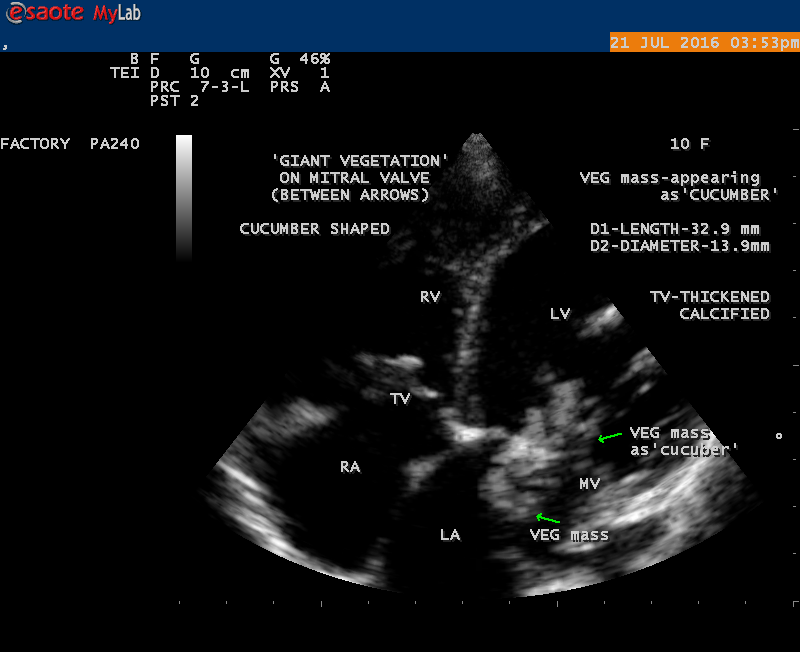
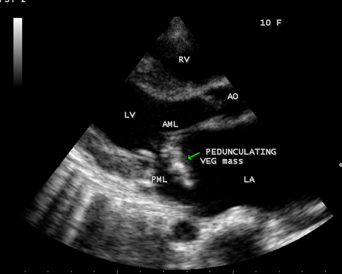
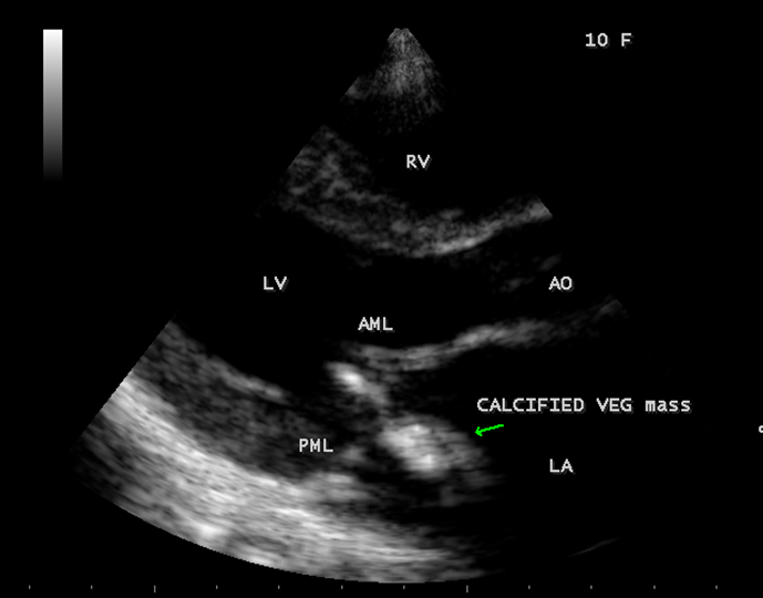
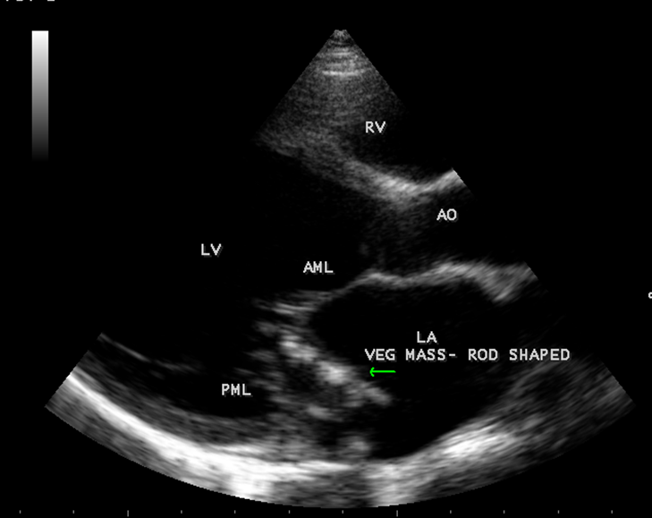
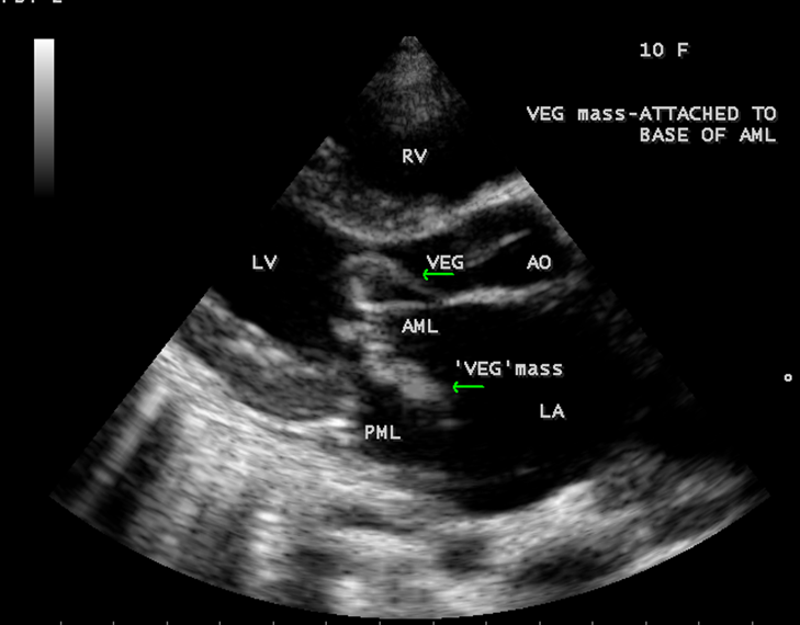
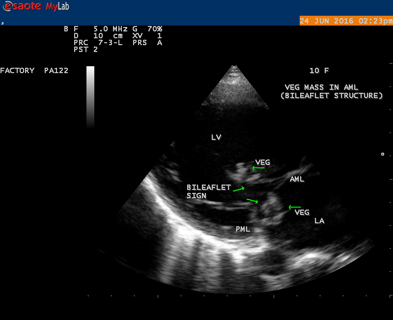
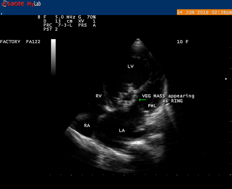
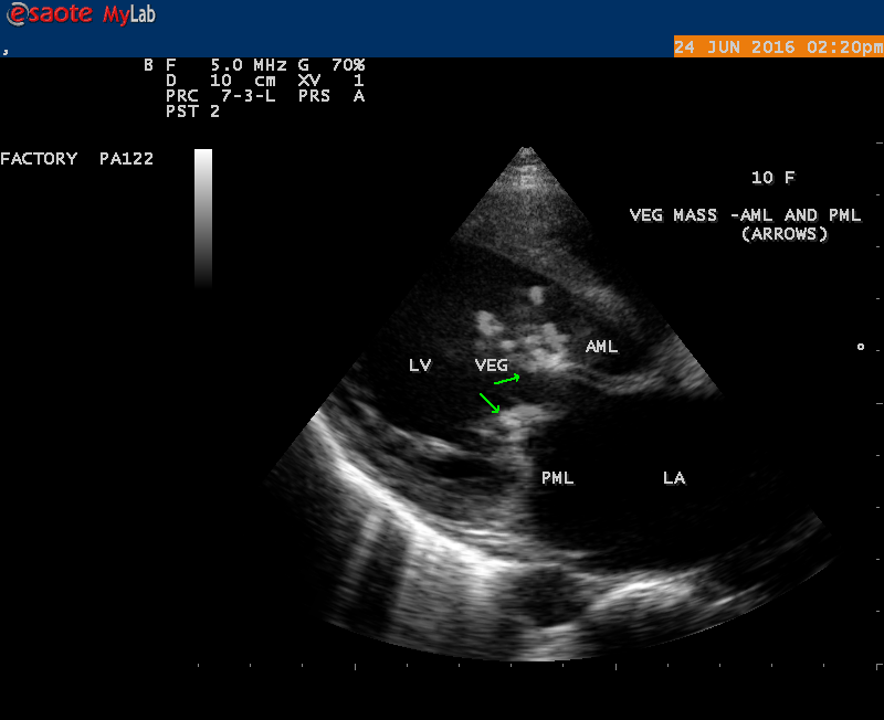
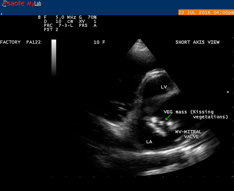
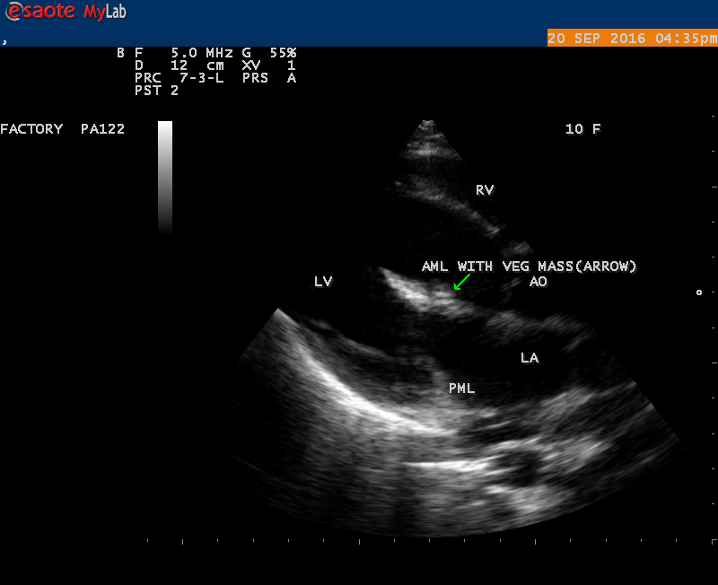
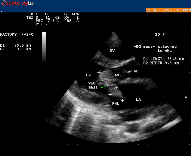
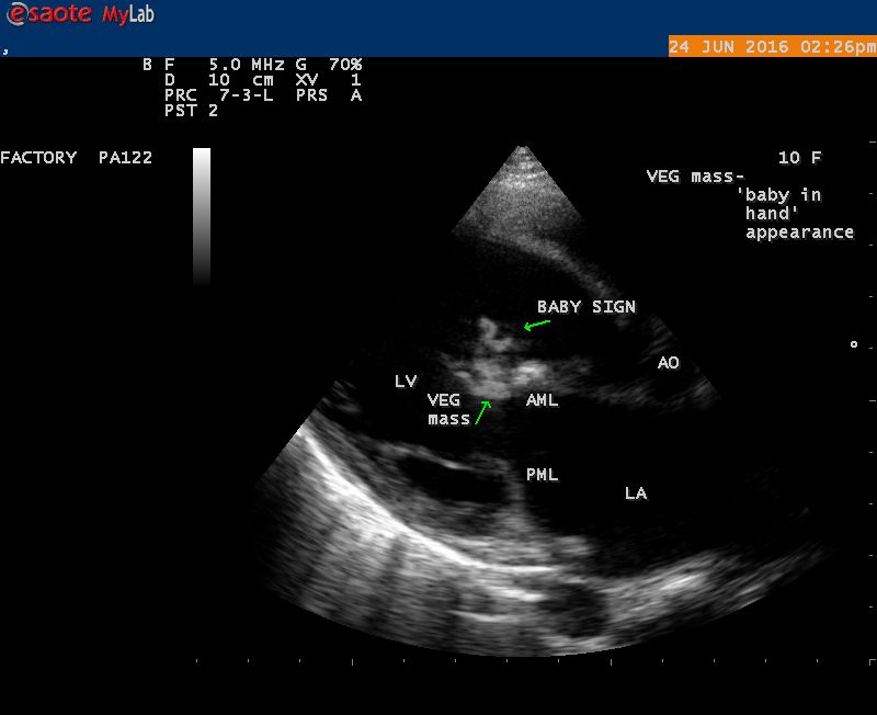
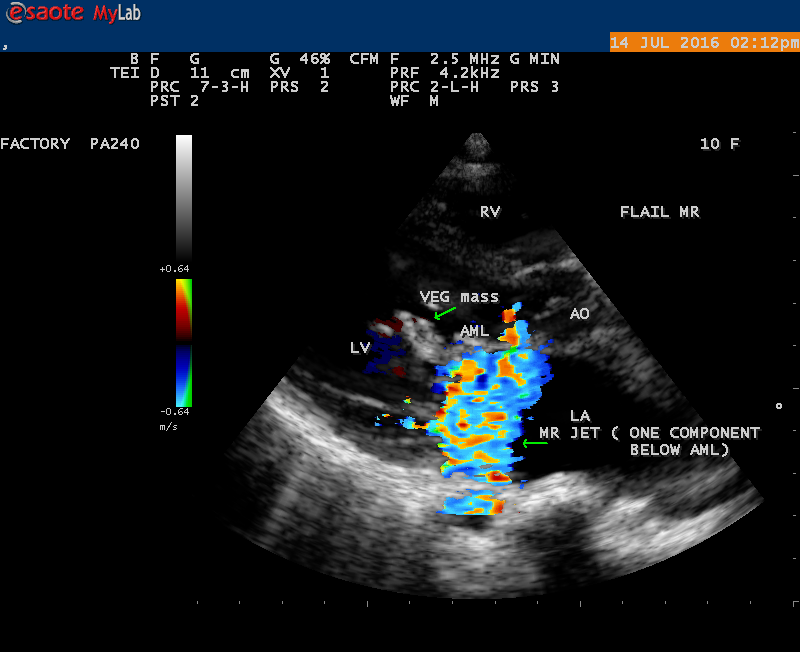
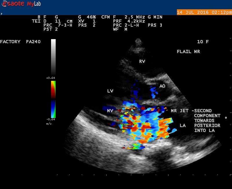
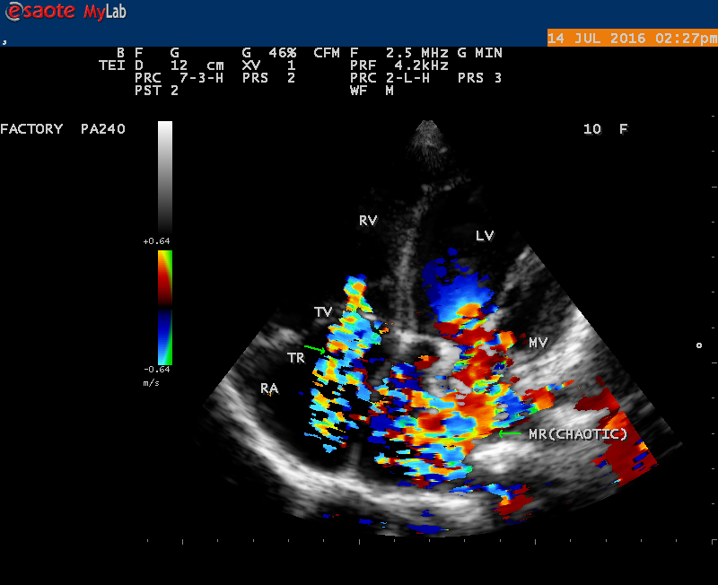
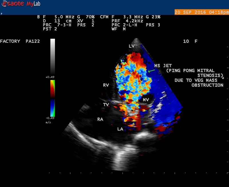
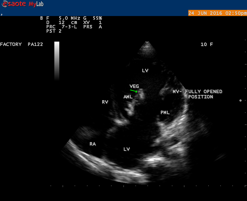
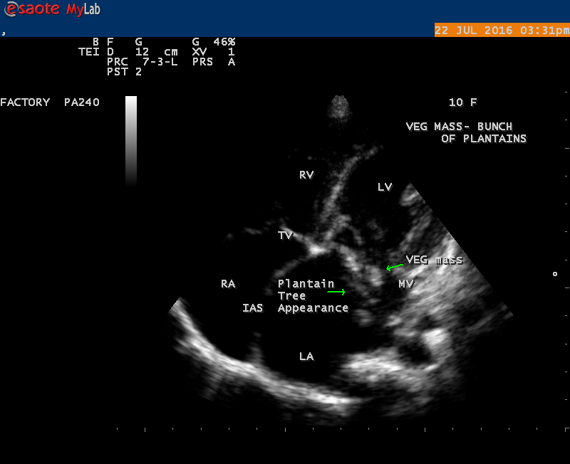
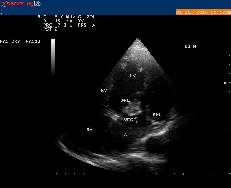
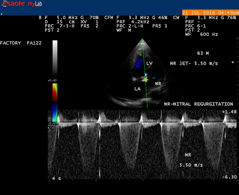
A 10 year female child was referred for echocardiographic evaluation with an apical systolic murmur. The child was having recurrent episodes of rheumatic fever (febrile illness with joint pains) at the age of 5-6 years and taken some treatment from the local medical practitioner, but she was not taken penicillin prophylaxis earlier. The child was remained afebrile for long period and no precipitating factors of infective endocarditis such as dental or genitourinary procedures in the past. General examination revealed normal growth and development, no cyanosis and clubbing and peripheral signs of infective endocarditis such s Osler’s nodes, Janeway lesions, Roths spots and splinter haemorrhages are not present and they are relatively rare in children. Physical examination revealed a grade 3/6 , blowing, high pitched, holosystolic murmur with a constant intensity and duration on dynamic auscultation and loudest at the apex with a radiation to left axilla and transmitted to the left infrascapular area and vertebral coloumn and it is due to the flow generating the murmur is directed posterolaterally within the left atrial cavity, suggesting the murmur of mitral regurgitation due to the rupture of chordae tendineae of anterior mitral leaflet. Blood culture revealed normal. Blood chemistry revealed the positive serum ASO titer, suggesting a recent streptococcal infection and other parameters are normal. X- ray chest reveled moderate cardiomegaly and ECG revealed a left ventricular volume overload pattern of eccentric hypertrophy due to LV dilatation as a result of severe mitral regurgitation and a normal rhythm. Transthoracic echocardiography revealed a giant vegetation ‘popcorn’ like in Figures 1,3 and 4 and ‘cucumber’ like in Figure 2, mainly attached to base and apical portion of anterior mitral leaflet as shown in Figure 31 and manifested in various size and shapes as shown in Figures 1 to 33. A flail anterior leaflet with a disorganized mitral regurgitation jet as shown in Figure 16 and 21 and the posterior leaflet is embedded with vegetation and resulting in ‘kissing forms’ as shown in Figures 13, 14 and 15 in echocardiography imaging. Tricuspid valve is also thickened and calcified as shown in Figure 2 in addition to thickened and calcified mitral leaflets, suggesting an underlying rheumatic etiology predisposing to the formation of vegetation. The child was given 1.2 million units of intramuscular benzathine penicillin injection as a therapeutic and initial prophylaxis dose for rheumatic fever and advised every 3 weeks for life long. Small doses of digoxin and diuretics are also prescribed and advised early surgery (mitral valve replacement) Transthoracic 2D images are as in Figures 1 to 33 are given below


3.Discussion
Etiopathogenesis
The mitral valve apparatus is a very complex structure and all of its components must work together for proper valve function. Mital regurgitation can occur when any one of these elements fails through different mechanisms that affect leaflet coaptation. Intrinsic valvular involvements by degenerative, rheumatic and infective endocarditis produce organic (primary or structural) mitral regurgitation whereas abnormal function of normal leaflets due to impaired ventricular function caused by ischemic heart disease and dilated cardiomyopathy [11], produce functional (secondary)mitral regurgitation as a result of an imbalance between tethering forces ( annular dilatation, LV (left ventricular) dilatation, papillary muscle displacement, LV sphericity) and closing forces (reduced LV contractility, global LV and papillary muscle dyssynchronies and altered mitral systolic annular contraction). Papillary muscle rupture secondary to myocardial infarction defined an organic ischemic MR (mitral regurgitation). A descriptive classification of mechanism was developed by Carpentier in 1983 based upon the movement of leaflets as normal, excessive or restricted [12] as shown in Table-1 by understanding the papillary muscle-chordal-leaflet scaliop relationship which has been defined anatomically by Lam et al and it is modified by Carpentier himself in 1995 to include a mechanism of leaflet restriction during systole causing regurgitation as Type IIIb.
Types | Leaflet motion | Mechanism |
Type 1
Type II
Type III Type IIIa
Type IIIb
| Normal motion
Excessive motion
Restricted motion Restriction during diastole and systole
Restriction during systole only | Annular dilation, clefts, leaflet perforation
Prolapse,flail chords, ruptured papillary muscle
Commissural fusion due to rheumatic heart disease
Chordal tethering, papillary musle displacement |
(Table-1-Mechanism of MR(mitral regurgitation)based on leaflet motion-Carpentier’s functional classification)
Kumar, et al [13] proposed a classification (Duran classification) based on chordal insertion from the two groups of papillary muscles as lateral half of both leaflets is designated with 1 and the medial half with 2.Thus, anterior leaflet is divided into A1 with chordal crossing from the anterolateral papillary muscle and A2 with chordal crossing from the posteromedial papillary muscle. The posterior leaflet scallops are designated as P1(anterolateral) and P2(posteromedial) and a large middle scallop as PM. The PM is a larger scallop and subdivided as PM1 and PM2 based on chordal origin.
When a part of the mitral valve body protrudes into the left atrium beyond the mitral annulus and producing mild regurgitation with a preservation of coaptation is termed as “ billowing valve”. It is stated that the mitral valve billows slightly into the left atrium normally and an exaggerated finding should be termed as “ billowing mitral valve”, extreme form of billowing is “floppy valve” and when chordal rupture, the prolapsed mitral valve is “ flail”. A morphologic abnormality with thickened leaflet ( diastolic thickness > 5 mm) due to redundant tissue is termed “ floppy valve”. When the coaptation line is beyond the annular plane, it is termed as mitral valve prolapse, the leaflet tip is directed towards LV and its most common phenotype is diffuse myxomatous degeneration (Barlow’s disease) in which the middle spongiosa component of the leaflet is unusually prominent and the quantity of acid mucopolysaccharide is increased. When the free edge of the leaflet is completely reversed into the left atrium (LA), the leaflet tip directed towards LA, it is termed as “flail leaflet”, usually as a consequence of chordal rupture[13].. In moderate prolapse, when the leaflet tip remains in the left ventricle, it is called as ‘billowing valve’ and in severe prolapse when the leaflet tips bulges into the left atrium, it is called as ‘flail leaflet”.
Rheumatic valvulitis is characterized by variable thickening of the leaflets at their free edge, chordal fibrosis, rigidity and reduced motion of posterior leaflet in diastole and in some patients, the posterior leaflet remains in a semi-open position throughout the cardiac cycle and the friction of anterior leaflet in systole produce a false aspect of prolapse. This friction causes endothelial damage and results in platelet-fibrin deposition which is more susceptible to colonization by microorganisms. The initially sterile platelet-fibrin nidus called as non bacterial thrombotic endocarditis (NBTE) become secondarily infected by microorganisms circulating in the blood, either from distant source of focal infection as result of transient bacteremia from a mucosal or skin source [14] and establishing an infectious nidus or vegetation on the endocardium and most commonly they are found at the valve closure-contact ( coaptation) line on the atrial surface of the mitral valve [15]. Following successful medical therapy, the vegetative lesions may heal by a process of endothelialization of the surface phagocytes of bacterial debris, sometimes with calcification and subsequent organization by fibroblasts. Vegetation can be attached to any part of the valve and move with the leaflet but in a more chaotic (oscillating) manner and prolapsed through the valve when it opens. Vegetation can prevents leaflet coaptation, and valvular retraction during the healing phase of endocarditis and result in mitral regurgitation. Large vegetation particularly at the mitral valve may result in functional valve stenosis and hemodynamic deterioration [15]. When the infection extends beyond the valve leaflet, distortion of leaflet and chordal rupture may occur, leading to severe regurgitation [16]. Ruptured mitral chordae tendineae (RMCT) are increasingly reported as an important cause of mitral regurgitation [17]. Anterior chordae tendineae rupture of the mitral valve was common in chronic rheumatic valvulitis (CRV) and infective endocarditis. In myxomatous degeneration, the posterior leaflet chordal rupture is more common and the posterior leaflet become ‘flail’ with a ‘saloon door effect’.
Rheumatic mitral regurgitation has a benign course as stated by Bland and James [18] in the analysis of the cases of rheumatic valvular lesions over a 20-year period. Levine and Friedberg have suggested that there are two groups of patients with mitral regurgitation, one group showing rapidly progressive and the other group showing a prolonged, stable and benign course. In most patients with severe primary MR (mitral regurgitation), left ventricular compensation is maintained for years, but ultimately the prolonged hemodynamic overload leads to myocardial decompensation.
Echocardiographic Features
Infective endocarditis is the microbial infection of the endocardial(endothelial) surface of the heart and the formation of vegetation ( a variably sized amorphous mass of platelets and fibrin in which abundant microorganisms and scant inflammatory cells are enmeshed) is the hallmark of the disease and the vast majority of vegetations, however, occur on valve leaflets. The classic approach to the diagnosis of endocarditis by von Reyn does not include echocardiographic findings and it is mainly focused on pathological, clinical and laboratory evidence (positive blood cultures). In 1994, the Duke criteria for the diagnosis of infective endocarditis [20] defined ‘positive echocardiogram’ as vegetation , an oscillating intracardiac mass, a new valvular regurgitation and microbiological evidence was established with a high sensitivity and specificity ( generally about 80%). Steckelberg and Wilson[21] suggest that the incidence of endocarditis in the general population is approximately 5 cases per 100,000 persons-years. Valve redundancy and thickened leaflets (>5mm) by echocardiography identify a population at increased risk for infective endocarditis. The relative risk of infective endocarditis among mitral valve prolapse ranges from 3.5 to 8.2. Rheumatic heart disease is the predisposing cardiac lesion in 20 to 25% of cases in 1970’s and 1980’s[22], in Europe, it is 7-18%[23]. In children, it is less than one in ten cases of infective endocarditis [24] and the mitral valve is most frequently affected in women while the aortic valve in men.
Echocardiography is the only noninvasive method available for direct visualization of endocarditis-induced lesions. Echocardiographic finding in patients with infective endocarditis was initially observed by Dillon [25] and Spangler, et al [26]. The vegetation will grow in size, either as a sessile clump or a highly mobile and even pedunculated mass with the potential for embolization. Vegetation can be detected when the valve attached mass reach a diameter of ≥ 2 to 3 mm [27]. In both children and adults, 2-D echocardiography is usually the more sensitive technique with sensitivity in children up to 80% [28]. Valvular dysfunction due to tissue disruption or large obstructing vegetation can be visualized and quantitated by echocardiogram with Doppler [29]. The detection of a large eccentric jet adhering, swirling, and reaching the posterior wall of the LA is in favour of significant MR (mitral regurgitation) as shown in Figures 22, 24 and 25.
Vegetation
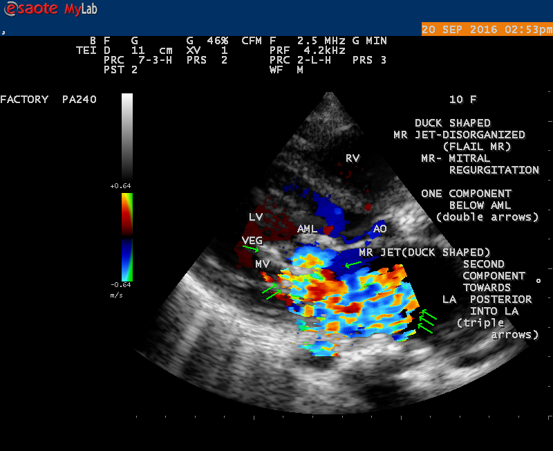
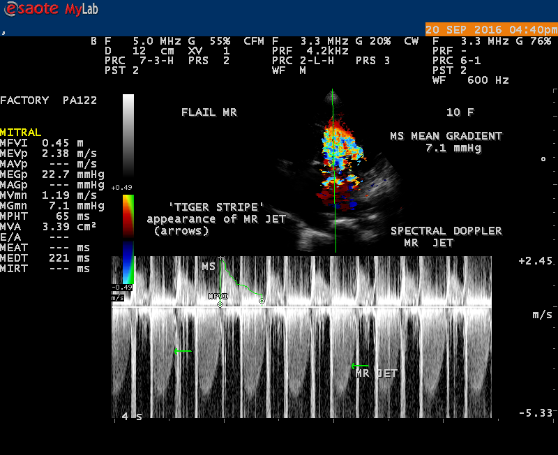
The most common and direct evidence of infective endocarditis is the vegetation and it begins as a microscopic focus of infection and gradually grows into a conspicuous mass. It is typically an irregularly shaped, highly mobile, echogenic mass attached to the free edge of a valve leaflet ( most commonly at the coaptation line) and tends to develop on the ‘upstream’ side of the valve leaflets ( ie, the ventricular side of aortic valve and the atrial side of mitral and tricuspid valves. They may be seesile or pedunculated, but usually has an oscillating or fluttering motion, a typical feature of most vegetations. Vegetation move with the leaflet in a more chaotic (‘oscillating’) manner and it may prolapse through the valve into the LV (left ventricle) as it opens as shown in Figures 3, 4 and 16 and into LA (left atrium) as it closing (Figure 5 and 6) . The mass of vegetation is typically homogeneous with echogenicity similar to that of the myocardium. The infectious process often alter the valvular structure and function. Extensive involvement of the leaflet may result in chordal rupture, leading to severe regurgitation as shown in Figure 21 . Direct and typical signs of RMCT (ruptured mitral chordate tendineae) were chain-flail or whiplash-like changes and had an incidence of 86.7%, causing severe regurgitation and mitral chordal rupture is the leading cause of flail mitral leaflet[30]. A large vegetation may obstruct the valve orifice as shown in Figure 1 and 2 , sometimes termed as “obstructive-type bacterial endocarditis” and producing a functional valve stenosis ( Ping-Pong mitral stenosis [31]) similar to left atrial myxoma as shown in Figure 29.
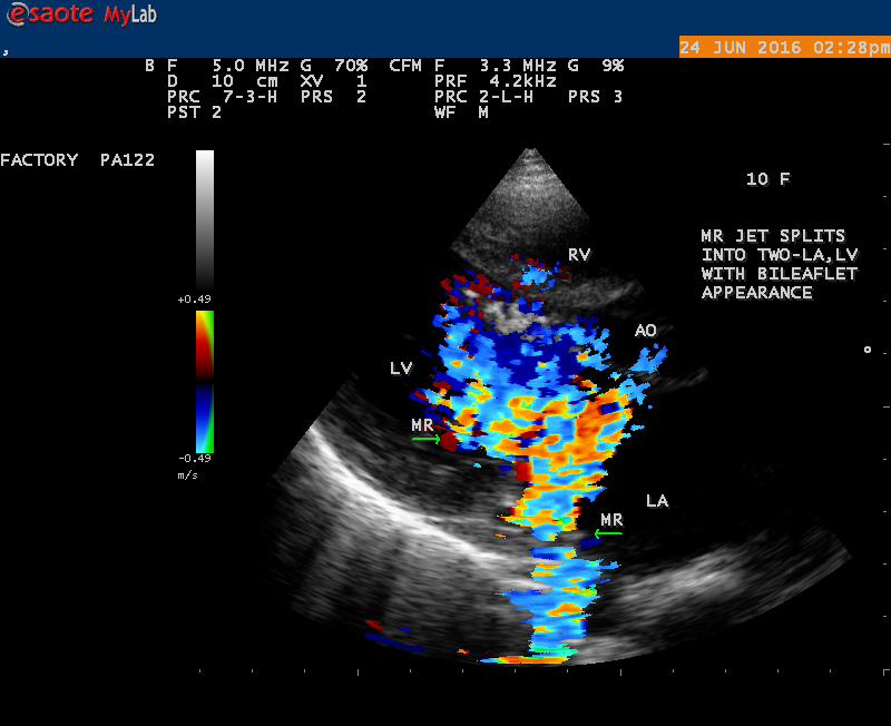
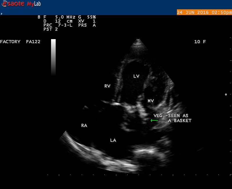
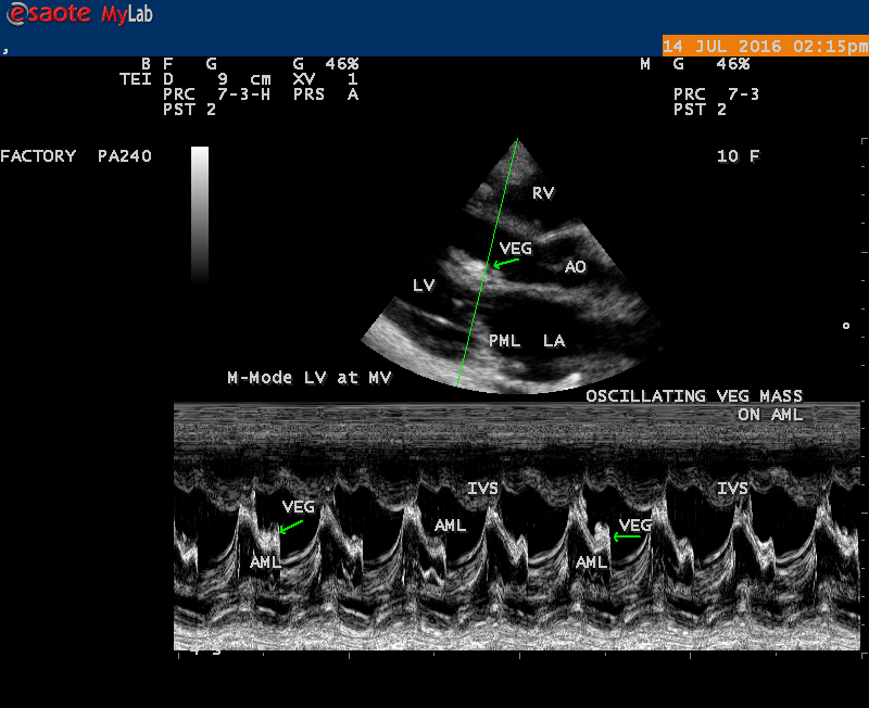
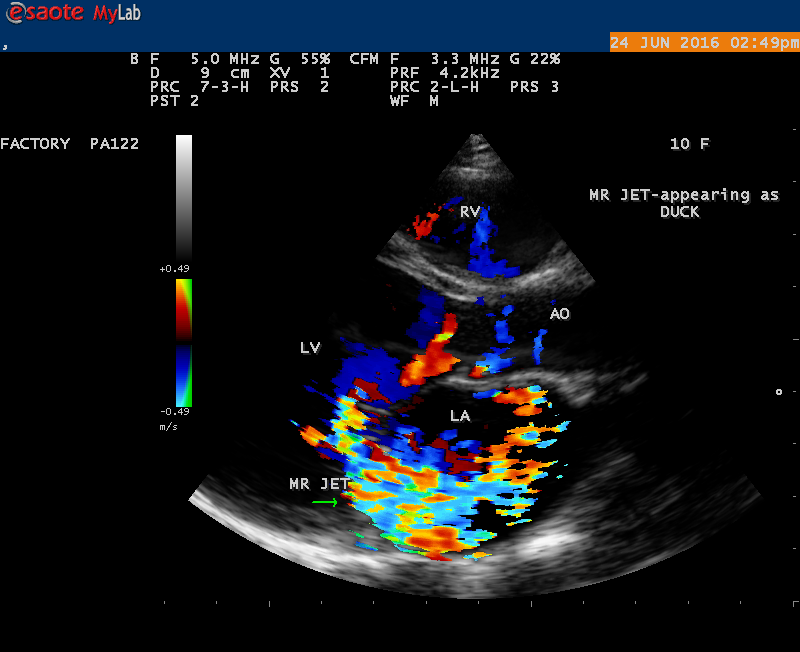
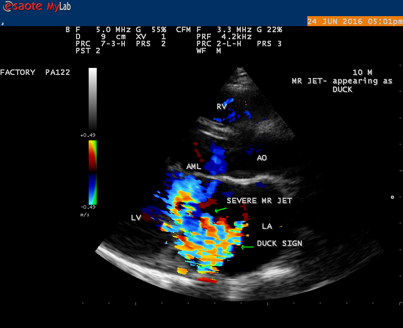
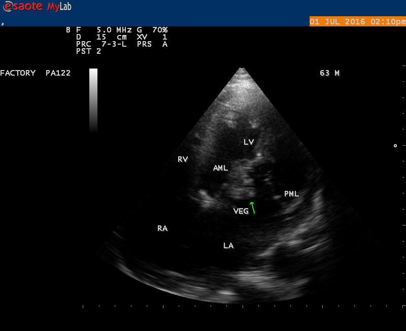
The shape and size of vegetation are quite variable and mostly it is polypoid [32]. The typical vegetation is a ‘sessile’ or ‘ pedunculating’ valve – attached mass. A ‘sessile’ vegetation had to be completely attached to the valve as shown in Figures 34 and 35 in a 63- year old male, in which a large vegetation is attached to the atrial side of anterior mitral leaflet [33-Figure 13.3-A], producing severe mitral regurgitation as shown in Figure 36 and a mobile vegetation showed a pedunculating part prolapsing into the ventricle as shown in Figure 3 and 4 or atrium as shown in Figure 5 [33-Figure 13.1] in a 10-year old female child. A vegetation was considered as ‘definite’ when shaggy echoes in the M-mode study as shown in Figure 20 . and a corresponding mass without restricted valve motion in the two-dimensional echocardiogram were found as shown in Figure 16 and 30 [33-Figure 13.7]. The vegetation vary in size, often being just a few mm and sometimes reaching to 2-3 cm. A vegetation must be atleast 3 to 6 mm in size to be reliably seen. The mean size of vegetation was 0.6 mm (range 3 to 28) and vegetation > 10 mm in diameter was defined as ‘large’ and those ≤ 10 mm in diameter was defined as ‘small’ and ≥ 15 mm is ‘very large’. Vegetations resulting from fungal infections (candida, aspergillus) are usually much bigger than bacterial vegetations and can be so big to be mistaken for a cardiac tumor. The large vegetations are at increased risk for embolic complications [34], especially on the anterior leaflet of the mitral valve with mobility [35]. A vegetation size of 3.2 x 4.4 cm is called as ‘giant vegetation’ on the mitral valve with a fibrillary appearance of the mass [36- Figure 3] as shown in Figure 1 is an important predictor of embolic phenomena in patients with infective endocarditis causing severe mitral regurgitation as ‘Duck’ shaped jets (Figures 24 and 25 ), disorganized (Figure 21) and sometimes the regurigitant jet splits into two components as one into LA and the second one into LV simultaneously as a bileaflet jets (Figure 10 ) similar to bileaflet structure of AML with vegetation masses (Figure 9 ) . The size of the largest vegetation reported on the mitral valve in the literature in patients with bacterial endocarditis is 7x4 cm[37]. In a study of Nunes, et al[38], vegetation size >13 mm was the only independent predictor of mortality, but some studies [39],[40] did not had an increased embolic risk in patients with vegetation focused only on its presence and size and not on their location. Embolic complications may occur in infective endocarditis(20.6%)and were not more prevalent in the groups with large vegetations [41]. However, Wong, et al [42] found an increased need for surgery in patients with a large vegetation (>10 mm).
The size and shape of vegetation vary due to curling of vegetation. The size of vegetation in this child is 35.6 x 9.3 mm as in Figure 17 , 20 x 23.7 mm as in Figure 1 , 32.9 x 13.9 mm as in Figure 2 .
The shape of vegetation varies in this child as ‘popcorn’ like (Figures 1,3 and 4 ), rod-shaped (Figure ), basket shaped (Figure 7 )[33-Figure 13.3], ‘baby in hand’ appearance (Figure 18), ‘cucumber shaped (Figure 2 ) and a ‘bunch of plantain’appearance (Figure 33 ), ring shaped (Figure 19 }, bileaflet structure (Figure 9 )with bileaflet MR jet as shown in Figure 10 . and kissing forms (Figure 13 - parasternal long axis view, Figure 14 - apical four chamber view and Figure 15 - short axis view)
‘Flail’ Mitral Regurgitation (MR)
The anatomic disruption of a portion of the mitral valve apparatus dueto the underlying rheumatic valvulitis with predisposing infective endocarditis which form a vegetation , resulting an eccentric regurgitation jet with orientation opposite in direction of the leaflet having the anatomic defect such as ‘flail’. In the presence of ‘flail leaflet’, the mitral regurgitant spectral signal may have an atypical appearance and the flail portion oscillate in the spectral signal of regurgitant flow stream to produce a ‘tiger stripe’ appearance as shown in Figure 27. associated with ‘whistling’ sound on auscultation[33-Figure 11.85]. The mitral regurgitation (flail MR) jet is chaotic as shown in Figure 26, highly eccentric (Figure 22) and disorganized with one component behind the anterior mitral leaflet and the second component directed towards posterior immediately as in Figure 21 [33- Figure 11.79].
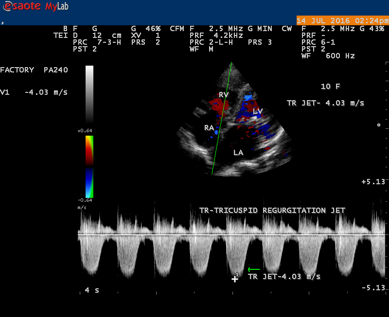
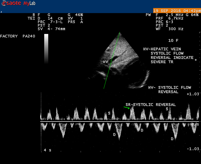
The severity of eccentric MR is underestimated because of coanda effect. If the regurgitant jet area fills < 20> 40% indicate severe regurgitation. The vena contracta ( the neck or narrowest portion of the jet), typically imaged perpendicular to the commissural line in parasternal long axis and apical four chamber views is well defined in both central and eccentric jets, but not in chaotic, disorganized jets due to flail leaflets. Its width < 3> 7 mm defines severe MR and a mean value of > 8mm indicates severe functional MR. The flow convergence method based on PISA (proximal isovelocity surface area) may not applicant for eccentric and multiple jets or complex and elliptical regurgitant orifices to assess the severity of mitral regurgitation.. The adaptation of LV to the increased volume overload is reflected by LV dimensions and ejection fraction.. In chronic compensated phase, the forward stoke volume is maintained through an increase in LV ejection fraction >65% and the patient could be asymptomatic. In chronic decompensated phase of MR, the forward stroke volume decreases and the LA pressure increase significantly. The patient may be still asymptomatic and the LV ejection fraction may be in the low normal range despite the presence of significant muscle dysfunction. The contractile function decreases silently and become irreversible. In the current guidelines, surgery is recommended in asymptomatic patients with severe organic MR when the LV ejection fraction is ≤ 60%. However, in acute stage, the LV ejection fraction increases in response to the increased preload. The end-systolic diameter is less preload dependent than the ejection fraction and it may be more appropriate to monitor the global LV function. The end-systolic diameter > 45 mm also indicate the need for mitral valve surgery [43]. In this child, the LVESD (end-systolic diameter) is 30.7 mm and the ejection fraction (EF) is 66% as shown in Figure 19. New parameters are currently available for a better assessment of LV function. A systolic tissue Doppler velocity measured at the lateral annulus <10>40-50mm) may predict the onset of atrial fibrillation and poor prognosis in patients with organic MR[48]. The excess regurgitant blood entering in the LA may induce acutely or chronically a progressive rise in pulmonary pressure and the presence of TR (tricuspid regurgitation) as shown in Figures 26 and 28 permits the estimate of systolic pulmonary arterial pressure and mitral valve surgery is recommended when it is > 50 mmHg at rest and LA reverse remodeling may occur after surgery. The severe TR may cause a decrease in hepatic vein systolic velocity and systolic flow reversal may occur as shown in Figure 32 and its sensitivity is 80% [49]. The TR (tricuspid regurgitation) jet velocity in this child is 4.03 m/s as shown in Figure 28 which corresponds to a systolic pulmonary artery pressure of 65 mmHg.
Treatment
Pharmacological Therapy
Pharmacological therapy aims to alleviate symptoms and to prevent the progression of LV dysfunction. Afterload reduction is of particular benefit in the management of both acute and chronic forms of mitral regurgitation [50]. Afterload reduction with sodium nitroprusside is life saving in acute conditions such as chordal or papillary muscle rupture. It stabilizes the patient by reducing the impedence to the LV ejection and thus, the regurgitant volume and LA pressure decrease. Dobutamine may be administered along with nitroprusside if hypotension exists. Afterload reduction in chronic, severe MR with vasodilators such as angiotensin converting enzyme inhibitors are beneficial in presence of heart failure, but not beneficial in its absence [51]. In addition to diuretics, digitalis glycosides are indicated in patients having severe MR and heart failure, particularly with established atrial fibrillation in addition to anticoagulants, IE prophylaxis was advised during surgical procedures in these patients.
Surgical Therapy
Asymptomatic patients with severe MR having excellent ventricular function (EF > 70%, ESD(end-systolic diameter) < 40>
Surgery may be considered in mobile vegetation >10 mm in size since the incidence of systemic embolization are increased (33%) compared to those with smaller size (19%) and particularly those on anterior mitral leaflet are uniquely associated with embolic episodes. Urgent surgery is indicated when a large vegetation is associated with embolic events, heart failure and persistent infection[59]. In patients with valvular dysfunction and in whom the infection is controlled with antibiotic therapy and cardiac function is compensated, surgery may be delayed until antimicrobial therapy has been completed. Early surgery is appropriate in patients with large vegetation who may need valve replacement in future since larger vegetations are associated with increased risk of mortality and embolization[60],[61]. Some authors suggest that the causative microorganism is associated with embolic complications rather than the size of vegetation [62]
Since this child is having a giant vegetation with disorganized severe mitral regurgitation due to flail anterior mitral leaflet as the result of chordal rupture, the child was advised MVR(mitral valve replacement) with mechanical prostheses along with removal of vegetation and to continue penicillin prophylaxis with anticoagulant therapy for life long. Since the vegetation is healed, partially calcified, organized mass, the antimicrobial regimen for infective endocarditis was not administered. The anticoagulant therapy is not preferred at this moment since the child had no embolic episodes and the role of prophylactic anticoagulant therapy is questionable in presence large vegetation in the absence of atrial fibrillation, The child was advised small doses of digoxin and diuretics along with penicillin prophylaxis.
Outcome
Several factors play a role in the prognosis of IE(infective endocarditis). Costa,et al described the clinical and echocardiographic scores as the predictors of mortality in infective endocarditis. Age > 40 years ( 4 points), class IV heart failure ( cardiogenic shock)(4 points), uncontrolled sepsis (6 points), conduction disease(5 points), arrhythmias(8 points), a valve with excessive damage (5 points), large and mobile vegetation(4 points)[63]. Mortality rates for scores below 10 were 5.26% and the scores over 20 were 78.9%. The child is having a large and mobile vegetation (4 points), extensive valve damage (5 points) and according to this study, the mortality rate of this child is 5.26%.
4.Conclusion
A giant vegetation attached to the anterior mitral leaflet and presented with various shapes in a 10-year old female child was described by Transthoracic 2D echocardiography imaging. The child had underlying rheumatic involvement of the mitral valve and the infective endocarditis causing chordal rupture leading to flail anterior mitral leaflet with severe mitral regurgitation. The vegetation functionally obstructing the mitral orifice and results in’ Ping- Pong’ mitral stenosis due to mass effect. The child remained asymptomatic without any embolic episodes and heart failure symptoms on follow up of two years.
References
- Oosthock, P.,W.,Wernik, A.,C.,Wisse, L.,J.,Gittenberger-de Groot, A.,C.,(1998) Development of The Papillary Muscles of The Mitral Valve. The Journal of Thoracic And Cardiovascular Surgery,116, 36-46
View at Publisher | View at Google Scholar - Silver,M.D.,Gotlieb,A.,I., Shoen,F.,J.,(2001) Examination of The Heart And Cardiovascular Specimens in Surgical Pathology, Cardiovascular Pathology, 3rd Edition, Churchill Livingstone Publishers, New York, Edinburgh.
View at Publisher | View at Google Scholar - Cole,W.,G.,Chan,D.,Hickey,A.,J.,Wilcken,D.,E.,L (1984) Collagen Composition of Normal And Myxomatous Human Mitral Heart Valves, The Biochemical Journal, 219(2), 451-460.
View at Publisher | View at Google Scholar - Grande-Allen, K.,J., Glabro, A., Gupta, V., Wight, T.,N.,Hascall, V.,C., Vesely,I.,(2004) Glycosaminoglycans And Proteoglycans in Normal Mitral Valve Leaflets And Chordae Associated With Regions of Tensile And Compressive Loading. Glycobiology, 14, 621-533.
View at Publisher | View at Google Scholar - Marron,K.,Yacoub, M.,H., Polak,J,,M.,et al (1996) Innervation of Human Atrioventricular And Arterial Valves, Circulation, 94, 368-375.
View at Publisher | View at Google Scholar - Lam, J.,H.,C.,Ranganathan, N., Wigle,E.,D.,Silver, M.D.,(1970)Morphology of The Human Mitral Valve: Chordae Tendineae: A New Classification, Circulation, 41, 449-458
View at Publisher | View at Google Scholar - Allen,W.,B.,Karp,R.,B.,Louchoukos,N.,T.,(1974) Mitral Valve Replacement, Archives of Surgery,109, 642-645
View at Publisher | View at Google Scholar - Hanson,T.,P., Edwards, B.,S., Edwards, J.,E.,(1985) Pathology of Surgically Excised Mitral Valves, One Hundred Consecutive Cases. Archives of Pathology & Laboratory Medicine,109, 823-829.
View at Publisher | View at Google Scholar - Olson,L.,J., Subramanian,R.,Ackermann, D.,M.,Edwards,W.,D.,(1987). Surgical Pathology of The Mitral Valve, A Study of 812 Cases Spanning 21 Years. Mayo Clinic Proceedings, 62, 22-30.
View at Publisher | View at Google Scholar - Waller,B.,F.,Morrow,A.,G.,Maron,B.,J.,Mclntosch,C.,Roberts,W.,C.,(1982),Etiology of Clinically Isoiated , Severe, Chronic, Pure, Mitral Regurgitation. Analysis of 97 Patients Over 30 Years of Age Having Mitral Valve Replacement. American Heart Journal, 104, 188-196.
View at Publisher | View at Google Scholar - Marwik, T.,H.,Lancellotti,F.,Pierard,L.,(2009)Ischemic Mitral Regurgitation: Mechanisms And Diagnosis. Heart, 95, 1711-1718.
View at Publisher | View at Google Scholar - Carpentier,A.,(1983) Cardiac Valve Surgery: the” French Correction”, Journal of Thoracic And Cardiovascular Surgery, 86, 323-337.
View at Publisher | View at Google Scholar - Kumar,N.,Kumar,M., Duran, C.,M.,G.,(1995) A Revised Terminology for Recording Surgical Findings of the Mitral Valve. Journal of Heart Valve Disease, 4,70-75
View at Publisher | View at Google Scholar - Zoghbi,W.,A., Enriquez-Sarano,M.,Foster,E.,et al(2003) Recommendations for Evaluation of the Severity of Native Valvular Regurgitation With Two-Dimensional And Doppler Echocardiography, Journal of American Society of Echocardiography,16,777-802
View at Publisher | View at Google Scholar - Durack,D.,T., Beeson,P.,B.,(1972) Experimental Bacterial Endocarditis:1. Colonization of a Sterile Vegetation, British Journal of Experimental Pathology, 53!1),44-49.
View at Publisher | View at Google Scholar - Douglas,J.,L., Dismukes,W.,E.,(1992) Surgical Therapy of Infective Endocarditis on Native Valves. In Kaye,D.,(edition); Infective Endocarditis, 2nd edition, New York, Raven Press, Page 397.
View at Publisher | View at Google Scholar - Croft, C.,H., Woodward,W., Elliot, A. et al (1983) Analysis of Surgical Versus Medical Therapy in Active Complicated Native Valve Infective Endocarditis, American Journal of Cardiology, 51, 1650.
View at Publisher | View at Google Scholar - Sedransk,K.,L., Grande-Allen,K.,J.,Vesely,I.,(2002) Failure Mechanics of Mitral Valve Chordae Tendineae, Journal of Heart Valve Disease,11(5),644-650.
View at Publisher | View at Google Scholar - Bland,E.,F., Janes, T.,D.,(1951) Rheumatic Fever And Rheumatic Heart Disease, A Twenty Year Report in 1000 Patients Followed since Childhood, Circulation, 4, 836.
View at Publisher | View at Google Scholar - Durack,D.,T.,Lukes,A.,S.,Bright, D.,K.,(1994)New Criteria for Diagnosis of Infective Endocarditis; Utilization of Specific Echocardiographic Findings, Duke Endocarditis Service, American Journal of Medicine, 98, 200-209.
View at Publisher | View at Google Scholar - Steckelberg, J.,M.,Wilson,W.,R.,(1993) Risk Factor for Infective Endocarditis, Infectious Disease Clinics of North America,7,9-19
View at Publisher | View at Google Scholar - Michel,P.,L.,Acar,J.,(1995) Native Cardiac Disease Predisposing to Infective Endocarditis, European Heart Journal, 16(Supplement B),2.
View at Publisher | View at Google Scholar - Shandre,R.,M.,Shafron,S.,D.,(1996) Infective Endocarditis, Review of 135 Cases Over 9 Years, Clinical Infectious Diseases, 22, 276-286.
View at Publisher | View at Google Scholar - Berkowitz, F.,E.,(1995) Infective Endocarditis, Critical Heart Disease in Infants And Children, 961-986.
View at Publisher | View at Google Scholar - Dillon, J.,C., Feigenbaum,H., Konecke, L.,L., Davis, R.,H.,Chang,S.,(1973) Echocardiographic Manifestations of Valvular Vegetations, American Heart Journal, 86, 698-714.
View at Publisher | View at Google Scholar - Spangler,R.,D., Johnson,M.,C., Holmes, J., Blount,G.,(1973)Echocardiographic Demonstration of Bacterial Vegetations in Active Infective Endocarditis, Journal of Clinical Ultrasound,1,126-128.
View at Publisher | View at Google Scholar - Kavey,R.,E.,W., Frank,D.,M., Byrum,C.,J.,et al(1983) Two-Dimensional Echocardiographic Assessment of Infective Endocarditis in Children, American Journal of Dis ease of Children,137, 851-856.
View at Publisher | View at Google Scholar - Aragam,J.,R.,Weyman, A.,E.,(1994) Echocardiographic Findings in Infective Endocarditis, In Weyman,A.,E.,(ed) Principles And Practice of Echocardiography, 2nd Edition, Philadelphia, Lea & Febiger, Page 1178.
View at Publisher | View at Google Scholar - Yuan,S.,(2009)Clinical Significance of Mitral Leaflet Flail, Cardiology Journal,16(2), 151-156.
View at Publisher | View at Google Scholar - Anandan,et al (2015)Ping-Pong Mitral Stenosis, Cardiology Research, 6(3), 286-288.
View at Publisher | View at Google Scholar - Kradin,R.,L.,(2007) Pathology of Infective Endocarditis, New York Infectious Health Care,101.
View at Publisher | View at Google Scholar - Harvey Feigenbaum, William F.Armstrong, Thomas Ryan(2005)Infective Endocarditis, Feigenbaum’s Echocardiography, 6th Edition, Philadelphia, Chapter 13, Page 342 (Figure 11.79), Page 345 (Figure11.85)
View at Publisher | View at Google Scholar - Page 376(Figure13.1), Page 377(Figure 13.3 A, 13.3 B), Page 379-Figure 13.7A.
View at Publisher | View at Google Scholar - Mugge,A.,(1993) Echocardiographic Detection of Cardiac Valve Vegetations and Prognostic Implications. Infectious Disease Clinics of North America, 7, 877.
View at Publisher | View at Google Scholar - Bayer,A.,S.,Bolger,A.,F.,Taubert,K.,A.,et al(1998) Diagnosis And Management of Infective Endocarditis and its complications, Circulation,98, 2936-2948.
View at Publisher | View at Google Scholar - Tiikenmez Tigens, et al (2014) Giant Mitral Valve Vegetation, Marmara Medical Journal,27,213-215(Figure 3)
View at Publisher | View at Google Scholar - Wachowiak-Baszyriska, H., Kazmierczak,E., Oko-Savnowska,Z., Cieslinski,A., Grajek,S.,(1991) Unusually Large Vegetation on the Mitral Valve in a Patient with Bacterial Endocarditis. Kardiologial Polska(Polish Heart Journal),34(6),367-370
View at Publisher | View at Google Scholar - Nunes, M.,C., Gelape,C.,L.,Ferrari,T.,C.,(2010) Profile of Infective Endocarditis at a Tertiary Care Centre in Brazil During a Seven-Year Period: Prognostic Factors and In-Hospital Outcome, International Journal of Infectious Diseases, 14,e394-398
View at Publisher | View at Google Scholar - Stewart,J.,A.,Silimperi,D.,Harris,P.,Wose,N.,K.,Fraker,T.,D.,Kisslo,J.,A(1980)Echocardiographic Documentation of Vegetative Lesions in Infective Endocarditis:Clinical Implications, Circulation,61,374-380
View at Publisher | View at Google Scholar - Lutas,E.,M.,Roberts,R.,B.,Devereux,R.,B., Prieto,L.,M.(1986) Relation Between the Presence of Echocardiographic Vegetations And the Complication Rate in Infective Endocarditis, American Heart Journal,112,107-113.
View at Publisher | View at Google Scholar - Marina Leitman, Mael Dreznik, Madimir Tyomkin, Theresa Fuchs, Ricardo Krakover, Zui Vered(2012) Vegetation Size in Patients with Infective Endocarditis, European Heart Journal-Cardiovascular Imaging,13,330-338.
View at Publisher | View at Google Scholar - Wong,D.,Chandraratna,P.,A.,N., Wishnow,R.,N., Dusitnanound, V., Nimalasuriya,A., (1983)Clinical Implication of Large Vegetations in Infective Endocarditis, Archives of Internal Medicine, 143, 1874-1877.
View at Publisher | View at Google Scholar - Tribouilloy,C.,Grigioni,F., Avierinos,J.,F Barbieri,A., Rusinaru,D., Szymanski,C., et al(2009)Survival Implication of Left Ventricular End- Systolic Diameter in Mitral Regurgitation Due to Flail Leaflets, A Long-Term Follow-Up Multicenter Study, Journal of American College of Cardiology,54, 1961-1968.
View at Publisher | View at Google Scholar - Agricola,E.,Galderisi,M.,Oppizzi,M., Schinkel,A.,F., Maisano,F., De Bonis, M., et al (2004)Pulsed Tissue Doppler Imaging Detects Early Myocardial Dysfunction in Asymptomatic Patients with Severe Mitral Regurgitation, Heart, 90, 406-410.
View at Publisher | View at Google Scholar - Marciniak,A., Claus,P., Sutherland,G.,R., Marciniak,M.,Karu,T.,Baltabaeva,A., et al (2007) Changes in Systolic Left Ventricular Function in Isolated Mitral Regurgitation. A Strain Rate Imaging Study, European Heart Journal, 28, 2627-2636.
View at Publisher | View at Google Scholar - Lee,R., Marwick, T.,H., (2007) Assessment of Subclinical Left Ventricular Dysfunction in Asymptomatic Mitral Regurgitation, European Journal of Echocardiography,8, 175-184.
View at Publisher | View at Google Scholar - Lancellotti, P., Cosyns,B., Zacharakis,D., Attena, E., Van Camp,G., Gach,O., et al (2008) Importance of Left Ventricular Longitudinal Function and Functional Reserve in Patients with Degenerative Mitral Regurgitation: Assessment by 2-D Speckle Tracking. Journal of American Society of Echocardiography,21,1331-1336
View at Publisher | View at Google Scholar - Messika-Zeitoun,D., Bellamy,M., Avierinos, J.,F, Breen,J., Eusemann,C., Rossi,A., et al(2007) Left Atrial Remodelling in Mitral Regurgitation,Methodologhical Approach, Physiological Determinants, and Outcome Implications: A Prospective Quantitative-Doppler Echocardiographic and Electron Beam- Computed Tomographic Study, European Heart Jopurnal,28, 1773-1781.
View at Publisher | View at Google Scholar - Gonzalez-Vilchez,F., Zarauza,J., Vazquez de Prada,J.,A. Martin Duran,R., Ruano,J., Delgado,C., et al (1994) Assessment of Tricuspid Regurgitation by Doppler Color Flow Imaging: Angiographic Correlation, International Journal of Cardiology, 44,275-283,
View at Publisher | View at Google Scholar - Tischler,MD, Rowan, M.,LeWinfer, M.,M.,(1998) Effect of Enalapril Therapy on Left Ventricular Mass and Volume in Asymptomatic, Chronic, Severe Mitral Regurgitation Secondary to Mitral Valve Prolapse, American Journal of Cardiology, 82, 242.
View at Publisher | View at Google Scholar - Carabella, B.,A.,(2008)The Current Therapy for Mitral Regurgitation, Journal of American College of Cardiology,52, 319-326.
View at Publisher | View at Google Scholar - Shuhaiber,J, Anderson ,R.,J.,(2007)Meta-Analysis of Clinical Outcome Following Surgical Mitral Valve Repair or Replacement, European Journal of Cardiothoracic Surgery,31,267-275.
View at Publisher | View at Google Scholar - Reardon, M.J., David, T.,E.,(1998) Mitral Valve Replacement with Preservation of the Subvalvular Apparatus, Cuurrent Opinion in Cardiology, 14,104
View at Publisher | View at Google Scholar - Ling,L.,H., Enriquez-Sarano,M., Seward,J}j;Jf;Fb,R(1997) Early Surgery in Patients with Mitral Regurgitation due to Flail Leaflets. A Long-Term Outcome Study, Circulation,96,1819.
View at Publisher | View at Google Scholar - Feldman,T.,(2007)Percutaneous Mitral Valve Repair, Journal of Interventional Cardiology,20,488-494
View at Publisher | View at Google Scholar - Grossi,E.,A.,Golloway,A.,C.,Miller,J.,S.,et al (1998) Valve Repair Versus Replacement for Mitral Valve Insufficiency, Journal of Thoracic and Cardiovascular Surgery,115,389.
View at Publisher | View at Google Scholar - Peterson,K.,L(1983)The Timing of Surgical Intervention in Chronic Mitral Regurgitation, Catheter And Cardiovascular Diagnosis, 9,433
View at Publisher | View at Google Scholar - Akins,C.,W.,Hilgenberg,A.,D.,Buckley,M.,J.,et al(1994) Mitral Valve Reconstruction Versus Replacement for Degenerative Ischemic Mitral Regurgitation, Annals of Thoracic Surgery,58,668
View at Publisher | View at Google Scholar - Habib, G.,Hoen,B., Tornos,P.,Thuny,F.,Prendergast,B.,Vilacosta, L., et al(2009)Guidelines on the Prevention, Diagnosis, and, Treatment of Infective Endocarditis, European Heart Journal,30,2369-2413.
View at Publisher | View at Google Scholar - De Casto,S., Magni,G.,Beni,S., et al(1997) Role of Transthoracic And Transesophageal Echocardiography in Predicting Embolic Events in Patients With Active Infective Endocarditis Involving Native Cardiac Valves, American Journal of Cardiology, 80, 1030-1034
View at Publisher | View at Google Scholar - Tischler,M.D, Vaitkus,P.,T.,(1997) The Ability of Vegetation Size on Echocardiography to Predict Clinical Implications: A Meta Analysis, Journal of American Society of Echocardiography,10,562-568
View at Publisher | View at Google Scholar - Cunha,B.,A., Gill,M.,V.,, Lazar,J.,M.,(1996) Acute Infective Endocarditis: Diagnosis And Therapeutic Approach, Infectious Disease of Clinics of North America,10,811-834
View at Publisher | View at Google Scholar - Costa,M.,A., Wollmann,D.,R.,Campos,A.,C., et al(2007) Risk Index for Death by Infective Endocarditis, A Multivariate Logistic Model, Brazilian Journal of Cardiovascular Surgery, 22, 192-200.
View at Publisher | View at Google Scholar
