

Research Article | DOI: https://doi.org/10.31579/2835-8325/046
Corroborating The Toxicological Activity of Lead Acetate in vivo: Using the Liver as The Organ of Keen Interest
Department of Human Anatomy, Faculty of Basic Medical Sciences, University of Uyo, Nigeria.
*Corresponding Author: Edem G.D, Department of Human Anatomy, Faculty of Basic Medical Sciences, University of Uyo, Nigeria.
Citation: Ekanem A.U, Edem G.D, Okon K.A, Archibong E.I, (2023), Corroborating the Toxicological Activity of Lead Acetate in vivo: Using the Liver as The Organ of Keen Interest, Clinical research and Clinical reports 3(1) DOI: 10.31579/2835-8325/046
Copyright: © 2023, Edem G.D. This is an open-access article distributed under the terms of the Creative Commons Attribution License, which permits unrestricted use, distribution, and reproduction in any medium, provided the original author and source are credited.
Received: 11 November 2023 | Accepted: 27 November 2023 | Published: 12 December 2023
Keywords: liver; lead acetate; hepatocytes; portal triad; pathology; sinusoids; leucocytosis
Abstract
Lead acetate has been widely used in the cosmetic industry for a long time but due to its toxicity, presently its use has been limited. This study assessed the effect of lead acetate on hematological indices and the histomorphology of the liver. A total of 20 adult male Albino Wistar rats weighing between 138g to 326g were divided into 4 groups designated as group 1, 2, 3 and 4. Group 1 served as control and was given feed and water ad libitum, while group 2 to 4 served as experimental groups. Group 2 was administered 60mg/kg of lead acetate; group 3 was administered 120mg/kg of lead acetate and group 4 was administered 180mg/kg of lead acetate respectively. Administration of lead acetate was carried out orally within 21 days through the use of orogastric tube. By the end of administration, all the rats were sacrificed after chloroform inhalation and the liver harvested, fixed in 10% buffered formalin, processed and stained using Haematoxylin and eosin staining technique. Blood was collected and stored in EDTA bottles to determine their full blood count. The tissues were observed under a light microscope to observe any pathology. Histologically, the control group showed normal cellular histostructure as there were no pathological changes. Group 2 showed lobular architecture of the liver with normal hepatocytes and average sized central vein. Groups 3 and 4 showed significant pathological changes ranging from inflamed hepatocytes, dilated sinusoids, degenerated portal triad etc. There was no significant change in the hematological indices except in the white blood cells within the group given the highest dosage. In this group, the serum levels of WBC were elevated (leucocytosis).
Introduction
Lead is a poisonous, heavy, bluish-gray metal that exists naturally in the earth crust. Unlike certain other metals like zinc and manganese, which are needed as necessary minerals, lead has no recognized positive functions in the human body [1]. In addition, it cannot be biodegraded nor can it be detoxified by living organisms [2]. Human exposure to lead is common and this may be due to its use in plumbing supplies, lead alloys, lead acid batteries, cable sheathing, paints, dyes, ceramic glazes, leaded gasoline, ammunitions and soldering materials owing to its extraordinary and distinctive qualities [3]. Inhalation, ingestion, and skin contact from a variety of sources such as air, dusts, food, and water are the primary pathways of lead exposure [4]. Inhalation and dermal contact are routes of exposure, more typical of occupational settings, whereas the primary route of exposure for the general population is ingestion [4]. Environmental pollution is the presence of a pollutant in an environment such as air, water, soil, and consequently in food which may be poisonous or toxic and will cause harm to living things in the polluted environment [5]. The excessive number of pollutants such as heavy metals in animal feed and feedstuffs are often due to human actions, resulting from either agricultural or industrial production or accidental or deliberate misuse. Lead is translocated through the food chain to man and animals. Its toxicity depends on its chemical form administrated to the animal, the route of administration and the frequency and duration administered to animals [6]. It is one of the toxic metals and is very dangerous to most organs of the body if exposure exceeds tolerable levels. Accumulation of lead produces damaging effects on the hematopoietic, hepatic, renal, and gastrointestinal systems [7]. The toxicity of lead is closely related to age, sex, route of exposure, level of intake, solubility, metal oxidation state, retention percentage, duration of exposure, frequency of intake, absorption rate, mechanisms and efficiency of excretion. Lead has been associated with various forms of cancer, nephrotoxicity, central nervous system effects and cardiovascular diseases in humans [8]. Animal tissues with the highest concentration are the liver, kidneys and bone. Lead concentrations in milk are usually much lower than blood levels [9]. According to WHO, exposure to lead is estimated to account for 143,000 deaths per year with the highest burden witnessed in developing countries. In recent times, there has been an increase in reported cases of lead poisoning in Nigeria [10, 11]. Similar to other lead compounds, lead acetate is very poisonous and soluble in water [12] and is used as a secret ingredient to sweeten wine and preserve fruit, as a mordant in textile printing and dyeing, as a lead coating for metals, as a drier in paints, varnishes and pigment inks, and as a colorant in hair dyes. It is also used in anti-fouling paints, waterproofing, insecticides, and the gold cyanidation process. Lead acetate has been widely used in the cosmetic industry for a long time. Nowadays it is mainly used in the production of hair coloring products. Although in many places such as Canada, European Union, and California, lead acetate is completely banned in food items and cosmetic products due to its carcinogenicity and reproductive toxicity. It was also used as a remedy for sore nipples [13]. The liver regulates most chemical levels in the blood and excretes a product called bile. This helps carry away waste products from the liver [14]. All the blood leaving the stomach and intestines passes through the liver. The liver processes this blood and also metabolizes drugs into forms that are nontoxic to the body. More than 500 vital functions have been identified in the liver [14]. Therefore, the present work aims to evaluate the effect of different doses of lead acetate on the hematological indices (Red blood cells (RBC), white blood cells (WBC), Platelet count (PLT), and haemoglobin) and the histomorphology of the liver of adult albino Wistar rats in order to corroborate the existing knowledge concerning the toxicity of lead acetate.
Materials and methods
2.1 Materials
The materials used includes twenty adults male Wistar rats, clean wooden cages, water bowls, weighing balance, vital feeds, sawdust, masking tapes, syringe, dissecting set, beaker, dissecting board, universal and EDTA sample bottles, normal saline, phosphate buffered formalin, chloroform, lead acetate, and distilled water.
2.2. Animal Care and Use
A total of 20 male Albino Wistar rats were obtained from the Animal House Unit of the Faculty of Pharmacy, University of Uyo, Akwa-Ibom State, Nigeria. The animals were divided into four groups (5 each), housed in wooden cages with stainless grill tops, and maintained under room temperature and hygienic conditions for seven days. The animals were maintained on standard feeds with water given ad libitum.
2.3. Chemical Preparation and Administration
Lead acetate was dissolved in distilled water to form the trihydrate, Pb (CH3COO)2·3H2O. It was administered according to the concentration gradient for each group. The second, third and fourth groups were ingested orally with sub-lethal doses of lead acetate which were 60 mg/kg, 120 mg/kg and180 mg/kg of the already established oral LD50 of 600mg/kg, respectively for 21 days. The administration was done orally using orogastric tubes.
2.4. Experimental Design
The standard regimens of the chemical for each group are as follows; Group 1 (control), group 2 (60 mg/kg) per day, group 3 (120 mg/kg) per day and group 4 (180mg/kg) per day.
Groups | Regimen | Duration |
Group 1
Group 2
Group 3
Group 4
| Distilled water
10% of LD50 (60 mg/kg)
20% of LD50 (120 mg/kg)
30% of LD50 (180 mg/kg) | -
Once daily for 21 days
Once daily for 21 days
Once daily for 21 days |
Table 1: Administration Regimen of Lead Acetate
2.5. Termination of Experiment
24 hours after the stoppage of administration, the animals were sacrificed after chloroform inhalation. Blood was collected and stored in EDTA bottles to determine their full blood count. The liver was harvested for histological studies.
2.6. Morphometric Analysis
The weight of the liver was measured with the aid of a weighing balance.
2.7 Histological Analysis
The organs were stained with H&E and viewed under the light microscope.
2.8 Statistical Analysis
Data obtained from the study were analyzed using graph prism (version 8.0.2) application and expressed as mean ± standard error of mean.
Results
3.1. Effect of Lead Acetate on Body Weight
There was no significant difference in the body weight of rats among groups except in group 4 given 180 mg/kg @p<0>
| Groups | Before Administration (g) | After Administration (g) | Changein body weight(g) | % Change in body weight(g) |
| Control | 176.6 ± 14.8 | 188.4 ± 14.6 | 11.8 | 6.26 |
| 60 mg/kg | 245.4 ± 25.2 | 256.0 ± 42.9 | 10.6 | 4.14 |
| 120 mg/kg | 188.4 ± 22.3 | 195.2 ±29.4 | 6.8 | 3.48 |
| 180 mg/kg | 184.2 ±6.2 | 175.0 ± 9.9+ | -9.2 | -5.26 |
Values are expressed in mean ± standard error of mean + indicates significance difference between group 4 and others @p<0>
Table 2: Difference in Body Weight Before and After Administration of Lead Acetate
3.2. Effect of Lead Acetate on Liver Weight
There was no significant difference in the liver weight of all groups administered with lead acetate.
| Group | Liver Weight (g) |
| Control Group | 6.47 ± 0.7 |
| 60 mg/kg | 8.70 ± 0.8 |
| 120 mg/kg | 7.45 ± 0.6 |
| 180 mg/kg | 6.21 ± 0.4 |
Values are expressed in mean ± standard error of mean.
No significant difference among groups @p<0>
Table 3: Effect of Lead Acetate on Liver Weight
3.3 Effect of Lead Acetate on Hematological Indices
There was no significant difference for red blood cell (RBC), procalcitonin (PCT) and Hemoglobin count (HGB). However, in WBC, there was a significant difference between control compared with group administered with 180 mg/kg of lead acetate @p<0>
| Groups | RBC | WBC | PCT | HGB |
| Control | 8.06 ± 0.42 | 6.00 ± 0.8 | 0.334 ± 0.03 | 15.7 ± 0.50 |
| 60 mg/kg | 6.78 ± 0.68 | 9.20 ± 1.53 | 0.459 ± 0.05 | 13.1 ± 0.98 |
| 120 mg/kg | 7.92 ± 0.20 | 8.83 ± 1.09 | 0.491 ± 0.06 | 14.8 ± 0.31 |
| 180 mg/kg | 6.93 ± 0.57 | 14.10 ± 1.13 ** | 0.423 ± 0.01 | 12.4 ± 1.13 |
Values are expressed in mean ± standard error of mean
** indicates significance from control group @p<0>
Table 4: Effect of Lead Acetate on Hematological Indices.
3.4. Effect of Lead Acetate on Hepatic Morphology
The effect of lead acetate on the hepatic morphology is shown in the photomicrographs below.
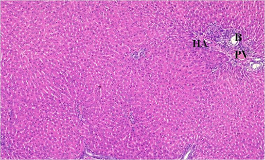
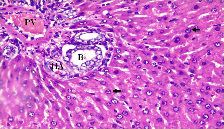
Figure 1: Micrographs of liver section showing normal hepatocytes (arrows), portal triad (B= bile duct, PV=Portal vein & HA= Hepatic artery). No pathological changes seen, 10x and 40x magnifications.
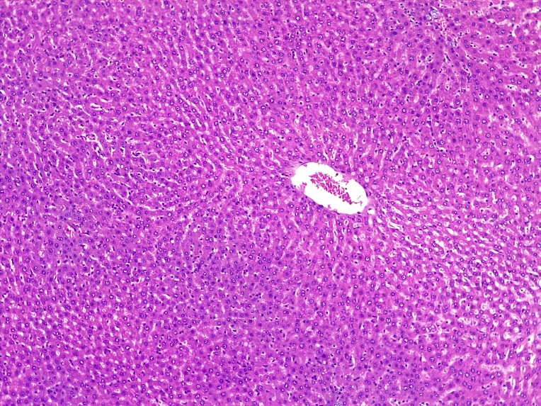
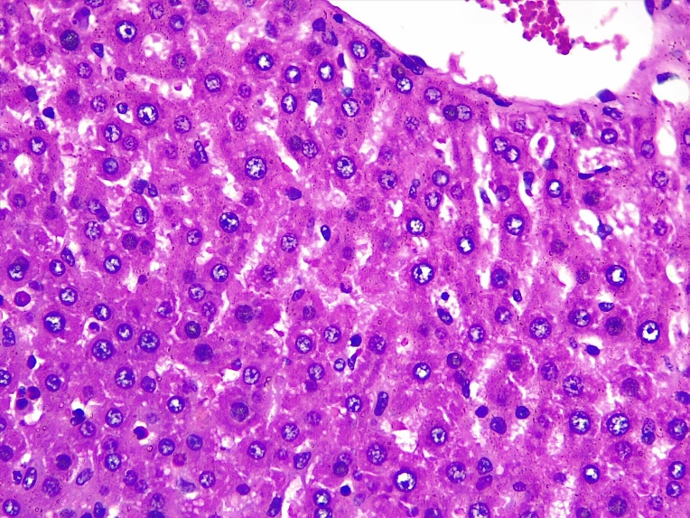
Figure 2: Micrographs of group 2 animals given 60mg/kg of lead acetate showing lobular architecture of the liver with normal hepatocytes (arrows) and average sized central vein (CV) and sinusoid (arrow head). Haematoxylin and Eosin (H&E Stain), 10x and 40x magnifications.
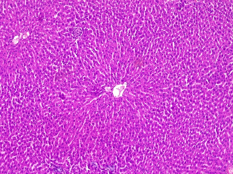
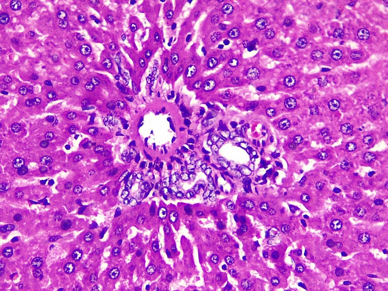
Figure 3: Micrographs of group 3 animals given 120mg/kg of lead acetate showing lobular architecture of the liver with inflamed hepatocytes (arrow) and average sized central vein (CV), dilated sinusoid (arrow head) and portal triad (PT). H&E Stain, 10x and 40x magnifications.
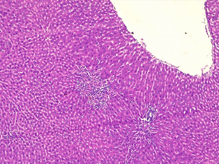
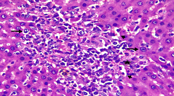
Figure 4: Micrographs of group 4 animals given 180mg/kg of lead acetate showing inflamed hepatocytes (arrows), dilated central vein (DCV), degenerated portal triad (double headed arrow), focal parenchymal inflammation (star) showing inflammatory infiltrates and dilated sinusoids (arrow head), 10x and 40x magnifications.
Discussion
Lead acetate has been reportedly linked to damage to several organs. Zhang et al. (2022) [15] investigated the effect of lead acetate exposure on body weight and glucose homeostasis in mice. The study found that mice exposed to lead acetate had increased body weight gain compared to control mice, as well as disruptions in glucose homeostasis. The researchers also observed changes in the expression of genes involved in lipid metabolism and inflammation in the liver and adipose tissue of the exposed mice. The study suggested that lead acetate exposure may contribute to weight gain and metabolic dysfunction. Li et al. (2021) [16] investigated the effect of chronic exposure to low-dose lead acetate on body weight and glucose homeostasis in rats. The study found that rats exposed to lead acetate had increased body weight gain and disruptions in glucose homeostasis, as well as changes in the expression of genes involved in lipid metabolism and inflammation. The researchers also observed structural changes in the liver and adipose tissue of the exposed rats. The study suggests that chronic exposure to low-dose lead acetate may contribute to weight gain and metabolic dysfunction. These reports are in accordance with our present investigation which indicated that lead acetate dose dependently increased the body weights of the experimental animals administered with 60mg/kg and 120mg/kg of lead acetate respectively and a decrease in the body weight of animals given 180mg/kg of lead acetate. In many instances, weight gain may affect one’s appetite. Al-Saleh et al. (2013) [17] suggested that lead exposure may affect the appetite and energy balance of an individual. Kumar et al. (2021) [18] investigated the effect of lead acetate on hematological parameters in mice. The study found that lead acetate exposure caused significant alterations in the levels of red blood cells, white blood cells, and platelets, as well as changes in hematocrit and hemoglobin levels. The researchers also observed histopathological changes in the spleen, liver, and bone marrow of the exposed mice which suggested that lead acetate exposure may lead to hematological abnormalities. Yang et al. (2022) [19] investigated the effect of lead acetate on hematological disorders and inflammation in rats. The study found that lead acetate exposure caused significant alterations in the levels of red blood cells, white blood cells, and platelets, as well as changes in hematocrit and hemoglobin levels. The researchers also observed increases in inflammatory markers and histopathological changes in the spleen and bone marrow of the exposed rats. The study reported that lead acetate exposure may contribute to hematological disorders and inflammation. According to Gao et al. (2018) [20], lead exposure has been shown to affect full blood count which includes various parameters such as red blood cells, white blood cells, procalcitonin and hemoglobin. There was a recorded decrease in the levels of RBC, WBC, and HGB and a consequent increase in procalcitonin. However, the mechanism by which lead exposure decreases the RBC WBC HGB and increases PCT is not fully understood, leaving researchers to believe that involvement in lead induced-oxidative stress and inflammation might sponsor this [20]. From our findings, there was no significant difference for red blood cell (RBC), procalcitonin (PCT) and hemoglobin count (HGB). This could be a result of the route of chemical administration and the duration of administration. However, in WBC, there was a significant difference in control group compared with group administered 180 mg/kg of lead acetate @p<0>et al. (2021) investigated the histomorphological effects of lead acetate on the liver and kidneys of mice. The study reported that lead acetate exposure caused significant histological changes in the liver, including cellular degeneration, necrosis, and infiltration of inflammatory cells. The study reported that lead acetate exposure could lead to toxic effects on the liver. Ojewole et al. (2022) [21] investigated the histomorphological and biochemical effects of lead acetate on the liver of mice. The study reported that lead acetate exposure caused significant histological changes in the liver, including cellular degeneration, necrosis, and infiltration of inflammatory cells. The researchers also observed an increase in oxidative stress markers and a decrease in antioxidant enzyme activity in the exposed mice. The study suggested that lead acetate exposure may lead to liver damage through oxidative stress. Wang et al. (2019) [22] posited that lead acetate exposure caused significant histological changes in the liver, including vacuolation, necrosis, and infiltration of inflammatory cells.
Conclusion
It is no doubt that lead acetate poses significant risks of hepatic damage. In our study, lead acetate impacted some deleterious burden on the liver tissues by inducing inflammation, etc. We conclude that lead acetate, regardless of the way it is consumed, has detrimental effects on the liver and hematological indices of an individual. Hence, prolonged consumption of lead acetate would lead to hepatic damage.
Competing Interest
Authors declare that no competing interests exist
References
- Ahmed, M, Singh, S, Behari, J. R, Kumar, A. and Siddiqui, M. K. J. (2007). Interaction of leadwith some essential trace metals in the blood of anemic children from Lucknow, Indina. ClinicaChimica Acta, 377: 92-97.
View at Publisher | View at Google Scholar - Brajesh, K, Kumari, S. and Cumbal, F.L. (2014). Plant mediated detoxification of mercury and lead. Arabian Journal of Chemistry, 10(2):2335-2342.
View at Publisher | View at Google Scholar - Gagan, F., Deepesh, G., and Archana, T. (2013). Toxicity of lead: A review with recent updates. Interdisciplinary Toxicology, 5(2):47-58.
View at Publisher | View at Google Scholar - WHO. (2019). Lead poisoning and health. Retrieved on 7th may 2019 from: www.who.int.
View at Publisher | View at Google Scholar - Duruibe, J.O, Ogwuegbu, M.O.C, and Egwurugwu, J.N. (2017). Heavy metal pollution and human biotoxic effects. International Journal of Physical Sciences, 2(5):112-118.
View at Publisher | View at Google Scholar - Baht, R.V. and Moy, G.G. (2015). Monitoring and assessment of dietary exposure to chemical contaminants. WHO: Geneva, 132-149.
View at Publisher | View at Google Scholar - Correia, P.R.M, Oliveira, E, and Oliveira, P.V. (2018). Simultaneous determination of Cd and Pb in foodstuffs by electro-thermal atomic absorption spectrometry. Analytica Chimica Acta; 405(1-2): 205-211.
View at Publisher | View at Google Scholar - Pitot, C.H, and Dragan, P.Y. (2016). Chemical carcinogenesis. Casarett and Doull’s toxicology. 5th ed. New York: McGraw Hill, 201-260.
View at Publisher | View at Google Scholar - Donia, A.M.A. (2018). Lead concentrations in different animal musclesand consumable organs at specific localities in Cairo. Global Veterinary, 2(5):280-284.
View at Publisher | View at Google Scholar - Yahaya, S. (2010). Lead poisoning from mining kills 163 in Nigeria.
View at Publisher | View at Google Scholar - Zinggl, M. (2016). A silent killer: Lead poisoning in Nigeria.
View at Publisher | View at Google Scholar - Hussin, F, Aroua, M.K, and Szlachta, M. (2021). Biochar derived from fruit by-products using pyrolysis process for the elimination of Pb (II) ion: An updated review. Chemosphere, 287(2): 132250.
View at Publisher | View at Google Scholar - Sharma, N, Garg, V, and Arpita, P. (2010). Antihyperglycemic, antihyperlipidemic and antioxidative potential of Prosopis cineraria bark. Indian Journal of Clinical Biochemistry, 25(2): 193-200.
View at Publisher | View at Google Scholar - Hopkins, R. O, Suchyta, M. R, Kamdar, B. B, Darowski, E, Jackson, J. C, and Needham, D. M. (2017). Instrumental Activities of Daily Living after Critical Illness: A Systematic Review. Annals of the American Thoracic Society, 14(8): 1332-1343.
View at Publisher | View at Google Scholar - Zhang, L, Wu, X, Zhang, Y, Ma, J, Ma, Y, Wang, J, and Zhao, J. (2022). Lead acetate exposure induces weight gain and disrupts glucose homeostasis in mice. Ecotoxicology and Environmental Safety, 229: 112974.
View at Publisher | View at Google Scholar - Li, Y, Wu, H, Tang, J, Zhang, Y, Zhao, X, and Wei, Y. (2021). Chronic exposure to low-dose lead acetate induces weight gain and disrupts glucose homeostasis in rats. Environmental Science and Pollution Research, 28(22): 28524-28534.
View at Publisher | View at Google Scholar - Al-Saleh, I, Neptune, S, Abdullah, M, and Abdullah, R. (2013). Birth Outcome measures and maternal exposure to heavy metals (lead, cadmium, and mercury) in Saudi Arabian population. International Journal of Hygiene and Environmental Health. 217:(2-3), 205-308.
View at Publisher | View at Google Scholar - Kumar, V, Singh, R, Singh, S, and Bhatnagar, P. (2021). Effect of lead acetate on hematological parameters in mice: A comparative study. Toxicology Reports, 8:1487-1491.
View at Publisher | View at Google Scholar - Yang, C, Zeng, X, Dong, Y, Liu, D, Wang, J, Zhang, M, and Wang, Z. (2022). Lead acetate exposure induces hematological disorders and inflammation in rats. Environmental Science and Pollution Research, 29(9):12609-12620.
View at Publisher | View at Google Scholar - Gao, X, Guo, Q, and Wang, X. (2018). Effects of lead on the expression of procalcitonin in human umbilical vein endothelial cells. Environmental Science and Pollution Research, 25(7): 6573-6580.
View at Publisher | View at Google Scholar - Ojewole, J. A, Owolabi, J. O, Ojo, O. A, and Abatan, O.M. (2022). Histomorphological and biochemical evaluation of lead acetate-induced liver and kidney damage in mice. Journal of Basic and Clinical Physiology and Pharmacology 33(1).
View at Publisher | View at Google Scholar - Wang, J, Zhang, X, Wang, L, Hu, L, Gao, L, and Li, G. (2019). Effects of lead acetate on the histology, oxidative stress, and apoptosis of the liver in rats. Environmental Science and Pollution Research, 26(12):11715-11723.
View at Publisher | View at Google Scholar
