

Case report | DOI: https://doi.org/10.31579/2834-796X/096
Constrictive Pericarditis and its Therapeutics
Ramachandran Muthiah, President of all Nations, Morning star hospital, Enayam Thoppu, Kanyakumari District, Tamil Nadu state, India.
*Corresponding Author: Ramachandran Muthiah, President of all Nations, Morning star hospital, Enayam Thoppu, Kanyakumari District, Tamil Nadu state, India.
Citation: Ramachandran Muthiah, (2025), Constrictive Pericarditis and its Therapeutics, International Journal of Cardiovascular Medicine, 4(2); DOI:10.31579/2834-796X/096
Copyright: © 2025, Ramachandran Muthiah. This is an open access article distributed under the Creative Commons Attribution License, which permits unrestricted use, distribution, and reproduction in any medium, provided the original work is properly cited.
Received: 24 February 2025 | Accepted: 10 March 2025 | Published: 14 March 2025
Keywords: aortoiliac disease;iliac artery; wrapsody endoprosthesis
Abstract
Aim: To present a case of ‘end-stage’ constrictive pericarditis with clinical manifestations such as ascites mimicking as cirrhosis of liver.
Introduction: ‘End-stage’ constrictive pericarditis has been readily confused with cirrhosis of liver in which there may be ascites, but venous pressure is normal and the neck veins are not engorged. There may be cardiac enlargement in other causes of heart failure. Etiology remains unknown in majority of case and inflammatory process play a central role in its development.
Case Report: A 67-year-old male presented with sudden onset of tachycardia. Clinical examination revealed right-sided heart failure, ‘Egg-shell’ calcification in Chest X-ray and a characteristic echocardiographic feature of pericardial constriction such as septal bounce and dynamic respiratory changes in mitral inflow velocity. The patient was advised medical measures since it is in advanced stage.
Conclusion: When clinical signs of right heart failure become unresponsive to increased doses of diuretics, constrictive pericarditis is more likely the underlying disease since severe, right-sided failure develops in very advanced, the end-stage of the disease.
1.Introduction
The normal pericardium is a fibroelastic sac surrounding the heart and consists of two layers. The visceral pericardium (serous pericardium) is a single layer of mesothelial cells contiguous with epicardium and reflects on itself over the origin of great vessels up to 1 to 2 cm and pulmonary veins. A tough, fibrous layer as a parietal pericardium and the sac created by these layers contain a small amount of fluid (<25 to 50 ml), composed mostly of an ultrafiltrate of plasma. The pericardium increases the cardiac efficacy by limiting the distance of cardiac chambers, protects the heart by reducing external friction and provide a barrier to extension of infection and malignancy. Stretch-sensitive mechanoreceptors sense the changes on cardiac volume and the pericardium is richly innervated with sympathetic and somatic afferents. When the pericardium limits the heart’s ability to function normally either due to accumulation of fluid (pericardial effusion) or scarred and inelastic (constriction), the pericardial compression syndromes such as cardiac tamponade, constrictive pericarditis and effusive-constrictive pericarditis may occur.
In constrictive pericarditis, the thickened, fibrous pericardium that form a non-complaint shell around the heart, inhibits the diastolic filling and it is typically chronic, but variants including acute, subacute, transient, occult and end-stage may occur. Historically, the eponym “ Pick’s disease” was given to constrictive pericarditis with ascites and hepatomegaly [1] and it was diagnosed as having chronic liver disease and so this case had been reported.
2. Case Report
A 67-year old male was admitted with sudden onset of palpitations in the emergency room. ECG revealed tachycardia with a heart rate of 150 bpm as in Figure 4 and blood pressure 110/70 mmHg. Blood chemistry revealed normal. Physical examination showed an engorged neck vein as shown in Figure 2 which fails to decrease with inspiration (Kussmaul’s sign) with a deep Y descent (Freidreich’s sign) reflecting the predominant ventricular filling during early diastole, ascites and pedal edema as shown in Figure 1 suggesting a right-sided heart failure. Auscultation revealed pericardial knock, an early diastolic sound occurs due to cessation in diastolic filling and retraction of apical impulse in systole. X-ray chest revealed “egg-shell’ calcification as shown in Figure 3. Transthoracic echocardiography revealed the features of constrictive pericarditis as in Figures 7 to 12. Since the patient is in end-stage disease, he was given conservative medical measures such as diuretics, antibiotics and anti-inflammatory drugs and the rhythm was controlled with calcium channel antagonist, verapamil 40 mg three times daily as shown in Figures 5 and 6.
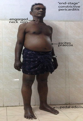
Figure 1. Showing the clinical features of ‘end-stage’ Constrictive pericarditis.
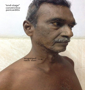
Figure 2. Showing the ‘engorged neck vein’ as a feature of elevated venous pressure in ‘end-stage’ constrictive pericarditis
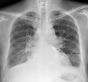
Figure 3. X-ray chest PA (postero-anterior) view showing the ‘egg-shell’ calcification’ – ‘tortoise-shell’ like and flattening of right heart border in ‘end-stage’ constrictive pericarditis.
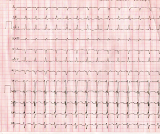
Figure 4. ECG showing tachycardia (rate 150 bpm) in a 67 year old male with ‘end-stage’ constrictive pericarditis.
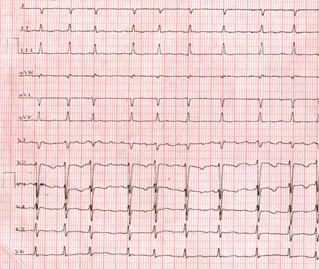
Figure 5. ECG showing atrial fibrillation after controlling the heart rate with verapamil
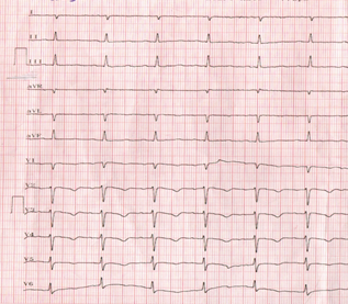
Figure 6. ECG normalizing on continuation of verapamil
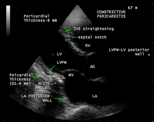
Figure 7. Showing the ‘acute angle’ between the LA (left atrium) and LV (left ventricle) posterior walls and a pericardial thickness of 8 mm.
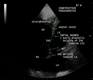
Figure 8. Apical view showing the ‘septal bounce’ as a sign of ventricular interdependence and bulging of IAS (interatrial septum) towards LA (left atrium).
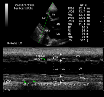
Figure 9. M-mode LV study showing the ‘septal notch’ and ‘ dip and flattening’ of LV posterior wall
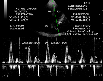
Figure 10. Pulsed Doppler imaging showing ‘the dynamic respiratory change’ of the mitral inflow velocity of constrictive pattern.
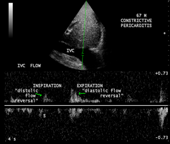
Figure 11. Pulsed Doppler imaging showing the expiratory increase in diastolic IVC flow reversal in both phases of respiration, but more prominent in expiration.
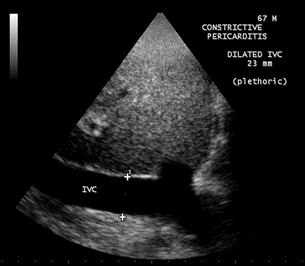
Figure 12. Subcostal view showing the dilated IVC (inferior vena cava- plethoric ) with no respiratory variation.
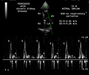
Figure 13. Pulsed Doppler imaging showing the mitral inflow velocity with no respiratory variation of restrictive pattern.
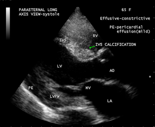
Figure 14. Showing the IVS (interventricular septum) calcification in effusive-constrictive pericarditis as evidenced by mild pericardial effusion with an associated disease of Endomyocardial fibrosis.
3. Discussion
Review of literature
In 1669, Lower [2] described the clinical effects of interference of cardiac diastole by a constricting fibrous pericardium. In 1756, Morgagni [3] contributed to the understanding of pathophysiology of constrictive pericarditis. In 1828, Lancisi described the characteristic syndrome of constrictive pericarditis and in 1842 [4], Chevers was the first to present clearly the clinical picture of chronic constrictive pericarditis. In 1870, Wilks [5] further emphasized this syndrome. In 1896, Pick [6] described the postmortem evidence of adhesive pericarditis and an atypical fibrosis (pseudocirrhosis) of the liver and its capsule in patients with constrictive pericarditis. The thickened peritoneum over the liver is not to be the engorgement of liver itself in so-called Pick’s disease, but rather to the occurrence of acute peritonitis at the time of acute pericarditis followed by residual fibrosis. Concato described the effusion in serous cavities (polyserositis) in patients with constrictive pericarditis is due to the result of cardiac compression, and inflammation of serous membranes is absent or occur secondarily.
Etiopathogenesis
Constrictive pericarditis is most commonly caused by conditions or events that cause inflammation to develop around the heart. Inflammatory process of the pericardium typically causes pain and fluid accumulation and more chronically results in fibrosis and calcification of pericardium with pericardial constriction, the process that inhibit diastolic filling of the heart. The most common antecedents are idiopathic and tuberculosis. In many cases, the etiology is not identified, however, pericardial fibrosis and calcification are often idiopathic in origin [7]. The tuberculosis accounted for 49% of cases of constrictive pericarditis in a series reported in 1962 [8] and it was found to be most common cause in third-world countries such as India [9],[10]. Viral pericarditis is more common in the west and in Europe and North, it is often a sequelae of cardiac surgery and mediastinal irradiation.
Constrictive pericarditis can occur after many pericardial disease process. All causes of pericarditis can lead to subsequent constriction [11]. Rheumatic fever, although frequently accompanied by pancarditis, does not result in chronic constrictive pericarditis and may have pericardial adhesion which are not maximally constricting. The pericarditis associated with uremia and with myocardial infarction is not of the constricting type and most cases of effusive-constrictive pericarditis are often idiopathic, can occur in malignancy of breast and lung, tuberculosis and hypothyroidism (cholesterol pericarditis or ‘gold paint’ pericarditis)
The causes of constrictive pericarditis are shown in Table 1
Most common | Less common |
Idiopathic -42 to 49 % [12] Mediastinal irradiation (5-10 years duration) Following Cardiac surgery-11 to 37 % (post-pericardiectomy-10 to 40 %, previous cardiac surgery-0.3 %) Radiotherapy- 9 to 31 % Post-infectious- 3 to 6 % Tuberculosis-49 % (the most common cause In developing countries) Viral infections (coxsackie virus A and B, Adenovirus, echovirus Pyogenic infections | Other infections Neoplasms -5-17 % (lung-33 %, breast-25 %, adenocarcinoma of intestine [13]) Connective tissue disorders (rheumatoid Arthritis, systemic lupus erythematosis, Scleroderma) Drugs(procainamide,hydralazine,methysergide) Trauma Hereditary-Mulibrey nanism (Mu-muscle, Li-liver, br-brain, ey-eyes, nanism-dwarf) in Finland and United states [14] |
(Table 1- showing the causes of constrictive pericarditis)
Hemodynamic changes
The normal pericardium can stretch to accomodate the physiological changes in cardiac volume. In constrictive pericarditis, the visceral and parietal pericardium are fibrosed and fused together [15], although not necessarily always thickened [16], prevent the heart from expansion and resulting in minimal ability to adapt to volume changes and significant dynamic respiratory variation in blood flow in the chambers of the heart attributed to isolation of the cardiac chambers from intrathoracic respiratory pressure changes, ie, dissociation between intrathoracic and intracardiac pressures with enhanced ventricular interaction as reported by Hatle et al in 1989 [17].
In the heart with a normal pericardium, inspiration causes a decrease in intrathoracic pressure, which is reflected in the cardiac chambers as decrease in intracardiac pressures simultaneously and there is no change in the driving pressure from the lungs across the pulmonary veins into the left atrium and across the mitral valve into the left ventricle. There is some increase in the filling of the right ventricle because of enhanced venous return, but filling of the left ventricle is unaffected throughout the cardiac cycle. In patients with constrictive pericarditis, the rigid pericardium does not allow the decrease in intrathoracic pressure to be transmitted to the left -sided chambers and there is a lower driving force from the lungs into the left side of the heart and the left ventricle becomes underfilled with a reciprocal increase in the filling of the right ventricle and therefore a septal shift occurs [18]. Conversely, during expiration, there is decreased filling of the right ventricle and increased filling of the left ventricle. As both ventricles are sharing the same limited space, the chamber size and function of one ventricle affect the other ventricle and this interaction is known as ‘ventricular interdependence’ since the amount of blood flow into one ventricle is dependent on the amount of blood flow into the other ventricle and it is enhanced in constrictive pericarditis with a discordance in right and left heart fillings.
Once the ventricular diastolic filling reaches the limitations of the pericardial restraint, the pressure and volume in the cavity rise, filling ceases, and congestion occurs [19]. If the right heart chambers are predominantly constricted, there is naturally an engorgement of neck veins as shown in Figure 2, which is constantly engorged in 86% of patients with constrictive pericarditis. The decreased compliance of right ventricle causes a rise in right atrial pressure that is greater than the fall in pleural pressure, ultimately leading to distended neck veins during inspiration [20], called as ‘Kussmaul’s sign’, which may be seen in right ventricular failure, right ventricular infarction, tricuspid stenosis and restrictive cardiomyopathy. It is nonspecific for constrictive pericarditis and reflects an elevation of Jugular venous pressure (JVP) on inspiration rather than the expected decrease in JVP. When the right heart fails because of constriction of left heart chambers, it may simulate the effect of tricuspid valve disease. With more severe constriction, the peripheral edema and ascites occur. In constrictive pericarditis, the ascites occurs early, disproportionately prominent as well as recurrent and appropriately called as ‘ascites praecox’, followed by minimal edema as a manifestation of later part ( end-stage) of the disease and it is usually confined to lower extremities and sacrum whereas in congestive heart failure, the edema appears first and ascites much later.
In isolated constrictive pericarditis, the myocardium is unaffected and therefore the systolic function and early diastolic filling are normal. In the mixed form (constrictive-restrictive- mainly due to radiation –induced, post cardiac surgery), atrophy of myocardial cells and fibrosis may develop during long-term compression by the pericardium. Both the irritation of the heart by the actual process involving the myocardium and the constricting effect of left heart chambers on the right ventricle and right atrium result in atrial arrhythmias such as atrial fibrillation as shown in Figure 5 and less commonly atrial flutter as complications in chronic constrictive pericarditis. With diminution in the output of heart, the blood pressure, especially the pulse pressure tends to be low and the blood pressure decreasing even to the point of disappearance during inspiration, manifested as absence of pulse, an important sign called as ‘paradoxical pulse’ in some of the more advanced cases. Pulsus paradoxus is an exaggeration of normal decrease in systolic blood pressure during inspiration and is formally defined as an inspiratory decrease in systolic blood pressure greater than 10 mmHg during quiet breathing (readily detected by sphygmomanometer, when the reduction is >20 mmHg- it can be detected by simple palpation of brachial artery, severe cases-inspiratory disappearance of pulse) and it is commonly associated with cardiac tamponade, but can be seen occasionally in other conditions such as effusive-constrictive pericarditis, acute severe asthma, acute pulmonary embolism and right ventricular infarction. Normally, the systolic pressure varies with respiratory cycle, but not to the extent seen in pulsus paradoxus. During inspiration, the right ventricle distends due to increased venous return and causes the interventricular septum to bulge into the left ventricle, decreasing the capacity for left ventricular filling and causing a pooling of blood into the pulmonary vessels, which in turn results in a decrease in left ventricular stroke volume, manifested as an exaggerated decrease in the systolic blood pressure [21]. ‘Reversed pulsus paradoxus’(an inspiratory rise in arterial pressure) may occur in hypertrophic obstructive cardiomyopathy [22] and aortic regurgitation tends to prevent the development of pulsus paradoxus despite the presence of cardiac tamponade and it may be absent when the LV and RV diastolic pressure is high or decompression of respiratory changes in pressures as in atrial septal defects. The venous pressure, on the other hand, is very much elevated and frequently exceeding 200 mm of H2O, even exceeding 300 mm of H2O. The salient features of end-stage constrictive pericarditis are shown in Table 2.
High venous pressure Diminution of blood pressure Paradoxical pulse Atrial fibrillation Ascites Low cardiac output (cardiac index = ≤ 1.2 L/m2/minute) Pseudocirrhosis Pedal edema |
(Table 2. showing the salient features of constrictive pericarditis)
Diagnostic Methods
Radiological
The plain radiograph is frequently abnormal in patients with hemodynamically significant constrictive pericarditis [23]. A typical X-ray chest of a patient with constrictive pericarditis shows a normal sized heart (47%) or only mildly enlarged (16%) and moderate to marked enlargement (37%) in effusive-constrictive pericarditis [24]. Cardiac contour abnormalities, particularly the flattening of right cardiac border is a characteristic feature of constrictive pericarditis, but infrequently present. The left atrium, which is covered only partly by the pericardium may be enlarged.
Calcification of pericardium on chest X-ray strongly suggests constrictive pericarditis in patients with features of heart failure ( especially right heart failure) and it is more obvious in regions where the normal fat is found, namely in atrioventricular and interventricular grooves. A localized form of constriction in the mid ventricular segments as a result of localized severe calcification resembling a ‘napkin’ ring shape is termed as “ napkin-ring” constrictive pericarditis [25]. Once calcification has developed, it represents chronic pericarditis and it was present in 40?ses in the Mayo clinic series [26], roughly visible on plain films in 50% of cases [27], [28] which, if present excludes restrictive cardiomyopathy with an overall incidence of 5 to 27 % [29], but it may be as high as 44 % in patients with tuberculous pericarditis [30]. Dalton, et al [31] have reported that only 90 % of patients reveal calcification radiologically compared with those found at postmortem. Three classic patterns of pericardial calcification have been found.
- The calcium can be thin and linear, and appears as “ eggshell calcification” around the margins of the heart ( tortoise shell like) and it is suggestive of constrictive pericarditis as shown in Figure 3
- The calcification also can appear as thick, shaggy, amorphous and historically believed to be specific for tuberculous pericarditis.
- Less dense and patchy calcification is a feature of ‘adhesive pericarditis’ and may occur in the absence of constriction.
Occasionally, extensive calcification involving the interventricular septum [32] may occur in constrictive pericarditis which indicate an associated disease such as endomyocardial fibrosis as shown in Figure 14 . Patients with pericardial calcification are more likely due to idiopathic with features of pericardial knock, atrial arrhythmias as in this patient and have ahigh perioperative mortality.
The normal pericardium is less than 3 mm ( 1-2 mm) thick. Thickening of pericardium occurs heterogeneously with some areas more thicker than other ( thinnest over the left ventricle (0.7-1.2 mm) and the upper limit of the normal for the thickest pericardium is 2 mm). Abnormal pericardial thickening is the most important radiologic diagnostic feature in patients with clinical suspicion of underlying constrictive pericarditis. A thickened pericardium ( > 4 mm) on its own does not indicate constrictive pericarditis [33] and 18-20 % of patients showed normal thickness in proven constrictive pericarditis surgically [34]. Tuberculous constrictive pericarditis is almost always associated with pericardial thickening. Some patients may have pericardial thickening without evidence of constriction as in radiotherapy or open heart surgery due to some pericardial reaction , which can manifest as an increase in pericardial thickness, an expected finding in weeks to months later [35].
In constrictive pericarditis, the parietal pericardium is 4 to 20 mm thick [36] and in those patients showing the normal thickness pericardium on imaging studies such as CT (computed tomography) or MRI (magnetic resonance imaging), the constrictive process may be caused by epicardial constriction rather than pericardial constriction. Mostly, the pericardial thickening is > 3 mm by the time the patient becomes symptomatic and > 6 mm when the patient is clinically in heart failure.
Echocardiography
In most patients with suspected constrictive pericarditis, two-dimensional imaging, M-mode and Doppler are capable of diagnosing the anatomic and pathophysiologic features of constrictive pericarditis.
Two-dimensional imaging
The anatomic features of constrictive pericarditis may be recognized by two-dimensional imaging. The thickened, constricting pericardium affects the posterior left ventricle (pericardial thickness is 8 mm) more than the posterior left atrium, which then expands at a more acute angle respected to the LV wall [37] as shown in Figure 7 . Abrupt anterior or posterior motion of IVS (interventricular septum) in early diastole is common in patients with constrictive pericarditis (CP). In classic CP, the IVS shows a brisk, early diastolic motion towards the left ventricle during inspiration, followed by a rebound in the opposite direction during expiration [38]. This septal bounce (shivering septum), a paradoxical bouncing motion of IVS , reflects the exaggerated interventricular dependence, ( ie, increase in volume of one ventricle causes a decreased volume in the opposite ventricle, caused by reduced ventricular compliance due to a fixed pericardial volume [39]), combined with forceful early diastolic filling , a classic feature of abnormal diastolic septal motion caused by abrupt termination of ventricular filling. IVS bounce (septal shudder) is the most commonest two-dimensional echocardiographic sign of CP as shown in Figure 8 with a sensitivity of 62% and specificity of 93% [40]. Displacement of interatrial septum towards left atrium during inspiration is an another sign of constrictive pericarditis [41] as shown in Figure 8 . The inferior vena cava is dilated (plethoric—dilated with failure to collapse by > 50%) in constrictive pericarditis as shown in Figure 12 without any respiratory variation in its diameter. Dilatation of superior vena cava, right atrium, coronary sinus occur in constrictive pericarditis as a manifestation of elevated venous pressures, but right ventricle may show normal contour with tubular morphology (tubularization).
M-mode findings
A parallel motion of epicardium and parietal pericardium separated by 1 mm thick, echo-free space strongly suggests thickened pericardium [42] and a finding of calcification at the LV apex is more consistent with LV aneurysm [43}. In CP, the LV posterior wall rapidly expands posteriorly during early diastole, followed by abrupt cessation of such movement during mid and late diastole, which corresponds to abrupt termination of rapid ventricular filling [44],[45] and this lack of motion, termed “ flattening” can be best observed with M-mode echocardiography [46],[47]. This flattening of the LV endocardium is the most consistent M-mode finding of constrictive pericarditis. The ‘septal notching’ and ‘abrupt flattening’ of posterior wall is well seen in Figure 9 [48] and the septum is ‘sigmoid-shaped’ with a bulging towards left ventricle and distal straightening.
Doppler Echocardiography
The hallmark of Doppler examination is reciprocal respiratory variation of right and left heart filling [49] caused by interventricular dependence. Because the heart is encased in a rigid shell, when the right heart fills during inspiration, the left heart filling is restricted by the shift of the septum to the left and the opposite changes occur with expiration. Doppler echocardiographic findings include
- Prominent, usually > 25% increase in initial E velocity during expiration and decrease during inspiration as shown in Figure 10, the E wave is greater than A wave in both phases of respiration.
In patients with restrictive cardiomyopathy (an infiltrative process that leads to myocardial stiffening) as in Figure 13 and up to 20% of patients with CP may lack the typical respiratory changes in the mitral inflow in the presence of mixed constrictive-restrictive disease [50] and/or markedly increased left atrial pressure. In such situations, maneuvers that decrease preload (head –up tilt, sitting, semi-recumbent positions or diuretics in patients with markedly elevated left atrial pressure [51] or leg raising in volume depleted states can unmask the characteristic respiratory variation in early mitral inflow velocity since the absence of respiratory variation may be due to the volume status of patient
Atrial fibrillation may complicates the interpretation of respiratory variation of Doppler velocities , but respiratory variation can still be appreciated regardless of cardiac cycle length.
Phasic respiratory changes in the mitral inflow may also be present in patients with chronic obstructive pulmonary disease or severe RV dysfunction and these changes are more gradual and occur later in the respiratory cycle. In such conditions, a marked inspiratory increase in SVC (superior vena cava) systolic forward flow can be helpful to rule out CP [52}. It occurs due to augmentation of blood flow to the right atrium from SVC during inspiration as a result of greater decrease in intrathoacic pressure during inspiration, which generates greater negative pressure changes in the thoracic cavity in chronic obstructive pulmonary disease. In normal and constrictive pericarditis, there is little respiratory variation of systolic flow velocity, but in tamponade, there is marked respiratory augmentation of both systolic and diastolic flow velocities of SVC.
- Increase in diastolic flow reversal in the hepatic venous flow during expiration. The Figure 11 shows the expiratory diastolic flow reversal in IVC (inferior vena cava) flow in constrictive pericarditis.
In advanced constriction or mixed constrictive-restrictive physiology, significant hepatic vein diastolic flow reversal may be seen in both phases of respiration whereas in restrictive cardiomyopathy, it is more prominent during inspiration.
Cardiac catheterization
Cardiac catheterization is usually reserved for unclear cases of constrictive pericarditis and invasive hemodynamic evaluation is occasionally needed when the diagnosis is uncertain by non-invasive methods such as echocardiography. Normally, the systolic pressure in the right ventricle and pulmonary artery does not exceed 25 mmHg. If there is predominant constriction of left heart chambers, pulmonary pressure may be twice or thrice the normal.
Cardiac catheterization can yield classic findings of constrictive pericarditis, but these findings are also present in restrictive cardiomyopathy which include an increase and equalization of end-diastolic pressures in all four cardiac chambers, a ‘dip (or drop) and plateau’ pattern (square root sign) in the ventricular pressure curves as demonstrated by Hansen, et al [53] and a rapid X and Y descents in the atrial pressure curves. If the pressures are approximately equal on both sides on simultaneous recordings, a fluid bolus should theoretically increase LVEDP (LV end diastolic pressure) above RVEDP (RV end diastolic pressure) in restrictive cardiomyopathy [54]. Discordance between RV and LV peak systolic pressures (during peak inspiration, an increase in RV pressure occurs when LV pressure is lowest [55]), a sign of increased ventricular interdependence, can be detected by both invasive hemodynamic monitoring and Doppler echocardiography.
Tachycardia, which abbreviates diastasis, may abolish the plateau in mid and late diastole, but ‘dip’ persists in ventricular pressure tracing of constrictive pericarditis. In patients with occult constriction, saline infusion (1-2 L in 5-15 minutes [56]) may produce an elevation and equalization of ventricular filling pressures, but unequal pressure on both sides may indicate myocardial disease. In patients with effusive-constrictive pericarditis, a condition noticed by Wood [57], in which pericardial fluid accumulates between the thickened, fibrotic parietal and visceral pericardium and the scarred pericardium not only constrict the cardiac volume, but can also causes the pericardial fluid under increased pressure leading to signs suggestive of cardiac tamponade. It is characterized by persistence of elevated right atrial pressure and ventricular diastolic pressure, but previously small or absent atrial Y descent and early diastolic dip in RV pressure tracing become prominent on needle pericardiocentesis [58]. Pulsus paradoxus is often present in this condition, which is uncommon in classic constrictive pericarditis because the inspiratory decrease in intrathoracic pressure is not transmitted to the right heart chambers. In constrictive pericarditis, early diastolic ventricular filling is resistance free and inspiration causes a fall in pulmonary artery wedge pressure with no change in right atrial pressure., but tamponade imposes a pandiasrolic resistance to ventricular filling and equalization of pressures in all four cardiac chambers throughout the respiratory cycle since the chambers are in a finite space within the pericardium. When intrapericardial pressure exceeds the intracardiac pressure, there is interference in diastolic filling and subsequent decrease in cardiac output [59].
Although there are significant differences between these conditions, there is such overlap that these criterias are difficult to apply in an individual case [60],[61].
Management
The treatment of chronic constrictive pericarditis is very much discouraging, but both medical and surgical treatments have been improved greatly. Patients with mild constriction, advanced disease, or mixed constrictive-restrictive disease may not benefit from pericardiectomy and medical measures may be helpful in such situations.
Medical therapy
A subset of patients with constrictive pericarditis undergoes spontaneous resolution or responds to medical therapy [62],[63] and the constriction may be transient or reversible. In these conditions, the constriction is due to inflammation (acute inflammatory pericarditis), mostly caused by prior cardiovascular surgery (after pericardiectomy-25%) and infections (viral, bacterial, or tuberculosis), idiopathic, trauma, malignancy, collagen vascular diseases are accounting for the remaining etiologies. Non-steroidal anti-inflammatory drugs (NSAIDs) are the most frequent treatment and follow-up studies showing resolution within 2 months to 2 years ( usually responding in an average of 8 weeks) and typically effective in idiopathic cases. Ibuprofen is preferred by some experts because it has lower incidence of sideeffects than the other agents [64]. Aspirin is useful in patients with a recent history of myocardial infarction since other NSAIDs tend to impede scar formation [65]. Indomethacin is an acceptable alternative, but it should be avoided in patients with coronary artery disease since it reduces the coronary blood flow by steel effect.
In chronic constrictive pericarditis, the efforts are made to keep the systemic congestion under control. Diuretics should be used sparingly with the goal of reducing , not eliminating the elevated venous pressure, ascites and edema. Loop diuretics ( torsemide if bowel edema is suspected or intravenous furosemide), thiazides and aldosterone antagonists (especially if ascites is present) may be used as a temporary measure and for patients who cannot undergo surgery. Signs of cardiac compression may disappear by the use of suitable antituberculous drugs as in cases of tuberculous etiology and the use of adjunctive corticosteroids remain controversial. The published trials showed a reduction in mortality and no significant decrease in pericardial fluid reaccumulation or progress to constriction [66],[67]. Patients with tachycardia due to auricular flutter or auricular fibrillation may become better with the control of heart rate by digitalis [68] or may prove of limited value [69].
A trial of conservative management for 2 to 3 months is usually advised in hemodynamically stable patients with constrictive pericarditis in the absence of chronic disease [70] as evidenced by cachexia, atrial fibrillation, hepatic dysfunction and pericardial calcification. However, the overzealous use of diuretics is not recommended and may lead to sudden death [71].
Surgical therapy
The surgical treatment of chronic constrictive pericarditis was first recommended by Delorme in 1898 [72] and some years later, the first pericardiectomy for constrictive pericarditis was performed by Franz Volhard collaborates with Viktor Schmieden in 1923. Decreased cardiac output resulting from a chronic constrictive process may require surgical intervention. Currently, pericardiectomy is the only accepted curative treatment for improving cardiac hemodynamics in constrictive pericarditis [73]. Surgery should be undertaken as soon as possible before clinical manifestations become worse. It is better to rely on the stage of pericarditis when considering the pericardiectomy since pericardiectomy is technically difficult if pericardium is still in the effusive or adhesive state. In prolonged constriction, the response may be less dramatic due to the development of extensive atrophy and fibrosis and lower extremity edema may persists even after the relief of systemic venous hypertension because of deep vein incompetence.
Pericardium resection (or pericardial stripping) is a surgical procedure where the entire pericardium is pealed away from the heart, is a delicate time-consuming procedure and somewhat hazardous. It was the custom to approach the heart anteriorly and to free the right heart chambers and if the whole heart is affected or elevated pulmonary pressure, it would be wise to decorticate the left heart chambers posteriorly first, ‘Total pericardiectomy’ (radical resection) was defined as resection of the anterior pericardium between the two phrenic nerves, the basal and posterior aspects of the pericardium over the diaphragm lying on both ventricles, pericardium over the great vessels and both atria. ‘Partial pericardiectomy’ was defined as any pericardial excision that did not meet the criteria for total pericardiectomy and is preferred in patients with high risk of coronary artery disease or myocardial disease. In patients with heavy calcification penetrating the myocardium, the pericardium could be resected in patches and some islands of epicardium and pericardium were left intact with multiple turtle shell incisions.
It is difficult to predict preoperatively which patients are likely to respond to total pericardiectomy. Plasma concentration of cardiac hormone, the brain natriuretic peptide (BNP) is increased in patients with myocardial disease such as restrictive cardiomyopathy (> 100 to 150 ng/L), but not in constrictive pericarditis (< 150>
Waffle procedure
Constrictive pericarditis is somewhat associated with constrictive epicarditis especially in Japanese people. Constrictive epicardial thickness might leads to repeat surgery in some cases. Attempts are made to decorticate the white, fibrous, thickened layer of epicardium over the ventricles. The waffle procedure performed by incising the tight, fibrotic epicardium in a crosshatched manner, releases the epicardial constriction [80],[81],[82].
In effusive-constrictive pericarditis, it is the visceral layer of pericardium, not the parietal layer, that constricts the heart and visceral pericardiectomy must be performed in these cases [83].
Amniotic membrane patches (amniotic stem cell therapy)
In advanced (end-stage) constrictive pericarditis, pericardiectomy may not offer a cure or desired long term results [84]. Current treatment focused on targeting inflammation cental to this disease process and has shown overall positive outcomes [85]. Amniotic stem cell therapy consisting of either stem cells with extracellular matrix or extracellular matrix alone in the form of human amniotic membrane allograft, an emerging anti-inflammatory and antifibrotic treatment [86],[87], applied intraoperatively in patients presenting with constrictive pericarditis [88] and four human allograft membranes were topically placed over the right atrium, right ventricle and left ventricle prior to closure.
Outcome
Long-term clinical outcome varies after pericardiectomy according to the etiology of constrictive pericarditis [89]. Idiopathic cases are related to better survival (88%) followed by post cardiac surgery (‘postoperative constriction’ following CABG, valvular surgery)(68%) and post-radiation (27%). Idiopathic and tuberculous patients showed good prognosis at 5 years after pericardiectomy [90].
Preoperative clinical status such as older age, pulmonary hypertension with concomitant myocardial dysfunction, multi-organ dysfunction, atrial fibrillation and high mitral inflow E velocity in Doppler study [91] are related to poor prognosis and 16% of cases may not show any postoperative improvement due to incomplete removal of the pericardial sac, involvement of visceral epicardium or myocardial atrophy [92]. Low-output syndrome during early postoperative period is the most common problem [93] seen especially in patients with long standing symptomatic pericardial constriction due to remodeling of ventricles and weakening of myocardium and it may gradually improves [94] on follow–up period.
4. Conclusion
“End-stage” constrictive pericarditis has posed a diagnostic dilemma since it presented with features of right-sided heart failure such as dyspnea, ascites, edema and elevated JVP (Jugular venous pressure) [95]. Approximately 9% of patients with acute pericarditis go on to develop constrictive physiology. The most important diagnostic tool is clinical suspicion and cardiac catheterization is no longer performed to diagnose it [96]. Two-dimensional echocardiography is used mainly to rule out other causes of right-sided heart failure and Doppler echocardiography may provide additional diagnostic information and confirm the presence of constrictive physiology. Medical therapy may be used as a palliative measure to control symptoms and to optimize hemodynamics in ‘end-stage’ disease, who are not candidates for surgery [97].
References
- Fowler,N.,O.,(1995) Constrictive Pericarditis: Its History And Current Status, Clinical Cardiology,18, 341-350
View at Publisher | View at Google Scholar - Lower,R.,(1669) Tractatus De Corde.
View at Publisher | View at Google Scholar - Morgagni,J.,B.,(1756) De Sedibus et Causis Morborum per Anatomen Indigatis, Especially Epistles, 16, 22, 53(3), 63 (4,5), Louvain: Typographica Academica.
View at Publisher | View at Google Scholar - Chevers,N.,(1842) Observations On The Disease of The Orifice And Valves of The Aorta. Guy’s Hospital Reports, 7, 387-392
View at Publisher | View at Google Scholar - Wilks,S.,(1870-1871) Adherent Pericardium as a Cause of Cardiac Disease. Guy’s Hospital Reports, 3, 16, 196.
View at Publisher | View at Google Scholar - Pick,F.,(1896) Ueber Chronische Unter Dem Bilde Der Lebercirrhose Verlaufende Pericarditis (Pericarditische Pseudolebercirrhose) Zeitschrift Klinische Medizin, 29, 385-410
View at Publisher | View at Google Scholar - Ling,L.,H., Oh,J.,K.,Schaff,H.,V.,Danielson,G.,K., Mahoney,D.,W.,Seward,J.,B.,et al (1999) Constrictive Pericarditis In The Modern Era: Evolving Clinical Spectrum And Impact On Outcome After Pericardiectomy, Circulation, 100, 1380-1386.
View at Publisher | View at Google Scholar - Robertson,R.,Arnold,C.,R.,(1962) Constrictive Pericarditis With Particular Reference To Etiology, Circulation, 26, 525-529.
View at Publisher | View at Google Scholar - Bawa, Y.,S.,Wahi,P.,L. Mehta,M.,C.,(1960) Pericarditis. A Clinical Survey of 35 Cases, Indian Journal of Medical Society,14, 111-121
View at Publisher | View at Google Scholar - Bashi, V.,V., John,S., Ravikumar, E., Jairaj,J.,Shyamsunder,K.,Krishnaswami,S.,(1988) Early And Late Results of Pericardiectomy Im 118 Cases of Constrictive Pericarditis, Thorax, 43, 637-641.
View at Publisher | View at Google Scholar - Hancock,E.,W.,(1990) Neoplastic Pericardial Disease, Cardiology Clinics, 8, 673-682
View at Publisher | View at Google Scholar - Bertong, S.,C., Thambidorai,S.,K.,Parakh,K., et al (2004) Constrictive Pericarditis; Etiology And Cause-Specific Survival After Pericardiectomy, Journal of American College of Cardiology, 43 (8), 1445-1452
View at Publisher | View at Google Scholar - Shane Patrick Flood, Omar Ayah, Satoshi Furukawa, Robert B Norris (2017) A Rare Cause of Constrictive Pericarditis, BMJ Case Reports
View at Publisher | View at Google Scholar - Voorhes,M.,L., Husson,G.,S., Blackman,M.,S.,(1976) Growth Failure With Pericardial Constriction, The Syndrome of Mulibrey Nanism, American Journal of Diseases of Children, 130, 1146-1148
View at Publisher | View at Google Scholar - White,P.,D., (1951) Chronic Constrictive Pericarditis, Circulation, 4, 288-294.
View at Publisher | View at Google Scholar - Talreja,D.,R., Edwards,W.,D.,Danielson,G.,K.,Schaff,H.,V.,Tajik, A.,J., Tazelaar,H.,D.,et al (2003) Constrictive Pericarditis In 26 Patients With Histologically Normal Pericardial Thickness, Circulation, 108, 1852-1857
View at Publisher | View at Google Scholar - Hatle,L.,K., Appleton,C.,P.,Popp,R.,L.,(1989)Differentiation of Constrictive Pericarditis And Restrictive Cardiomyopathy By Doppler Echocardiography, Circulation, 79, 357-370.
View at Publisher | View at Google Scholar - Camm, Demosthenes,G., Katritsis, Bernard, Gersh,J., John,A., (2013)Constrictive Pericarditis. Clinical Cardiology, Current Practice Guidelines (1st Edition), Page 388, Retrieved, 21 September, 2015.
View at Publisher | View at Google Scholar - Shabetai,R., Fowler,N.,O.,Guntheroth,W.,G.,(1970) The Hemodynamics of Cardiac Tamponade And Constrictive Pericarditis, American Journal of Cardiology, 26, 480-489.
View at Publisher | View at Google Scholar - Bilchik,K.,C., Wise,R.,A.,(2002)Paradoxical Physical Findings Described By Kussmaul: Pulsus Paradoxus And Kussmaul’s Sign, Lancet, 359, 1940-1942.
View at Publisher | View at Google Scholar - Khasnis,A., Lokhandwala,Y.,(2012)Clinical Signs In Medicine: Pulsus Paradoxus, Journal of Postgraduate Medicine, 48, 46-49.
View at Publisher | View at Google Scholar - Massumi,R.,A.,Mason,D.,T.,Zakauddin,V.,et al (1973)Reversed Pulsus Paradoxus, New England Journal of Medicine, 289, 1272.
View at Publisher | View at Google Scholar - Webb,W.,R., Higgins, C.,B.,(2005) Thoracic Imaging: Pulmonary And Cardiovascular Radiology, Philadelphia. (PA): Lippincott Williams And Wilkins
View at Publisher | View at Google Scholar - Hancock, E.,W., (1971) Subacute Effusive,Constrictive Pericarditis, Circulation, 43, 183-192
View at Publisher | View at Google Scholar - Anastasios Milkas, Carlos Van Mieghem, Lieven Van Hoe, Emanuele Barbato, Bernard De Bruyne (2016) The ‘Napkin-Ring’ Constrictive Pericarditis, European Heart Journal- Cardiovascular Imaging, 17(12), 1436
View at Publisher | View at Google Scholar - McCaughan,B.,C.,Shoff,H.,V.,Piehler,J.,M.,Danielson,O.,K.,Otszi,T.,A.,Puga,F.,J.,Pluth,J.,R.,Connolly,D.,C., McGoon,D.,C.,(1985) Early And Late Results of Pericardiectomy For Constrictive Pericarditis, Journal of Thoracic Cardiovascular Surgery, 8,340-350
View at Publisher | View at Google Scholar - Miller,S.,W.,(2005) Cardiac Imaging: The Requisites, 2nd Edition, Philadelphia (PA): Elsevier Mosby.
View at Publisher | View at Google Scholar - Masui,T., Finck,S., Higgins, C.,B.,(1992) Constrictive Pericarditis And Restrictive Cardiomyopathy: Evaluation With MR Imaging, Radiology, 182, 369-373.
View at Publisher | View at Google Scholar - Ling. L.,H., Oh, J.,K., Breen,J.,F., Schaff,H.,V., Danielson,G.,K., Mahoney,D.,W.,et al (2000) Calcific Constrictive Pericarditis: Annals of Internal Medicine, 132 (6),444-450.
View at Publisher | View at Google Scholar - Bozbuga,N.,Erentug,V.,Eren,E.,Erdogan,H.,B.,Kirali,K.,Antal,A.,et al (2003) Pericardiectomy For Chronic Constrictive Tuberculous Pericarditis: Risks And Predictors of Survival, Texas Heart Institute Journal, 30 (3), 180-185.
View at Publisher | View at Google Scholar - Dalton,J.,C., Pearson,R.,J.,Jr.,White, P.,D.,(1952) Constrictive Pericarditis: A Review And Long-Term Follow-Up of 78 Cases. Annals of Internal Medicine, 45, 445-458.
View at Publisher | View at Google Scholar - Marcelo Villaca Lima, Juliano Novaes Cardoso, Cristina Martins Dos Reis Cardoso, Euler Cristovan Ochiai Brancalhao Renan Prado Limaco, Antonio Carlos Pereira Barretto (2011) Constrictive Pericarditis With Extensive Calcification, Arquivos Brasileiros De Cardiologia, 96 (1).
View at Publisher | View at Google Scholar - O’ Leary, S.,M.,Williams,P.,L.,Williams, M.,P., et al (2010) Imaging The Pericardium: Appearances On ECG-Gated 64- Detector Row Cardiac Computed Tomography, British Journal of Radiology, 83 (987), 194- 205.
View at Publisher | View at Google Scholar - Nishimura,R.,A.,(2001) Constrictive Pericarditis In The Modern Era : A Diagnostic Dilemma, Heart, 86, 619-623.
View at Publisher | View at Google Scholar - Wang,Z.,F.,Reddy,G.,P.,Gotway, M.,B.,et al (2003) CT And MR Imaging of Pericardial Disease, Radiographics, 23, S 167-180.
View at Publisher | View at Google Scholar - Hoit, B.,D.,(1990) Imaging The Pericardium, Cardiology Clinics, 8 (4), Philadelphia, W.,B. Walter Burns) Saunders.
View at Publisher | View at Google Scholar - D’Cruz,I.,A.,Dick,A.,Gross,C.,M.,Hand,C.,R.,Lalmalani,G.,G.,(1989)Abnormal Left Ventricular-Left Atrial Posterior Wall Contour: A New Two-Dimensional Echocardiographic Sign In Constrictive Pericarditis, American Heart Journal, 118, 128-132.
View at Publisher | View at Google Scholar - Candell- Riera,J., Garcia Del Castillo,H.,Permanyer-Miralda,G., Soler-Soler,J.,)1978) Echocardiographic Features of The Interventricular Septum In Chronic Constrictive Pericarditis, Circulation, 57, 1154-1158
View at Publisher | View at Google Scholar - Giargi, B.,Mollet,N.,R.,Dymarkowski,S.,et al (2003) Clinically Suspected Constrictive Pericarditis: MR Imaging Assessment of Ventricular Septal Motion And Configuration In Patients And Healthy Subjects, Radiology, 228, 417-424.
View at Publisher | View at Google Scholar - Himelman,R.,B.,Lee, E., Schiller,N.,B.,(1988) Septal Bounce, Vena Cava Plethora, And Pericardial Adhesion: Informative Two-Dimensional Echocardiographic Signs In The Diagnosis of Pericardial Constriction, Journal of American Society of Echocardiography, 1, 333- 340.
View at Publisher | View at Google Scholar - Vilacosta, I., San Roman Calvar,J.,A., Iturralde Prieto,E., Gomez Recio, M., Martinez Elbal,L.,(1991) Transesophageal Echocardiographic Features of The Atrial Septum In Constrictive Pericarditis, American Journal of Cardiology, 68, 271-273.
View at Publisher | View at Google Scholar - Hoit,B.,D.,(1997) Pericardial Heart Disease, Current Problems in Cardiology, 79, 355-400.
View at Publisher | View at Google Scholar - Thomas, M. Bashore, Christopher,B.,Granger, Kevin,P. Jackson, Manesh R. Patel (2016), Heart Disease, Current Medical Diagnosis & Treatment, Chapter 10, Constrictive Pericarditis, Page 424-425.
View at Publisher | View at Google Scholar - Feigenbaum,H.,(1972) Echocardiography, Philadelphia, Lee & Febiger
View at Publisher | View at Google Scholar - Popp,R.,L.,(1976) Echocardiographic Assessment of Cardiac Disease, Circulation, 54, 538-552
View at Publisher | View at Google Scholar - Schnittger,I.,Bowden,R.,E., Abrams,J.,Popp,R.,L.,(1978) Echocardiography: Pericardial Thickening And Constrictive Pericarditis, American Journal of Cardiology, 42, 388-395.
View at Publisher | View at Google Scholar - Voelkel,A.,G.,Pietro,D.,A.,Folland, E.,D.,Fisher,M.,L.,Parisi, A.,F.,(1978) Echocardiographic Features of Constrictive Pericarditis, Circulation, 58, 871-875.
View at Publisher | View at Google Scholar - Dai-Biano,et al (2009) Role of Echocardiography In The Diagnosis of Constrictive Pericarditis, Journal of The American Society of Echocardiography, 22(1), 24-33.
View at Publisher | View at Google Scholar - Klein,A.,L.,Cohen, G.,I.,(1992) Doppler Echocardiographic Assessment of Constrictive Pericarditis, Cardiac Amyloidosis, And Cardiac Tamponade, Cleveland Clinical Journal of Medicine,59, 278-290.
View at Publisher | View at Google Scholar - Oh,J.,K.,Hatle, L.,K.,Seward,J.,B.,Danielson,G.,K.,Schaff,H.,V.,Reeder, G.,S.,et al (1994) Diagnostic Role of Doppler Echocardiography In Constrictive Pericarditis, Journal of American College of Cardiology, 23, 154-162.
View at Publisher | View at Google Scholar - Oh,J.,K.,Tajik, A.,J.,Appleton, C.,P.,Hatle, L.,K.,Nishimura,R.,A.,Seward,J.,B.,(1997) Preload Reduction To Unmask The Characteristic Doppler Features of Constrictive Pericarditis: A New Observation, Circulation, 95, 796-799.
View at Publisher | View at Google Scholar - Boonyaratavej,S.,Oh,J.,K.,Tajik, A.,J.,Appleton,C.,P., Seward,J.,B.,(1998) Comparison of Mitral Inflow And Superior Vena Cava Doppler Velocities In Chronic Obstructive Pulmonary Disease And Constrictive Pericarditis, Journal of American College of Cardiology, 32, 2043-2048
View at Publisher | View at Google Scholar - Hansen, A.,T., Eskildsen,P.,Gotzsche,H.,(1961) Pressure Curves From The Right Auricle And The Right Ventricle In Chronic Constrictive Pericarditis, Circulation, 3, 881-888.
View at Publisher | View at Google Scholar - Tyberg,T.,I.,Goodyer, A.,V.,Hurst, V.,W.,3rd et al (1981)Left Ventricular Filling In Differentiating Restrictive Amyloid Cardiomyopathy And Constrictive Pericarditis, American Journal of Cardiology, 47,791.
View at Publisher | View at Google Scholar - Hurrell,D.,G., Nishimura,R.,A., Higano,S.,T.,et al (1996) Value of Dynamic Respiratory Changes In Left And Right Ventricular Pressures For The Diagnosis of Constrictive Pericarditis, Circulation, 93, 2007-2013.
View at Publisher | View at Google Scholar - Bush,C.,A., Stang,J.,M., Wooley, C.,F.,Kilman,J.,W.,(1977) Occult Constrictive Pericardial Disease: Diagnosis By Rapid Volume Expansion As A Correction By Pericardiectomy, Circulation, 56, 924-930.
View at Publisher | View at Google Scholar - Wood,P.,(1961) Chronic Constrictive Pericarditis, American Journal of Cardiology, 7,48-61.
View at Publisher | View at Google Scholar - Spodick,D.,H., Kumar,S.,(1968) Subacute Constrictive Pericarditis With Tamponade, Diseases of The Chest, 54, 62-66.
View at Publisher | View at Google Scholar - Spodick, D.,H.,(1983) The Normal And Diseased Pericardium, Current Concepts of Pericardial Physiology , Diagnosis And Treatment, Journal of American College of Cardiology, 1, 240-251.
View at Publisher | View at Google Scholar - Shabetai,R.,(1992) Controversial Issues In Restrictive Cardiomyopathy, Postgraduate Medical Journal, 68 (Supplement-1), 547-551)
View at Publisher | View at Google Scholar - Vaitkus,P.,T., Kussmaul,W.,G.,(1991) Constrictive Pericarditis Versus Restrictive Cardiomyopathy: A Reappraisal And Update of Diagnostic Criteria, American Heart Journal, 122, 1431-1441.
View at Publisher | View at Google Scholar - Haley,J.,H.,Tajik,A.,J.,Danielson,G.,K.,Schaff,H.,V.,Mulwagh,S.,L.,Oh,J.,K.,et al (2004) Transient Constrictive Pericarditis: Causes And Natural History, Journal of American College of Cardiology, 43, 271-275.
View at Publisher | View at Google Scholar - Sagrista-Sauleda,J., Permanyer-Miralda,G.,Candel-Riera,J.,et al (1987)Transient Cardiac Constriction: An Unrecognized Pattern of Evolution In Effusive Acute Idiopathic Pericarditis. American Journal of Cardiology, 59,961.
View at Publisher | View at Google Scholar - Troughton,R.,W.,Asher,C.,R.,Klein,A.,L.,(2004)Pericarditis: Lancet, 363, 717-727
View at Publisher | View at Google Scholar - Lange,R.,A.,Hillis,L.,D.,(2004)Clinical Practice, Acute Pericarditis, New England Journal of Medicine, 351, 2195-2202.
View at Publisher | View at Google Scholar - Mayosi,B.,M., Burgess,L.,J.,Doubell,A.,F.,(2005)Tuberculous Pericarditis, Circulation,112, 3608-3616.
View at Publisher | View at Google Scholar - Ntsekhe,M., Wiysonge,C., Volmink,J.,A.,Commerford,P.,J.,Mayosi,B.,M.,(2003)Adjuvant Corticosteroids For Tuberculous Pericarditis, Promising, But Not Proven, Quarterly Journal of Medicine, 96, 593-599.
View at Publisher | View at Google Scholar - Paul D. White (1951) Chronic Constrictive Pericarditis, Circulation, 4,288-294.
View at Publisher | View at Google Scholar - Anurag Mehta, Mahaveer Mehta, Abnash C. Jain (1999) Constrictive Pericarditis, Clinical Cardiology, 22, 334-344.
View at Publisher | View at Google Scholar - Brain D. Hoit (2017)-Pericarditis-Cardiovascular Disorders-Merck Manuals Professional Edition, Merck Sharp & Dohme Corp, Kenilwork, NJ,USA.
View at Publisher | View at Google Scholar - Marshall,A.,Ring,N., Lewis,T.,(2006)Constrictive Pericarditis: Lessons From The Past Five Years Experience In The South West Cardiothoracic Centre, Clinical Medicine, 6, 592-597.
View at Publisher | View at Google Scholar - Delorme (1898) Sur Un Traitement Chirurgical De La Symphyse Cardiopericardique. Bull et Memb. Soe Chir, Paris, 24, 918: Gaz D. Hop, 71,1150.
View at Publisher | View at Google Scholar - Augustin,P.,Desmard,M.,Mordant,P.,et al (2011) Clinical Review. Intrapericardial FibrinolysisIn Management of Purulent Pericarditis, Critical Care, 15, 220.
View at Publisher | View at Google Scholar - Semrad, Michal (2014) Cardiovascular Surgery, Charles University,p 114. Retrived, 21, September, 2015.
View at Publisher | View at Google Scholar - Chowdhury,U.,K.,Subramanian,G.,K.,Kumar,A.,S.,et al (2006) Pericardiectomy For Constrictive Pericarditis: A Clinical, Echocardiography, And Hemodynamic Evaluation of Two Surgical Techniques, Annals of Thoracic Surgery, 81, 522-529.
View at Publisher | View at Google Scholar - DeValeria,P.,A., Baumqartnes,V.,V.,A.,Casale,A.,S.,et al (1991) Current Indications, Risks, And Outcome After Pericardiectomy, Annals of Thoracic Surgery, 52, 219-224.
View at Publisher | View at Google Scholar - Berlog, S.,C., Thambidorai,S.,K.,Parakh,K.,et al (2004) Constrictive Pericarditis: Etiology And Cause-Specific Survival After Pericardiectomy, Journal of American College of Cardiology, 43, 1445-1452.
View at Publisher | View at Google Scholar - Ling,L.,H.,Oh,J.,K.,Schaff,H.,V.,et al (1999)Constrictive Pericarditis In The Modern Era: Evolving Clinical Spectrum And Impact On Outcome After Pericardiectomy, Circulation, 100, 1380-1388.
View at Publisher | View at Google Scholar - Seifat, F.,C.,Miller,D.,C.,Oesterle,S.,N.,Oyer,P.,E.,StIinson,E.,B.,Shumway,N.,E.,(1985)Surgical Treatment of Constrictive Pericarditis: Analysis of Outcome And Diagnostic Error, Circulation, 72(Supplement II), 264-273.
View at Publisher | View at Google Scholar - Heimbecker,R.,O., Smith,D.,Shimizu,S., Kestle,J.,(1983) Surgical Technique For The Management of Constrictive Epicarditis Complicating Constrictive Pericarditis (the Waffle Procedure), Annals of Thoracic Surgery, 36(5), 605-606.
View at Publisher | View at Google Scholar - Shiraishi,M.,Yamaguchi,A., Muramatsu, Kimura,N., Yuri,K., Matsumoto,H., et al (2015) Validation of Waffle Procedure For Constrictive Pericarditis With Epicardial Thickening, General Thoracic And Cardiovascular Surgery, 63(1),30-37.
View at Publisher | View at Google Scholar - Matsuura,K., Mogi,K., Takahara,Y.,(2015)Off-Pump Waffle Procedure Using An Ultrasonic Scalpal For Constrictive Pericarditis, European Journal of Cardiothoracic Surgery, 47, e220-222.
View at Publisher | View at Google Scholar - Hancock,E.,W.,(2004) A Clear View of Effusive-Constrictive Pericarditis, New England Journal of Medicine, 350,435.
View at Publisher | View at Google Scholar - Little,W.,C., Freeman,G.,L.,(2006)Contemporary Review In Cardiovascular Medicine: Pericardial Disease, Circulation, 113, 1622-1632.
View at Publisher | View at Google Scholar - Syed,F.,F., Schaff,H.,V.,Oh,J.,K.,(2014)Constrictive Pericarditis-A Curable Diastolic Heart Failure, Nature Reviews Cardiology, 11(9), 530-544.
View at Publisher | View at Google Scholar - Hemphil,C.,Stavoe,K.,Khalpey,Z.,(2014)First In Man: Amniotic Stem Cell Injection Promotes Scar Remodeling And Healing Processes In Late-Stage Fibrosis, International Journal of Cardiology, 174(2), 442-443.
View at Publisher | View at Google Scholar - Khalpey,Z., Marsh, K.,M.,Ferng,A., Riaz,I.,B., Friedman,M.,Indik,J., Avery,R., Jokerst,C., Oliver,I.,(2015)First In Man: Amniotic Patch Reduces Post-operative Inflammation, American Journal of Medicine,128(1), e5-e6.
View at Publisher | View at Google Scholar - Marsh,et al (2017)Anti-inflammatory Properties of Amniotic Membrane Patch Following Pericardiectomy For Constrictive Pericarditis, Journal of Cardiothoracic Surgery, 12,6.
View at Publisher | View at Google Scholar - Avgerinos,D.,Rabitnokov,Y.,Worku,B.,Neragi-Miandoab,S., Girardi,L.,N.,(2014)Fifteen Year Experience And Outcome of Pericardiectomy For Constrictive Pericarditis, Journal of Cardiac Surgery, 29, 434-438.
View at Publisher | View at Google Scholar - Szabo,G.,Schmack,B.,Bulut,C., Soos,P.,Weymann,A., Stadtfeld,K.,et al (2013)Constrictive Pericarditis; Risks, Aetiology And Outcome After Total Pericardiectomy: 24 Years of Experience, European Journal of Cardiothoracic Surgery, 44, 1023-1028.
View at Publisher | View at Google Scholar - Kang,S.,H.,Song,J.,M.,Kim,M.,Choo,S.,J.,Chung,C.,H.,Kang,D.,H.,et al (2014)Prognostic Predictors In Pericardiectomy For Chronic Constrictive Pericarditis. Journal of Thoracic And Cardiovascular Surgery, 147, 598-605.
View at Publisher | View at Google Scholar - Lingh,L.,H.,Oh,J.,K.,Shaff,H.,V.,Danielson,G.,K., Mahoney,D.,W., Seward,J.,B.,et al (1999)Constrictive Pericarditis In The Modern Era. Evolving Clinical Spectrum And Impact On Outcome After Pericardiectomy, Circulation,100, 1380-1386.
View at Publisher | View at Google Scholar - Pankaj Gar., et al (2015) Mid-term Outcome of Surgery For Chronic Constrictive Pericarditis: A Single Center Experience, American Journal of Advanced Medicine & Surgical Research, 1(1), 9-14.
View at Publisher | View at Google Scholar - Omoto,J.,Minami,K., Varvaras, D.,Bothig,D., Korfer,R.,(2001) Radical Pericardiectomy For Chronic Constrictive Pericarditis, Asian Cardiovascular And Thoracic Annals, 9(4), 286-290.
View at Publisher | View at Google Scholar - Chinnaiyan,K.,M., Leff,C.,B.,Marsalese,D.,L.,(2004) Constrictive Pericarditis versus Restrictive Cardiomyopathy: Challenges In Diagnosis And Management, Cardiology Review, 12, 314-320.
View at Publisher | View at Google Scholar - Dae-Won Sohn (2012) Constrictive Pericarditis As A Never Ending Story: What’s New? Korean Circulation Journal, 42, 143-150.
View at Publisher | View at Google Scholar - Adler,Y.,Charron,P.,Imazio,M., Badano,L.,Baron-Esquivias, G., Bogaert,J.,et al (2015) ESC Guidelines For The Diagnosis And Management of Pericardial Disease, European Heart Journal, 36, 2921-2964.
View at Publisher | View at Google Scholar
