

case report | DOI: https://doi.org/10.31579/2835-7957/104
Congenital Malformation in The Embryonic Stage in Sheep and Surgical Removal of The Extra Mouth in The Lamb: A Case Report
Department of Anatomy, Histology and Embryology, College of Veterinary Medicine, Al-Furat University, Deirez-Zor, Syria.
*Corresponding Author: Ziad Ahmad Alabdallah, Department of Anatomy, Histology and Embryology, College of Veterinary Medicine, Al-Furat University, Deirez-Zor, Syria.
Citation: Ziad A. Alabdallah, (2024), Congenital Malformation In The Embryonic Stage In Sheep And Surgical Removal Of The Extra Mouth In The Lamb: A Case Report, Clinical Reviews and Case Reports, 4(1); DOI:10.31579/2835-7957/104
Copyright: © 2024, Ziad Ahmad Alabdallah. This is an open-access article distributed under the terms of the Creative Commons Attribution License, which permits unrestricted use, distribution, and reproduction in any medium, provided the original author and source are credited.
Received: 13 December 2024 | Accepted: 30 December 2024 | Published: 06 January 2025
Keywords: congenital defect; additional mouth; lower jaw; tongues; surgically
Abstract
Congenital malformations are structural and functional abnormalities that appear at birth. Abnormal development may be due to poor gene control, genetic manipulation and consumption of toxic plants during pregnancy leads to fetal abnormalities. Genetic manipulation can cause morphological and histological changes that cause malformation. Sometimes a deformed fetus needs a cesarean section or miscarriages, these viruses are able to penetrate the placenta and the blood-brain barrier of the fetus. The report of this case indicates a case of a congenital defect in the formation of an extra mouth in the vicinity of the ear in pregnancy at the age of 3 months. Through clinical observation, the health condition was good in terms of pulse and temperature and eating well, but it was found that there is an extra mouth that consists of the lower jaw and tongue, and it was noted that the additional oral cavity comes into contact with the esophagus through the passage of food residues through it. This condition is considered a birth defect caused by birth defects during fetal development and can affect part of the body. This growth defect was surgically removed and treated postoperatively with antibiotics.
Introduction
Congenital malformations are observed at birth and can affect the functional work of some organs [1]. Congenital malformations are rarely seen in horses, and occasionally in cats and dogs [2], human [4] and in ruminants, pigs [5], goats [6], sheep [3;7;8;22], cows [9] and buffaloes [10]. The rate of malformations in lambs was 0.5-2% [11] and in calves 0.2-5% [12]. Abnormalities of different parts are noted: the skeletal, muscular, respiratory, nervous, reproductive, skin, and other systems [13-15]. Congenital anomalies may be caused by genetic diseases [16;18] Abnormal developments may be due to poor gene control [4], disruption of cellular and tissue interactions [19], or local environmental influences on gene expression [20]. Genetic manipulation can lead to morphological and histological changes that leads to malformation and consumption of toxic plants during pregnancy leads to fetal abnormalities [21]. The malformations decreases with the age of the fetus, as the fertilized egg is susceptible to embryonic mutations (14 - 42 days), which leads to fetal malformation [1]. Anomalies can occur in the fetus at the age of 4-8 weeks due to an infection with some viral diseases, these viruses are able to penetrate the placenta and the blood-brain barrier of the fetus [2]. partial in the structure of the body [21]. Sometimes a deformed fetus needs a cesarean section because of the difficulty of removing it by natural delivery [16], miscarriages may occur in mothers due to malformations [11].
Case Report:
An owner of 3-month old lamb weighing 10 kg brought him to the veterinary clinic. it was born with an additional mouth .The additional mouth was in incomplete form, near the area of the ear. This extra mouth had lower jaw and tongue was in contact with the oesophagus. The owner confirmed that the lamb was in a normal behavioural state but noted that the parts of the food came out of the extra oral cavity.
Clinical examination:
The clinical examination of the lamb where the body vital indicators (rectal temperature, heart rate, respiratory rate) were found at normal limits. When examining the area near the ear, an oral cavity attached to the oesophagus and mouth consisted of a lower jaw was observed. The presence of a small tongue is shown in the picture (1). This condition is a congenital malformation resulting in the embryonic stage. However, it was decided to do excision surgically, and the extra mouth eradicated.
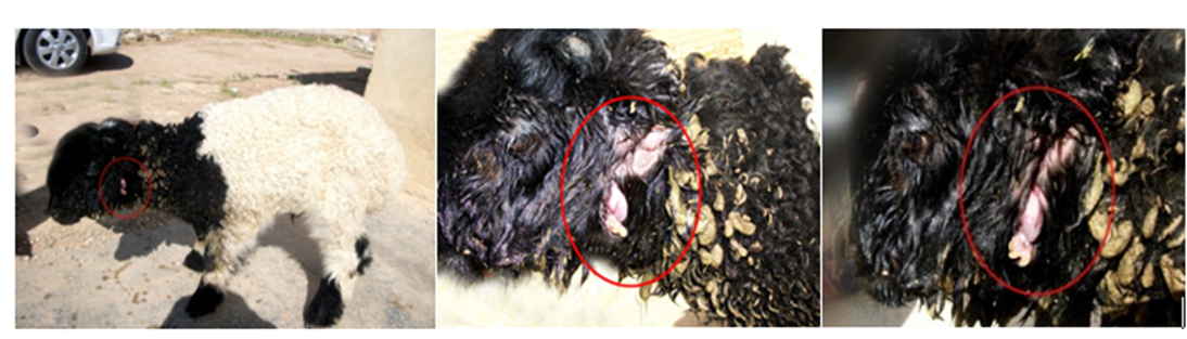
picture (1): illustrate the location of anatomical extra-mouth
Surgical intervention:
The site of the operation was prepared as usual. The wool was removed, shaved, and disinfected with Povidone Iodine solution. Local- rounded anaesthesia was performed around the extra mouth using lidocaine chloride. The lamb was placed on the table in the lateral position where the jaw was removed firstly as shown in the picture. After that, the tooth was connected from the contact point, and then removed by the scalpel including tongue and the lower jaw of extra-mouth shown in the picture (2,3). In this case, the jaw was disengaged and the oesophagus was stitched using a thread (PGA number 0.2). in the shape of a chamida and then Lambert, and then the muscle layer is sutured by a simple continuous stitch using a chromic thread number 1. The skin is stitched using a surgical silk number 1 picture (4).
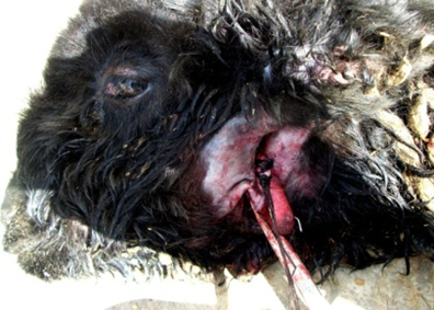
picture (2): surgical excision of extra mouth and lower jaw
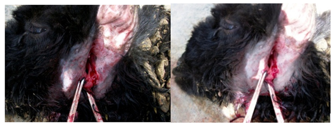
picture (3): complete eradication of all constituents of extra mouth including the toque
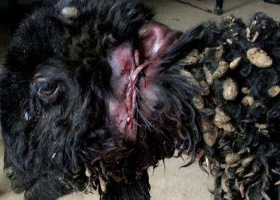
picture (4): surgical stitch of skin to close opening as the final step of operation
After the surgery, an antibiotic was administered benzyl penicillin, procaine, 200,000 IU and dihydrostreptomycin sulfate 250 mg once daily for 5 days.
Discussion:
This condition is rare for the removal of an extra mouth near the area of a lamb at the age of 3 months so you have to talk about and how to surgically remove. Skeletal defects are common, and the skeletal system may be affected in its entirety or with single, isolated defects [15]. Some researchers have found congenital malformations in the mouth itself [21] or Congenital anomalies with accessory limbs and digits occur very rarely in cattle and there are few, if any, reports on such incidences. The animals with supernumerary ectopic limbs and supernumerary digits can survive successfully with normal locomotion and better aesthesis if the surgical excision is performed under proper aseptic conditions and appropriate postoperative care is taken [8]. but in this case marked by the presence of congenital deformity of the formation of an addition mouth composed of the lower jaw and tongue and the result of the connection of the oral cavity additional pharynx, resulting in the exit of feed intake through extra mouth we found it better to remove it surgically because of the consumption of lamb feed too much because part of the extra mouth and also affects the growth of the lamb.
Conclusions:
The surgical eradication of extra mouth can be easily done although many tissues like salivary glands, lymph nodes and carotid artery surround the location.
Authors contribution:
The author did the entire work alone.
Conflicts of interests:
The author declares that there are no conflicts of interests.
Funding:
The study has not received any external funding.
Ethical approval:
Ethical guidelines were followed in this work.
Data and materials availability:
All data associated with this study are present in the paper.
References
- Alabdallah Z. A. et al. (2021). Prevalence and surgical treatment of swelling occurring in Syrian Awassi sheep .Iranian Journal of Ichthyology.8:68-77. DOI:10.1504/IJARGE.2021.10045099
View at Publisher | View at Google Scholar - Alabdallah Z., Nikishov A. A., Volkova N. A., Vetokh A. N., Rebouh N. Y. et al. (2020). Histological and morphometric characteristics of chicken embryos with different genotypes. EurAsian Journal of BioSciences. 14: 719-725.
View at Publisher | View at Google Scholar - AL-abdallah, DVM Ziad, and Taher Asaad. (2013).
View at Publisher | View at Google Scholar - Binanti D., Riccaboni P. (2011). Thoraco-omphalopagus conjoined twins in Chamoiscolored domestic goat kids. Anat. Histol. Embryol.41: 159-162. DOI: 10.1111/j.1439-0264.2011.01118.x
View at Publisher | View at Google Scholar - Bowers D. (2011). Growth in children with clefts: serial hand-wrist x-ray evidence. Cleft Palate Craniofac. J. 48: 762-772. DOI: 10.1597/09-129
View at Publisher | View at Google Scholar - Bryan L, Schmutz S, Hodges SD, Snyder FF. (1993). Bovine b–mannosidosis: Pathologic and genetic findings in salers calves. Vet. Pathol.30: 130–139. DOI: 10.1177/030098589303000205
View at Publisher | View at Google Scholar - Dennis SM. (1974). A suyvey of congenital defects of sheep. Vet. Rec. 95: 488 – 490. DOI: 10.1136/vr.95.21.488
View at Publisher | View at Google Scholar - Dennis SM. (1975). Perinatal lamb mortality in western Australia. 7. Congenital defects. Aust Vet J. 51: 80-82. DOI: 10.1111/j.1751-0813.1975.tb09410.x
View at Publisher | View at Google Scholar - Fisher KRS, Partlow GD and Walker AF. (1986). Clinical and Anatomical observations of a two-headed lamb. Anat. Rec. 214: 432-440. DOI: 10.1002/ar.1092140415
View at Publisher | View at Google Scholar - Hishinuma, M., M. Kohnose, Y. Takahashi, H. Kanagawa. (1987). Diprosopus in a Holstein calf. Jpn. J. Vet. Res. 35: 287-293. DOI: 10.14943/jjvr.35.4.287
View at Publisher | View at Google Scholar - Khoury G, Gruss P. (1983). Enhancer elements. Cell. 33:313–314. DOI: 10.1016/0092-8674(83)90410-5
View at Publisher | View at Google Scholar - Leipold H W, Hiraga T, Dennis S M. (1993). Congenital defects of the bovine central nervous system. Vet Clin North Am Food Anim Pract.9 (1):77-91. DOI: 10.1016/s0749-0720(15)31188-9
View at Publisher | View at Google Scholar - McGirr WJ, Partlow GD, Fisher KRS. (1987). Two-headed, two-necked conjoined twin calf with partial duplication of thoracoabdominal structures: Role of blastocyst hatching. Anat Rec; 217:196-202. DOI: 10.1002/ar.1092170212
View at Publisher | View at Google Scholar - Monfared AL, Sahar HN, Mohammad TS. (2013). Case Report of a Congenital Defect (Dicephalus) in a Lamb. Global Veterinaria.10 (1): 90-92. DOI: 10.5829/idosi.gv.2013.10.1.71180
View at Publisher | View at Google Scholar - Mukaratirwa S, Sayi ST. (2006). Partial facial duplication (diprosopus) in a goat kid. J S Afr Vet Assoc. 77(1):42-4. doi: 10.4102/jsava.v77i1.339
View at Publisher | View at Google Scholar - Ramadan RO. (1996). A dicephalus goat with other defects. J. Vet. Med. 43: 337-343. DOI: 10.1111/j.1439-0442.1996.tb00461.x
View at Publisher | View at Google Scholar - Robert, S.J. (2004). Veterinary Obstetrics and Genital Diseases. Indian Edition. Delhi: CBS Publishers and distributors; 73-74 p.
View at Publisher | View at Google Scholar - Saperstein G. (1981). Diprosopus in a Herford calf. Vet. Rec;108: 234- 235. DOI: 10.1136/vr.108.11.234
View at Publisher | View at Google Scholar - Scott FW, Kahrs RF, Lahunta A de, Broen TT, Mc Entee K. et al. (1993). Virus induced congenital anomalies of the bovine fetus. I. Cerebellar degeneration (hypoplasia), ocular lesions and fetal mummification following experimental infection with bovine viral diarrhea–mucosal disease virus. Cornell Vet. 63: 536 – 560. PMID: 4748881
View at Publisher | View at Google Scholar - Staley GP, Lugt JJ, Vander–Axsel G, Loock AH, Vander LJJ. (1994). Congenital skeletal malformations in Holstein calves with putative manganese deficiency. J. South Afr. Vet. Assoc.65:73–78. PMID: 7776338
View at Publisher | View at Google Scholar - Szczerbal I., Stefaniak T., Dubiel A., Siembieda J., Nizanski W. et al. (2006). Chromosome instability in a calf with amelia of thoracic limbs. Vet Pathol. 43 (5):789-792. doi: 10.1354/vp.43-5-789.
View at Publisher | View at Google Scholar - Z AL-abdallah, T. A. (2012). The Surgical Treatment Of Caseous Lymphadenitis In Awass Syrian Sheep. Assiut Veterinary Medical Journal, 58(132), 1-21.
View at Publisher | View at Google Scholar
