

Review Article | DOI: https://doi.org/10.31579/ 2835-8147/019
Application of Nanotechnology in Cartilage Regeneration
1Division of Surgery, ICAR-Indian Veterinary Research Institute, Izatnagar, Bareilly, Uttar Pradesh, India
*Corresponding Author: Swapan Kumar Maiti, Division of Surgery ICAR-Indian Veterinary Research Institute, Izatnagar, Bareilly, Uttar Pradesh.
Citation: Merlin Mamachan, Swapan K. Maiti, Amitha Banu S, Khan Sharun, Mamta Mishra, Manjusha K.M(2023), Application of Nanotechnology in Cartilage Regeneration, Clinics in Nursing, 2(3); DOI:10.31579/ 2835-8147/019
Copyright: © 2023, Swapan Kumar Maiti, This is an open access article distributed under the Creative Commons Attribution License, which permits unrestricted use, distribution, and reproduction in any medium, provided the original work is properly cited.
Received: 01 June 2023 | Accepted: 08 June 2023 | Published: 15 June 2023
Keywords: Cartilage injury; Extracellular matrix (ECM); Biocompatibility; Articular Cartilage; hyaline cartilage
Abstract
The reconstruction of articular cartilage injury is one of the major challenges dealt in tissue engineering field. The avascular and aneural anatomical features of the cartilage itself impede the regenerative process. The presently available biomimetic scaffolds fail to recapitulate the original characteristics of hyaline cartilage. The advent of nanotechnology and it’s wide application in tissue engineering has brought a lot of innovations in the construction of scaffolds for chondral regeneration. The nano constructed scaffolds can very well simulate the natural property of the extracellular matrix (ECM) of hyaline cartilage and thereby creating a better microenvironment for regenerative mechanisms. It also acts as a better platform for stem cell attachment and proliferation. It can even direct the cellular function by taking the control of gene expressions. The nanotechnology has been extrapolated for growth factor delivery and tracking of implanted stem cells. The biocompatibility of nano-scaffolds and the results in preclinical studies reveal the importance of the nanotechnology in cartilage regeneration therapeutics.
Introduction
Nanotechnology (NT) is the branch of science that deals with the design and manufacture of systems, biomaterials as well as equipment in nanoscale dimensions. particle with a minimum of one of its components having a nanometric scale dimension is classified as a nanoparticle (NP) [24]. In our body, most of the cellular and biomolecular activity functions at the nanometric level. The immense potential of NP allows for the functional regulation of a cell even at the genomic level. This forged NT as a novel method to resolve the problems at the cellular level. Conventional treatment strategies often fail to give complete functional or structural restoration of diseased organs. Tissue engineering provides several biomimetic scaffolds to restore the natural characteristics of these diseased organs. Thus, combining NT with tissue engineering paves the way for the development of high-quality biomimetic scaffolds with appropriate therapeutic efficiency [38]. In veterinary orthopaedics, the treatment of osteoarthritis (OA) is still a major challenge as most treatment modalities yield futile results. Articular cartilage is avascular, aneural and alymphatic structure with few numbers of chondrocyte present in the cartilage (2%). Thus, these tissues inherently lack regeneration capacity [35]. For a long time, the therapeutic methods employed in the management of OA were arthroscopic debridement, microfracture technique, and marrow stimulation techniques. These techniques were having limited applicability due to the production of fibrocartilage instead of hyaline cartilage [5]. The usually employed scaffolds for tissue engineering also exhibited major drawbacks such as bio-incompatibility and imbalance in the scaffold degeneration and cartilage regeneration rate. The application of stem cells in cartilage regeneration resulted in superior-quality cartilage regeneration. However, it demanded a suitable biomaterial for promoting cell expansion, proliferation as well as migration [23]. The NP can very well simulate the natural characteristics of the extracellular matrix (ECM) of hyaline cartilage and thereby creating a better microenvironment for regenerative mechanisms. They also mimic the cell receptors and regulate gene expressions [20]. There are various techniques available for the production of nano scaffolds (NS) and nanoparticles (NPs). They are fabricated in particular structures and shapes to mimic the microscopic and molecular cell structures. Cell attachment, migration, and proliferation are greatly influenced by the structure in which cells are implanted. NP can be used for the delivery of various biomolecules and signaling factors into the cell Labelling with magnetic NPs like SPIONS has the advantage of post-implantation cell tracking [8]. The nanotherapeutic applications resulted in superior quality healing and regeneration of articular cartilage which is critical for better clinical outcomes.
Histology of Articular Cartilage
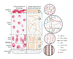
The structure and composition of articular cartilage are highly complex in nature. The structural complexity of cartilage restricts cartilage tissue engineering in constructing scaffolds mimicking cartilage. It is a highly specialized avascular, alymphatic, aneural and hypocellular structure located in between joints [17,43]. It is composed of chondrocytes and dense extracellular matrix (ECM) containing water, collagen fibers, proteoglycans and glycosaminoglycans. Chondrocytes are responsible for the synthesis and maintenance of ECM [39]. The synthesis of collagen is relatively slow and plays a crucial role in the homeostasis mechanism. The compressive force acting on the cartilage is resisted by the ECM whereas the tensile force is resisted by type II collagen fibers. Structure and biochemical composition of hyaline cartilage. Schematic representation of hyaline cartilage zonal structure, cellular distribution, organization, morphology and collagen orientation. Created with BioRender.com. (Trends in Articular Cartilage Tissue Engineering: 3D Mesenchymal Stem Cell Sheets as Candidates for Engineered Hyaline-Like Cartilage - Scientific Figure on ResearchGate. Available from: https://www.researchgate.net/figure/Hyaline-cartilage-structure-and- biochemical-composition-Schematic-representation-of_fig1_350068814 [accessed 24 May, 2023] The percentage of chondrocytes is few in articular cartilage. Any damage to the chondrocyte will invariably harm ECM synthesis. The anisotropic and pseudostratified structure of the cartilage along with the lack of vascularity is a major challenge in the reparative mechanisms. The current treatment modalities for reconstruction like allograft, autograft, prostheses and implants are unsuccessful in regenerating the actual hyaline cartilage. The major demerits of grafting include infections, limited autologous donor site, donor site morbidity, immunosuppression associated with allografts, and reduced functionality of prostheses [29]. The conventional therapeutic measures for cartilage injuries attempt to replicate the structure and function of native hyaline tissue.
Properties of Nano Scaffold
The NS used for cartilage regeneration should possess some minimum essential features to obtain a quality outcome. The surface architecture of NS has a high surface area- to-volume ratio. Hence provides more attachment sites for ligands and increases protein adsorption [47]. The biocompatibility of the scaffold can be increased by incorporating functional groups and natural polymers into it. This feature is very essential for super absorbent properties, cytocompatibility and chondrogenic differentiation of seeded stem cells. The porosity and pore size of the manufactured scaffold determines the rate of cell attachment, migration and the mechanical strength of the scaffold [38]. The scaffold with suitable porosity causes increased protein adsorption, collagen II and aggrecan expression and thereby helps to produce cartilage-specific ECM [18]. Moreover, it enhances the delivery of nutrients to the regenerating cells.
The mechanical strength of the scaffold has paramount importance in cartilage regeneration. The implanted scaffold and surrounding healthycartilage should have almost comparable mechanical strength to counteract the constant loading force into the joint. The mechanical strength of the scaffold cannot be compromised while selecting the pore size and porosity. There should be compatibility between the biodegradability rate of the scaffold and the cartilage regeneration rate. For a scaffold to be ideal, it should possess superior morphology suitable for cell growth and rapid degradation after four weeks of implantation. The degradation rate can be controlled by a proper material compounding strategy (Wu et al., 2004). For example, the use of nanohydroxyapatite along with the poly (lactide-co-E- caprolactone) (PLCL) scaffold has improved the biodegradability rate of the scaffold (Diaz and Puerto, 2005). The capacity for cell adhesion and expansion is governed by the nano topography (NT) of the scaffold. The malleability of the scaffold material should be flexible to conform to various sizes, shapes and 3D patterns (Peran et al., 2013). The geometry of the NS has a great influence on chondrocyte adhesion, function and phenotype regulation (Yang et al., 2020). The NT signals like nano grooves, nanogrids, nanoholes and nanopillars can induce receptor-mediated cellular responses (Lee et al., 2017). This nano-patterning provides cell adhesion receptors with robust ligands (eg. Integrin like receptors) which enhance receptor-mediated cell adhesion, proliferation and migration (Ye et al., 2015). NT has an impact on gene expression, microRNA expression and lineage-specific mesenchymal stem cell (MSC) differentiation (Izadpanahi et al., 2018). In a study conducted to investigate the effect of different NT on the chondrogenic differentiation of MSC, the nanoholes and nanopillar shows a better expression of chondrogenic differentiation marker when compared to the nanogrill pattern (Wu et al., 2014). Surface stiffness has some role in the chondrogenic differentiation of MSC. The soft substrate with mild stiffness can stimulate signaling pathway like the ROCK signal pathway. This is responsible for the regulation of chondrocyte phenotype and helps in the maintenance of round cell morphology of chondrocytes forming a suitable cytoskeletal tension (Zhang et al., 2016). The NS is to be designed in such a proper manner to incorporate all these properties to give the best functionality.
Fabrication Techniques for Nano Scaffold Construction
Photolithography is a technique used to design nanopatterns like nanopillar, nanogroove, and nano pits on the surface of a scaffold using a photon beam and a photoresist template. Nanoimprinting is used to create nanoscale deformation of a resist using a mold and it is later cured using heat energy or ultraviolet rays (Barcelo and Li, 2016).
Different Types of Nanomaterials-Assisted Cartilage Tissue Engineering
a) Nanofibrous
They are constructed utilizing electrospinning, 3D printing, molecular self-assembly and phase separation techniques. They are characterized by two nanoscales dimensions and three dimensions which are below 1000 nm (larger) (Jeevanandam et al., 2018). They have tunable porosity, large surface area to volume ratio, malleability and mechanical strength. These properties enable them to be a unique candidate for fabricating tissue engineering scaffolds [27]. Their nanofibrous structure can simulate the cartilage ECM and accelerate the regeneration process. They are highly biocompatible in nature. The surface of the nanofiber polymer can be treated with plasma to incorporate functional groups like hydroxyl, carboxyl, amino groups etc. These functional groups modify the physical properties of scaffolds like wettability, polarity and bio-adhesion (Asadian et al., 2020). The nanofiber improves cell attachment, adhesion and expansion and helps in maintaining chondrocyte phenotype. They provide more functional ligands or receptors for cells and growth factors to enhance cartilage regeneration. The nanoarchitecture has an impact on the multilineage differentiation of MSC [26]. The polycaprolactone (PCL) nanofibers induced chondrogenic differentiation of MSC, when compared to the normal PCL, based 3 D scaffold (Mellor et al., 2015). In a study, PLA/gelatin nanofibrous scaffold was seeded with rabbit bone marrow- derived mesenchymal stem cells (BM-MSC) to assess cartilage healing in rabbit models. The scaffold showed better biocompatibility, mechanical strength, chondrogenic differentiation and thereby improving cartilage regeneration [6]. In another study, chondral defects of rabbits were filled with PCL/PEO nanofibrous scaffold and rabbit synovial stem cells. The results revealed the filling of cartilage defect with hyaline cartilage and the scaffold exhibited a chondroprotective effect [41]. The mechanical property of hydrogels can be reinforced by incorporating suitable nanofibres into them. In a study, electrospun silk fibres were added to chitosan/glycerophosphate hydrogels to improve the mechanical properties of the hydrogel which revealed increased expression of chondrogenic phenotype [33]. Similarly, two or more nanofibres are blended to obtain composite mats to improve the physicochemical properties of the scaffold [34]. The electrospun polyvinylalcohol (PVA) and sulfated alginate nanofibrous mat were seeded with human BM- MSC demonstrating an increase in type-II collagen expression and chondrogenic differentiation of MSC [20]
b) Nanotubes
The nanotubes have cylindrical geometry with a diameter of 1-100 nm. Single-wall and multi-wall are two types of nanotubes. They are mainly derived from carbon, boron, silicon and halloysite compounds [16]. Carbon nanotubes (CNT) exhibit supreme mechanical, tensile, electrical and thermal properties when compared to other nanotubes. They are commonly used as fillers in composite scaffolds to give better mechanical strength. CNTs have to be functionalized by acid treatment or bioconjugation to remove their chemical inertness and increase biocompatibility [54]. In a study, 0.5% COOH-functionalized multiwalled CNTs (MWCNT) was incorporated into a scaffold to check the improvement in its physicochemical properties. There was an increase in hydrophilicity, and tensile strength, with a reduction in fiber diameter and no adverse effect on chondrocyte expression [57]. Vertically aligned MWCNT micropillar helps in the unidirectional orientation of chondrocytes. Young’s modulus of the material was comparable with the ECM of articular cartilage. These micropillars intensified ECM production, cell attachment and proliferation [21]. Silica nanotubes are an excellent choice for tissue-engineered scaffolds due to their high potential for integration in biomaterials, photoluminescence, ease of surface modification and biocompatibility (Wan et al., 2015). Titanium oxide nanotubes are also used in a wide variety of scaffolds in the biomedical field recently. They have good biocompatibility, large surface area and the inner space of the tubes can be filled with bioactive molecules [7]. Boron nitride nanotubes incorporated poly propylene fumarate nanocomposite expressed higher adsorption of collagen I, ECM production and cell attachment [14]. Halloysite is a better alternative for CNTs in tissue engineering, as it does not need to be functionalized and is highly biocompatible [13].
c) Nanoparticle
NP can be constructed in various forms like nanocapsules, liposomes, nanospheres and dendrimers. NPs mainly act as carriers of bioactive molecules and protect them from physiological degradation. They help in the controlled and consistent release of biomolecules for a particular time period and reduce the adverse side effect caused by overdosing [11]. The addition of NPs in the hydrogel has a tremendous effect on the mechanical property of hydrogel. Additionally, it helps in the sustainable release of bioactive molecules. Growth factors (GF) incorporated with polylactide co glycolide (PLGA) nanoparticles and silk fibroin/poly (ethylene glycol) methacrylate (PEGDMA) hydrogel found to improve an improved proliferation of dental pulp stem cells (DPSCs) with an enhanced rate of chondrogenesis [14]. Heparin-functionalized-NPs have a great role in cartilage tissue engineering field, because it helps in the well-controlled release of GF and stabilizes the structure and function of GF [36]. The use of GF is very essential for the trilineage differentiation of MSCs. Fibrin hydrogel containing TGF-loaded NPs was used in the chondrogenic analysis of rabbit BM-MSC. The result was a sustained release of GF for a long period and also helped in hyaline cartilage formation [22]. Magnetic NPs incorporated hydrogels are a promising tool in cartilage tissue regeneration, since they respond to external magnetic fields and relocate to defective cartilage sites for better regeneration [22]. Kartogenin (KGN) loaded PLGA NPs were used in the injectable hyaluronic acid hydrogel in a study of full-thickness osteochondral defect in a porcine model. The controlled release of KGN resulted in the efficient healing of full-thickness chondral defect in the porcine model [51].
d) Nanocomposite Hydrogels
Hydrogel forms an exceptional class of biomaterials used for cartilage regeneration. It is available in the injectable form and involves a minimally invasive procedure [30]. It mimics the ECM of cartilage due to its structural peculiarities. It efficiently fills the defects of any shape and polymerizes into gel after the application [48,49,50] The GF and cells can be entirely dispersed in the hydrogel. Chitosan, alginate, agarose, hyaluronic acid, collagen, and silk are the natural biomaterials used in the synthesis of hydrogels. Synthetic biomaterials are preferred over natural ones due to their superior properties in processing potential, less batch-to-batch variation, and improved mechanical strength. Polyethene glycol (PEG), polyvinyl alcohol (PVA), N-isopropyl acrylamide (NiPAAm) etc are the commonly used synthetic materials in the manufacture of hydrogels [38]. The incorporation of NP in the hydrogel increases its mechanical strength and improves the pore size. This enhances cell attachment and thereby increases the quality of tissue regeneration. Magnetic nanocomposite hydrogel with gelatin, β-cyclodextrin and Fe3O4 were used in in vivo cartilage regeneration studies in rabbits. The properties like superparamagnetism, biocompatibility and mechanical strength of the hydrogel resulted in better cartilage regeneration in rabbits. It also promoted the differentiation of BM-MSC into chondrocytes [19]. Carbon-based NPs improves the lubrication, biocompatibility and mechanical property of hydrogels. In a study, graphene oxide-coated hydroxyapatite (HA) particles encapsulated in PVA hydrogel showed a significant enhancement in the composite's lubrication property and compressive deformation resistance. It also improved the cytocompatibility and proliferation rate of rat BM-MSC, suggesting superior quality healing of chondral defects [5]. Three-dimensional alginate gel was developed using PCL-PEG-PCL microspheres as carriers for calcium gluconate. The hydrogel exhibited good pore connectivity, biodegradability and compressive modulus. These features demonstrated the hydrogel as a suitable matrix for cartilage regeneration studies [28].
Conclusion
Tissue engineering is a multidisciplinary area where there is a strong interplay between cell, scaffold and biological cues. The interaction between these three factors should Electrospinning, phase separation, self-assembly, photolithography and nano- imprinting are the major techniques used for the fabrication of NS [34]. In electrospinning, polymer fibres with nanoscale range diameter are produced using electrostatic force generated between a spinneret and ground target collector. The self- repulsive charges of the polymer solution overcome the surface tension of the solution using the electrostatic force. This results in an accelerating jet of charged polymer solution towards the zero-charged ground target. The solvent evaporates and charged polymer fiber gets deposited to the collecting target. This technique is very useful in the production of nanofibrous scaffolds which has a wide application in the field of tissue engineering [3,4,9]. The most important advantage of this technique is the ability to produce nanoarchitecture patterns of fibers in randomly oriented or parallel alignment form [12]. These patterns have a role in the cell attachment and orientation along the direction of fibers [52,53]. The nanofibrous scaffolds possess a high surface area to volume ratio which is very helpful in the uptake and diffusion of nutrients. The properties of scaffolds like hydrophilicity, mechanical strength, biocompatibility, and biodegradability are controlled by the chemical composition of the polymer. The electrospun scaffolds with desired functions are tailored by selecting the chemical components in appropriate combination and ratio [27]. Phase separation is a technique in which the polymer solution is segregated into solvent-rich and polymer-rich domains either by thermal induction or the addition of non- solvent polymer. The morphology is fixed by cooling and the solvent is removed by the freeze-drying process. A 3D fibrous network of porosity of 98% and fiber diameter below 500 nm is produced [31]. The advantage of this technique is that fiber diameter, porosity of scaffold, and mechanical properties can be controlled by manipulating processing parameters. The scaffold can be designed into any anatomical shape as it is fabricated utilizing suitable molds [42]. In self-assembly, the molecules undergo self-organisation into patterns and structures employing van der Waals, electrostatic and hydrophobic interactions [46]. Nanoscale scaffolds with a fiber diameter of around 10 nm are produced using synthetic polypeptides [55]. They have the potential to carry more biologically compatible motifs. Large-scale production is limited due to low yield and complexity be in an appropriate balance to result in a quality outcome. The incorporation of nanoscale features in biomimetic scaffolds will enhance the natural properties of articular cartilage in them. It improves cell adhesion, migration, and expansion and also provides more cues for cell-matrix interactions. The constant release of biological factors using NPs has given better results in cartilage regeneration. The nanomaterials have numerous advantages like design flexibility, high surface area to volume ratio, ease of surface modification, small size etc. However, the clinical translation of technology from bench to bedside is a perpetual challenge. The safety of nanomaterials for in vivo application is highly questionable. The rate of degradability, toxicity, composition, and side effects of the nano scaffold has to be well evaluated before any clinical translation. Clinical translations are restricted largely by economic investment and time involved in the production of NPs. The application of NT in the field of regenerative medicine can bring highly advanced and quality outcomes in treatment modalities. The replacement of functional articular cartilage in place of degenerated one using these technologies will be a giant leap in the field of orthopaedics in future
References
- Asadian M., Chan K.V., Norouzi M., Grande S., Cools P., et. al, (2020). Fabrication and plasma modification of nanofibrous tissue engineering scaffolds. Nanomaterials. 10(1): 119
View at Publisher | View at Google Scholar - Barcelo, S. and Li, Z., (2016). Nanoimprint lithography for nanodevice fabrication. Nano Convergence. 3(1): 1-9.
View at Publisher | View at Google Scholar - Becerra, J., Andrades, J.A., Guerado, E., Zamora-Navas, P., López-Puertas, J.M. at al., (2010). Articular cartilage: structure and regeneration. Tissue Engineering Part B: Reviews. 16(6): 617-627.
View at Publisher | View at Google Scholar - Cao, J., Meng, Y., Zhao, X. and Ye, L., (2020). Dual-anchoring intercalation structure and enhanced bioactivity of poly (vinyl alcohol)/graphene oxide–hydroxyapatite nanocomposite hydrogels as artificial cartilage replacement. Industrial & Engineering Chemistry Research. 59(46): 20359-20370.
View at Publisher | View at Google Scholar - Chen, S., Chen, W., Chen, Y., Mo, X. and Fan, C., (2021). Chondroitin sulfate modified 3D porous electrospun nanofiber scaffolds promote cartilage regeneration. Materials Science and Engineering. 118: 111312.
View at Publisher | View at Google Scholar - Cheng, Y., Yang, H., Yang, Y., Huang, J., Wu, K., at all., (2018). Progress in TiO 2 nanotube coatings for biomedical applications: a review. Journal of Materials Chemistry B. 6(13): 1862-1886.
View at Publisher | View at Google Scholar - Cromer Berman, S.M., Walczak, P. and Bulte, J.W., 2011. Tracking stem cells using magnetic nanoparticles. Wiley Interdisciplinary Reviews: Nanomedicine and Nanobiotechnology. 3(4): 343-355.
View at Publisher | View at Google Scholar - Deitzel, J.M., Kleinmeyer, J., Harris, D.E.A. and Tan, N.B., (2001). The effect of processing variables on the morphology of electrospun nanofibers and textiles. Polymer. 42(1): 261-272.
View at Publisher | View at Google Scholar - Díaz, E. and Puerto, I., 2015. In vitro degradation of PLCL/nHA biodegradable scaffolds. Polymer-Plastics Technology and Engineering. 54(6): 556-564.
View at Publisher | View at Google Scholar - Dyondi, D., Webster, T.J. and Banerjee, R., (2012). A nanoparticulate injectable hydrogel as a tissue engineering scaffold for multiple growth factor delivery for bone regeneration. International journal of nanomedicine. 47-59.
View at Publisher | View at Google Scholar - Dzenis, Y., (2004). Spinning continuous fibers for nanotechnology. Science. 304(5679): 1917- 1919.
View at Publisher | View at Google Scholar - Fakhrullin, R.F. and Lvov, Y.M., (2016). Halloysite clay nanotubes for tissue engineering. Nanomedicine. 11(17): 2243-2246.
View at Publisher | View at Google Scholar - Fathi Achachelouei, M., Keskin, D., Bat, E., Vrana, N. E., Tezcaner, A.J., (2020). Biomed. Mater. Res., Part B. 108: 2041-2062.
View at Publisher | View at Google Scholar - Federico, S. and Herzog, W., (2008). On the anisotropy and inhomogeneity of permeability in articular cartilage. Biomechanics and modeling in mechanobiology. 7: 367-378.
View at Publisher | View at Google Scholar - Fraczek, A., Menaszek, E., Paluszkiewicz, C. and Blazewicz, M., (2008). Comparative in vivo biocompatibility study of single-and multi-wall carbon nanotubes. Acta Biomaterialia. 4(6): 1593-1602.
View at Publisher | View at Google Scholar - Gadjanski, I., Spiller, K. and Vunjak-Novakovic, G., (2012). Time-dependent processes in stem cell-based tissue engineering of articular cartilage. Stem Cell Reviews and Reports. 8: 863-881.
View at Publisher | View at Google Scholar - He, L., Liu, B., Xipeng, G., Xie, G., Liao, S., (2009). Microstructure and properties of nano-fbrous PCL-b-PLLA scafolds for cartilage tissue engineering. Eur Cell Mater. 18:63- 74.
View at Publisher | View at Google Scholar - Huang, J., Jia, Z., Liang, Y., Huang, Z., Rong, Z., at al., (2020). Pulse electromagnetic fields enhance the repair of rabbit articular cartilage defects with magnetic nano-hydrogel. RSC advances. 10(1): 541-550.
View at Publisher | View at Google Scholar - Izadpanahi, M., Seyedjafari, E., Arefian, E., Hamta, A., Hosseinzadeh, S., (2018). Nanotopographical cues of electrospun PLLA efficiently modulate non- coding RNA network to osteogenic differentiation of mesenchymal stem cells during BMP signaling pathway. Materials Science and Engineering. 93:686-703.
View at Publisher | View at Google Scholar - Janssen L., Saranya, M., Leinonen, M., Pitkänen, O., Mobasheri, A. (2020). Vertically aligned carbon nanotube micropillars induce unidirectional chondrocyte orientation. Carbon. 158: 681-689.
View at Publisher | View at Google Scholar - Jung, Y., Chung, Y.I., Kim, S.H., Tae, G., Kim, S.H., (2009). In situ chondrogenic differentiation of human adipose tissue-derived stem cells in a TGF-β1 loaded fibrin–poly (lactide-caprolactone) nanoparticulate complex. Biomaterials. 30(27): 4657-4664.
View at Publisher | View at Google Scholar - Kaviani, A., Zebarjad, S.M., Javadpour, S., Ayatollahi, M. and Bazargan-Lari, R., (2019). Fabrication and characterization of low-cost freeze-gelated chitosan/collagen/hydroxyapatite hydrogel nanocomposite scaffold. International Journal of Polymer Analysis and Characterization. 24(3): 191-203.
View at Publisher | View at Google Scholar - Lawson, T. B., Mäkelä, J. T., Klein, T., Snyder, B. D., Grinstaff, M. W. (2021). Nanotechnology and osteoarthritis. Part 1: clinical landscape and opportunities for advanced diagnostics. Journal of Orthopaedic Research. 39(3): 465-472.
View at Publisher | View at Google Scholar - Lee, L.C., Gadegaard, N.,.C., Turner, L De Andrés, M.A., Burgess, M.J., (2017). Nanotopography controls cell cycle changes involved with skeletal stem cell self-renewal and multipotency. Biomaterials. 116: 10-20.
View at Publisher | View at Google Scholar - Li, W.J., Tuli, R., Huang, X., Laquerriere, P., Tuan, R.S., (2005). Multilineage differentiation of human mesenchymal stem cells in a three-dimensional nanofibrous scaffold. Biomaterials. 26(25): 5158-5166.
View at Publisher | View at Google Scholar - Liang, D., Hsiao, B.S. and Chu, B., (2007). Functional electrospun nanofibrous scaffolds for biomedical applications. Advanced drug delivery reviews. 59(14): 1392-1412.
View at Publisher | View at Google Scholar - Liao, J., Wang, B., Huang, Y., Qu, Y., Peng, J. (2017). Injectable alginate hydrogel cross-linked by calcium gluconate-loaded porous microspheres for cartilage tissue engineering. ACS omega. 2(2): 443-454.
View at Publisher | View at Google Scholar - Lim, E.H., Sardinha, J.P. and Myers, S., (2014). Nanotechnology biomimetic cartilage regenerative scaffolds. Archives of plastic surgery. 41(03):231-240.
View at Publisher | View at Google Scholar - Liu, M., Zeng, X., Ma, C., Yi, H., Ali, Z., Mou, X., Li, S., Deng, Y. and He, N., 2017. Injectable hydrogels for cartilage and bone tissue engineering. Bone research. 5(1): 1-20.
View at Publisher | View at Google Scholar - Ma, Z., Gao, C., Gong, Y. and Shen, J., (2005). Cartilage tissue engineering PLLA scaffold with surface immobilized collagen and basic fibroblast growth factor. Biomaterials. 26(11): 1253-1259.
View at Publisher | View at Google Scholar - Mellor, L.F., Mehendale, S., Mohiti-Asli, M., Taylor, M., Pedersen, (2015). Evaluation of Micro and Nano-Scale Scaffold Architectures for Osteochondral Tissue Engineering. Las Vegas, NV, USA: Orthopaedic Research Society.
View at Publisher | View at Google Scholar - Mirahmadi, F., Tafazzoli-Shadpour, M., Shokrgozar, M.A. and Bonakdar, S., (2013). Enhanced mechanical properties of thermosensitive chitosan hydrogel by silk fibers for cartilage tissue engineering. Materials science and engineering. 33(8): 4786-4794.
View at Publisher | View at Google Scholar - Nabizadeh, Z., Nasrollahzadeh, M., Daemi, H., Eslaminejad, M.B., Shabani, (2022). Micro-and nanotechnology in biomedical engineering for cartilage tissue regeneration in osteoarthritis. Beilstein Journal of Nanotechnology. 13(1): 363-389.
View at Publisher | View at Google Scholar - Nayyer, L., Patel, K.H., Esmaeili, A., Rippel, R.A., (2012). Tissue engineering: revolution and challenge in auricular cartilage reconstruction. Plastic and reconstructive surgery. 129(5): 1123-1137.
View at Publisher | View at Google Scholar - Park, J.S., Yang, H.N., Woo, D.G., Chung, H.M., Park, K.H., (2009). In vitro and in vivo chondrogenesis of rabbit bone marrow–derived stromal cells in fibrin matrix mixed with growth factor loaded in nanoparticles. Tissue Engineering Part A. 15(8): 2163-2175.
View at Publisher | View at Google Scholar - Perán, M., García, M.A., Lopez-Ruiz, E., Jiménez, G. and Marchal, J.A., (2013). How can nanotechnology help to repair the body? Advances in cardiac, skin, bone, cartilage and nerve tissue regeneration. Materials. 6(4): 1333-1359.
View at Publisher | View at Google Scholar - Qiao, K., Xu, L., Tang, J., Wang, Q., Lim, K.S., (2022). The advances in nanomedicine for bone and cartilage repair. Journal of Nanobiotechnology. 20(1): 1-42.
View at Publisher | View at Google Scholar - Responte, D.J., Natoli, R.M. and Athanasiou, K.A., (2007). Collagens of articular cartilage: structure, function, and importance in tissue engineering. Critical Reviews™ in Biomedical Engineering. 35(5): 363-411.
View at Publisher | View at Google Scholar - Rolauffs, B., Williams, J.M., Aurich, M., Grodzinsky, A.J., Kuettner, K.E. (2010). Proliferative remodeling of the spatial organization of human superficial chondrocytes distant from focal early osteoarthritis. Arthritis & Rheumatism: Official Journal of the American College of Rheumatology. 62(2): 489-498.
View at Publisher | View at Google Scholar - Shimomura, K., Rothrauff, B.B., Hart, D.A., Hamamoto, S., Kobayashi, M., (2019). Enhanced repair of meniscal hoop structure injuries using an aligned electrospun nanofibrous scaffold combined with a mesenchymal stem cell- derived tissue engineered construct. Biomaterials. 192: 346-354.
View at Publisher | View at Google Scholar - Smith, L.A. and Ma, P.X., 2004. Nano-fibrous scaffolds for tissue engineering. Colloids and surfaces B: biointerfaces. 39(3): 125-131.
View at Publisher | View at Google Scholar - Sophia Fox, A.J. and Bedi, A., (2009). Rodeo SA The Basic Science of Articular Cartilage. Sports Health. 1: 461-468.
View at Publisher | View at Google Scholar - Thorp, H., Kim, K., Kondo, M., Maak, T., Grainger, D.W. (2021). Trends in articular cartilage tissue engineering: 3D mesenchymal stem cell sheets as candidates for engineered hyaline-like cartilage cells. 10(3): 643.
View at Publisher | View at Google Scholar - Wan, Y., Liu, P., Zhang, C., Yang, Z., Xiong, G., Zheng, X. (2015). Synthesis of a three-dimensional network-structured scaffold built of silica nanotubes for potential bone tissue engineering applications. Journal of Alloys and Compounds. 647: 711-719.
View at Publisher | View at Google Scholar - Whitesides, G.M. and Grzybowski, B., (2002). Self-assembly at all scales. Science. 295(5564): 2418-2421.
View at Publisher | View at Google Scholar - Wimpenny, I., Ashammakhi, N., Yang, Y., (2012). Chondrogenic potential of electrospun nanofibres for cartilage tissue engineering. Journal of tissue engineering and regenerative medicine. 6(7): 536-549.
View at Publisher | View at Google Scholar - Wu, J., Chen, Q., Deng, C., Xu, B., Zhang, Z., (2020). Exquisite design of injectable hydrogels in cartilage repair. Theranostics. 10(21): 9843.
View at Publisher | View at Google Scholar - Wu, L. and Ding, J., (2004). In vitro degradation of three-dimensional porous poly (D, L- lactide-co-glycolide) scaffolds for tissue engineering. Biomaterials. 25(27): 5821-5830.
View at Publisher | View at Google Scholar - Wu, Y.N., Law, J. B. K., He, A. Y., Low, H. Y., Hui, J. H. P., Lim, C. T., Yang, Z., Lee, E. H., Nanomedicine. 10: 1507–1516.
View at Publisher | View at Google Scholar - Yan, W., Xu, X., Xu, Q., Sun, Z., Lv, Z., Wu, R (2020). An injectable hydrogel scaffold with kartogenin-encapsulated nanoparticles for porcine cartilage regeneration: a 12-month follow-up study. The American journal of sports medicine. 48(13): 3233-3244.
View at Publisher | View at Google Scholar - Yang, F., Murugan, R., Wang, S. and Ramakrishna, S., (2005). Electrospinning of nano/micro scale poly (L-lactic acid) aligned fibers and their potential in neural tissue engineering. Biomaterials. 26(15): 2603-2610.
View at Publisher | View at Google Scholar - Yang, K.C., Chen, H., Yang, Y.T., Hsiao, J.K. and Wang, C.C., (2020). Effects of scaffold geometry on chondrogenic differentiation of adipose-derived stem cells. Materials Science and Engineering. 110: 110733.
View at Publisher | View at Google Scholar - Zhang, M., Wang, W., Wu, F., Yuan, P., Chi, C, N., (2017). Magnetic and fluorescent carbon nanotubes for dual modal imaging and photothermal and chemo-therapy of cancer cells in living mice. Carbon. 123: 70-83.
View at Publisher | View at Google Scholar - Zhang, N., Lock, J., Sallee, A. and Liu, H., (2015). Magnetic nanocomposite hydrogel for potential cartilage tissue engineering: synthesis, characterization, and cytocompatibility with bone marrow derived mesenchymal stem cells. ACS applied materials & interfaces. 7(37).
View at Publisher | View at Google Scholar - Zhang, S., (2003). Fabrication of novel biomaterials through molecular self-assembly. Nature biotechnology. 21(10): 1171-1178.
View at Publisher | View at Google Scholar - Zhang, T., Gong, T., Xie, J., Lin, S., Liu, (2016). Softening substrates promote chondrocytes phenotype via RhoA/ROCK pathway. ACS Applied Materials & Interfaces. 8(35): 22884-22891.
View at Publisher | View at Google Scholar
