

Research Article | DOI: https://doi.org/10.31579/2835-7957/117
Anatomical And Biochemical Stress of The Small Intestine Caused by Castor Oil: An Insight on The Effect of Uvaria Chamae Root Extract in Ameliorating the Stress
1Department of Human Anatomy, Faculty of Basic Medical Sciences, University of Uyo, Nigeria.
2Department of Medical Biochemistry, Faculty of Basic Medical Sciences, University of Uyo, Nigeria.
3Department of Anatomy, School of Basic Medical Sciences, University of Benin, Nigeria.
*Corresponding Author: Gabriel Donatus Edem, Department of Human Anatomy, Faculty of Basic Medical Sciences, University of Uyo, Nigeria.
Citation: Gabriel D. Edem, Eno Uwah Ettetor, Vitalis Chukwuma Ezeuko, Solomon E. Ikono, (2025), Anatomical and Biochemical Stress of the Small Intestine Caused by Castor Oil: An Insight on The Effect of Uvaria Chamae Root Extract In Ameliorating The Stress, Clinical Reviews and Case Reports, 4(1); DOI:10.31579/2835-7957/117
Copyright: © 2025, Gabriel Donatus Edem. This is an open-access article distributed under the terms of the Creative Commons Attribution License, which permits unrestricted use, distribution, and reproduction in any medium, provided the original author and source are credited.
Received: 13 January 2025 | Accepted: 29 January 2025 | Published: 03 February 2025
Keywords: stress; castor oil; small intestine; superoxide dismutase; Uvaria chamae
Abstract
Background: Uvaria chamae is rich in antioxidants which is very useful in the management of stress related health issues thereby protecting the body from damage caused by free radicals. The purpose of this study was to determine the effect of ethanol root extract of Uvaria chamae in ameliorating stress in the small intestine induced by castor oil.
Method: Twenty-five (25) adult male albino rats, weighing between 146-375g were divided into five (5) groups of five (5) animals each. Group 1 served as control and was given feed and water ad libitum, while group 2 to 5 served as the experimental groups. Group 2 was administered 2ml/kg of castor oil for five (5) days; Group 3, Group 4 and Group 5 were administered 2ml/kg of castor oil for five days followed by the administration of 500mg/kg, 1000mg/kg, 1500mg/kg of ethanol root extract of Uvaria chamae respectively for 28 days. All administration were done orally. By the end of administration, all the rats were sacrificed after chloroform inhalation and their small intestine harvested, fixed in 10% buffered formalin, processed and stained with hematoxylin and eosin. Portions of the intestine were collected in plain sample bottles for analysis of tissue levels of superoxide dismutase (SOD) while the other portions were subjected to histological analysis and later observed under the light microscope.
Results: The biochemical analysis of oxidative stress in all the groups showed that ethanol extract of Uvaria chamae was able to significantly cause an increase in the tissue levels of superoxide dismutase when compared to control and group 2, Histologically, the following observations were made; the control group showed well defined finger-like intestinal villi with normal intestinal glands and well-defined crypts of Lieberkuhn. Group 2 showed prolapsed crypt of Lieberkuhn with eroded intestinal villi, muscle hypertrophy and presence of inflammatory cells in the muscle layer. Group 3 to 5 showed positive and significant dose-dependent changes in the intestinal villi, glands and crypts of Lieberkuhn.
Conclusion: The ameliorating effect of Uvaria chamae root extract, thus affirm the dose-dependent efficacy of the plant in ameliorating castor oil induced stress in the small intestine.
Introduction
The small intestine is a vital organ responsible for the digestion and absorption of nutrients in the human body. It consists of three main sections, namely the Duodenum, Jejunum, and Ileum, each playing a unique role in the breakdown of food particles, absorption of nutrients, and transport of digested materials to the bloodstream for distribution to different organs and tissues [1]. However, various factors can disrupt the normal functioning of the small intestine, leading to gastrointestinal disorders and compromised overall health. One of such factors is the administration of castor oil. Castor oil, derived from the seeds of the castor plant (Ricinus communis), has been traditionally used as a laxative due to its potent purgative effects [2]. It contains Ricinoleic acid, a hydroxy fatty acid that acts as a stimulant laxative by increasing the peristaltic movements of the intestine. While castor oil is known for its laxative properties, its prolonged and excessive use can lead to adverse effects on the gastrointestinal system, particularly the small intestine. The administration of castor oil can cause irritant laxative effects, leading to inflammation and damage to the intestinal mucosa. The mucosa of the small intestine is composed of a single layer of epithelial cells that line the inner surface of the organ. These cells are responsible for the absorption of nutrients and the secretion of digestive enzymes. When exposed to castor oil, the mucosal lining of the small intestine can undergo structural and functional alterations, leading to disturbances in nutrient absorption and digestive processes [3]. Histological analysis of the small intestine after castor oil administration has revealed various changes. Han et al (2019) conducted a study on rats and found that castor oil caused significant damage to the intestinal mucosa, characterized by increased infiltration of inflammatory cells, loss of villi structure, and disruption of the epithelial barrier [4]. The histological alterations observed in the small intestine as reported by several authors indicate the presence of anatomical stress caused by castor oil. In addition to the anatomical changes, castor oil-induced stress can also lead to biochemical alterations in the small intestine. Oxidative stress is one of the key mechanisms underlying the damaging effects of castor oil. Castor oil contains fatty acids that can undergo lipid peroxidation, leading to the generation of reactive oxygen species (ROS) and oxidative damage to the intestinal cells [5]. Increased oxidative stress can disrupt cellular functions and exacerbate inflammation in the small intestine. Inflammatory markers, such as tumor necrotic factor-alpha (TNF-α) and interleukin-6 (IL-6), are also elevated in the small intestine following castor oil administration [4]. These inflammatory mediators contribute to the recruitment of immune cells and the amplification of the inflammatory response, further exacerbating the stress on the small intestine. Considering the potential harmful effects of castor oil-induced stress on the small intestine, it is crucial to explore strategies to mitigate its adverse effects. One potential approach is the use of natural remedies with protective properties. Uvaria chamae is a plant native to Nigeria and has been traditionally used due to its medicinal properties [6,18]. Several studies have reported the various pharmacological activities of Uvaria chamae, including antioxidant, anti-inflammatory, and cytoprotective effects [7]. The potential ameliorating effect of Uvaria chamae on castor oil-induced stress in the small intestine remains largely unexplored. Investigating the ameliorative properties of Uvaria chamae root in the context of castor oil-induced stress can provide valuable insights into its potential as a natural remedy for mitigating intestinal stress. It is on this note that the study examines the effects of Uvaria chamae root extract in anatomical and biochemical stress of the small intestine caused by castor oil. The prolonged use of castor oil as a laxative has been associated with various adverse effects on the small intestine, including inflammation, structural damage, and altered biochemical profiles. Despite its widespread use, there is an inadequacy in the comprehensive understanding of the precise anatomical and biochemical changes that occur in the small intestine as a result of castor oil-induced stress. Furthermore, the potential of Uvaria chamae root, a medicinal plant known for its therapeutic properties, to ameliorate the stress on the small intestine caused by castor oil remains largely unexplored.
Materials and method
Materials
The materials that were used include twenty-five (25) adult Wistar rats, surgical gloves, sawdust, clean wooden rat cages, sample bottles, dissecting board, dissecting kits, water bowls, rat vital feed, sawdust, nose masks, syringes, plastic buckets, weighing balance, manual grinder, microscope, Uvaria chamae root extract, smooth glass pipette, automated tissue processor, staining rack, alcohol, xylene, hematoxylin, eosin, slides, cover slips, masking tapes, microtome, normal saline, 10% buffered formalin, and distilled water.
Plant collection and authentication
The plant was collected from a local farm at Ikot Efre Itak in Ikono Local Government Area, Akwa Ibom state, Nigeria. The whole plant was identified and authenticated by a certified taxonomist at the Herbarium Unit of the Department of Botany and Ecological Studies, Faculty of Biological Sciences, University of Uyo, Nigeria.
Preparation of Extract
The roots of the authenticated Uvaria chamae were properly rinsed and exposed to the atmosphere for a few days to dry after which they were pulverized using an electric blender. Extraction was carried out using cold maceration with occasional shaking for 72 hours (3days) by adding 70% ethanol to 500g of the grinded paste. The mixture was filtered with a Whatman No. 5 filter paper into a conical flask to obtain ethanol extract of Uvaria chamae. After filtration the extract was concentrated in a rotary evaporator (-60°C) and then stored in a bottle with a tight-fitting cover and preserved in a refrigerator until use.
Experimental animals
Twenty-five (25) male albino rats of Wistar strain with weight ranging from 146g-375g were obtained from the Animal House of the Faculty of Basic Medical Sciences, University of Uyo, Uyo, Nigeria.
Animal management
The animals were housed and randomly divided into five (5) groups (Groups 1-5) and placed in clean and well-ventilated animal cages (35cm to 40cm). The cages were cleaned regularly and dressed with sawdust shavings for comfort. The animals were kept under standard room temperature of 27°C - 30°C and acclimatized for 14 days while being fed standard pellet diet and water ad libitum. The animals were divided into five (5) groups with each group consisting of five animals. Animals in group one (1) served as the control group while two to five (2-5) were the experimental groups. The animals were handled in accordance with the National Academy of Science’s Guide for care and use of laboratory animals, published by the National Institute of Health.
Administration of Castor oil and Extract
The body weight of the rats was taken prior to administration in order to determine the dosage of the substances. Castor oil and the root extract of Uvaria chamae were administered orally. The dosage for the administration of castor oil was estimated as 2ml/kg for five (5) days to initiate diarrhea, followed by the administration of the extract for 28 days.
Experimental Design
Male albino Wistar rats with normal morphology and physiology were utilized to test the effects of ethanol root extract of Uvaria chamae in mitigating the stressful effect of castor oil on the small intestine. They were weighed and divided into five (5) groups with five rats per group. Group 1 (control) were left undisturbed in their cages and given water and standard pellet orally. Group 2 were administered only castor oil (2ml/kg) for five days. Group 3 animals were administered castor oil (2ml/kg) for five days followed by the administration of 500mg/kg of the extract for 28 days. Group 4 animals were administered 2ml/kg of castor oil prior to the administration of 1000mg/kg of the extract. Group 5 animals were administered castor oil (2ml/kg) for five days followed by the administration of 1500mg/kg of the extract for 28 days. The animals were observed for 1 to 3 hours after administration of the castor oil to confirm the induction of diarrhea by means of their stool.
Groups | Regimen | Duration |
|---|---|---|
Group 1 | Feed + distilled water only, no castor oil, no extract. | Ad libitum |
Group 2 | 2ml/kg of castor oil alone | Once daily for 5 days |
Group 3 | 2ml/kg castor oil for 5days + 500mg/kg of Uvaria chamae root extract | Once daily for 28days |
Group 4 | 2ml/kg castor oil for 5days + 1000mg/kg of Uvaria chamae root extract | Once daily for 28days |
Group 5 | 2ml/kg castor oil for 5days + 1500mg/kg of Uvaria chamae root extract. | Once daily for 28days |
Table 1: Experimental design
Termination of Experiment
Twenty-four (24) hours after the stoppage of administration, the animals were fasted overnight and subsequently sacrificed through dissection after chloroform inhalation. Intestinal tissues were collected and stored in plain sample bottles for biochemical analysis. The small intestines were removed from the dissected animals and fixed in 10% buffered formalin. The tissues were promptly processed and stained with hematoxylin and eosin (H&E).
Histological Analysis
Once the animals were sacrificed, the small intestines were extracted and fixed in 10% buffered formal saline for tissue processing.
Microscopy
Processed tissues on slides were viewed under the light microscope and photomicrographs from control and experimental groups were obtained using the microscope's camera attached to the computer.
Biochemical Analysis
Tissue samples obtained from rats in all the groups were blended using a homogenizer and preserved for biochemical analysis. Tissue levels of superoxide dismutase were measured. After the treatment period, tissue samples were collected from all rats, for the measurement of SOD levels. The SOD activity was assessed using a validated biochemical assay, which quantifies the ability of SOD to inhibit the reaction of superoxide radicals with a specific substrate.
Statistical Analysis
Data obtained from the studies were expressed as mean ± stand error (mean ± SE). Means were analyzed using one-way analysis of variance (ANOVA) with the use of GraphPad Prism (V.8.0.2). Values with P<0>
Results
Effect of Castor oil and Uvaria chamae root extract on tissue level of superoxide dismutase
From the chart shown below, castor oil was found to significantly decrease tissue SOD level in castor oil induced alone group, given 2ml/kg of castor oil while Uvaria chamae root extract dose-dependently increased the tissue levels of SOD. The extract was able to significantly caused an increase in the tissue levels of superoxide dismutase when compared to control and group 2 @P<0>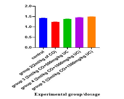
Figure 1: Bar chart indicating the effect of administration of 2ml/kg of castor oil and various doses of Uvaria chamae root extract on SODNote: CO – castor oil, UC- Uvaria chamae, SOD- superoxide dismutaseHisto-morphologic effect of castor oil and ethanol root extract of Uvaria chamae on the small intestine of male albino Wistar rat
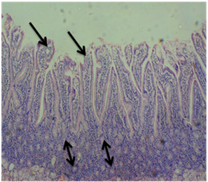
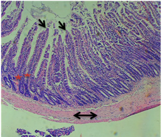
From the photomicrograph illustrated in figure 2, the result showed a well-defined intestinal villus, normal intestinal glands and well-defined crypts of Lieberkuhn.
A
B
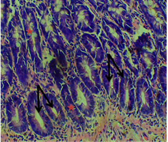
Figure 2: Photomicrograph of small intestine of control rats given water and feed alone showing well defined finger-like intestinal villi (single-headed arrow), normal intestinal glands (double headed arrow), 10x magnification and well-defined crypts of Lieberkuhn (single-headed arrow),40x magnification (H&E)
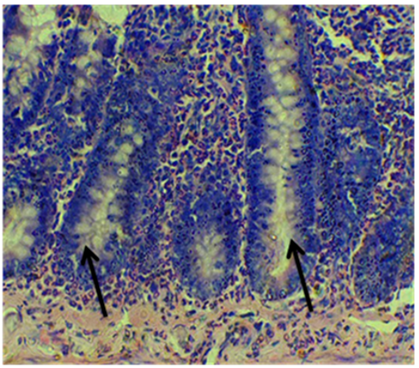
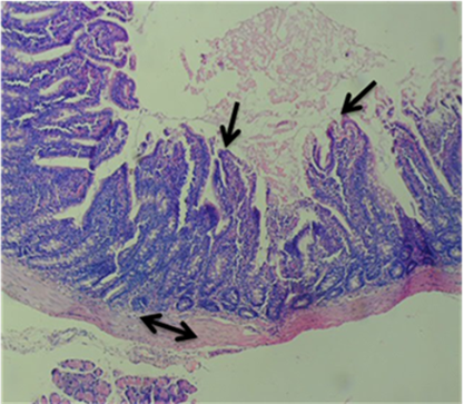
The photomicrograph below (figure 3) demonstrates eroded intestinal villi, muscle hypertrophy, prolapsed crypts of Lieberkuhn and inflammatory cells of the muscular layer following administration of castor oil in group 2.

A
B
Figure 3: Photomicrograph of small intestine of group 2 rats given 2ml/kg of castor oil alone showing eroded intestinal villi (single-headed arrow), muscle hypertrophy and presence of inflammatory cells in the muscle layer (double headed arrow), 10x magnification. Prolapsed crypt of Lieberkuhn (single-headed arrow),40x magnification (H&E)
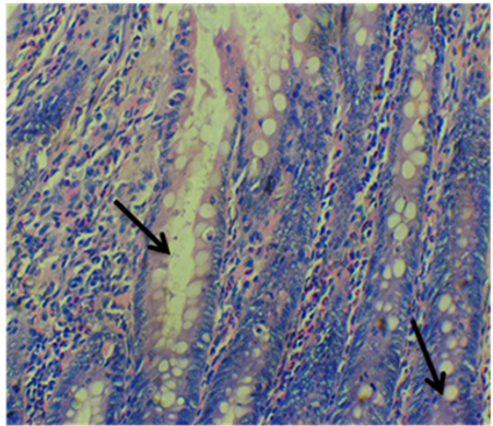
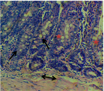
Photomicrograph of group 3 animals (figure 4) shown below depicts mild erosion of intestinal villi, reduced muscle hypertrophy, mild prolapsed crypts and presence of inflammatory cells following the administration of 500mg/kg of U. chamae after the administration of castor oil.
A
B
Figure 4: Photomicrograph of small intestine of group 3 rats given 2ml/kg of castor oil plus 500mg/kg of Uvaria chamae root extract showing mild erosion of the intestinal villi (single-headed arrow), reduced muscle hypertrophy and presence of inflammatory cells (double headed arrow), 10x magnification.
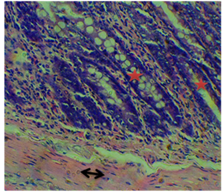
Mild prolapsed of the crypt’s ultrastructure (single headed arrow) but with the presence of few prolapsed crypts of Lieberkuhn (red star), 40x magnification (H&E). From the photomicrograph below, improved intestinal villi, crypts of Lieberkuhn, reduced muscle hypertrophy and inflammatory cell were shown following administration of 1000mg/kg of U. chamae after the administration of castor oil (figure 5).
A
B
Figure 5: Photomicrograph of small intestine of group 4 rats given 2ml/kg of castor oil plus 1000mg/kg of Uvaria chamae root extract showing improvement in the intestinal villi (single-headed arrow), reduced muscle hypertrophy and inflammatory cells (double headed arrow), 10x magnification. Improvement in the crypt’s ultrastructure (red star), 40x magnification (H&E).
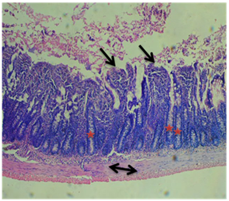
Photomicrograph of group 5 animals shown below showed improved intestinal villi, reduced muscle hypertrophy and inflammatory cells as well as improved crypt structure (figure 6).
A
B
Figure 6: Photomicrograph of small intestine of group 5 rats given 2ml/kg of castor oil plus 1500mg/kg of Uvaria chamae root extract showing improvement in the intestinal villi (single-headed arrow), reduced muscle hypertrophy and inflammatory cells (double headed arrow), 10x magnification. Improvement in the crypt’s ultrastructure (red star) and presence of well-defined intestinal glands ultrastructure (double arrow), 40x magnification (H&E).
Discussion
Castor oil, from Ricinus communis, has been used since ancient times as a safe and reliable laxative. The pharmacologically active molecule in the oil (ricinoleic acid) has multiple effects on the intestinal mucosa, resulting in fluid secretion [8]. Castor oil ingestion also induces biochemical stress in the small intestine. It stimulates the release of prostaglandins and cytokines, leading to the activation of inflammatory pathways. This inflammatory response disrupts the intestinal barrier function and increases intestinal permeability [9]. Furthermore, castor oil administration can elevate oxidative stress markers and induce lipid peroxidation in the small intestine [10]. Early studies indicated that the mucosal effects were due to enteritis or interference with cellular metabolism. Later studies revealed that fatty acid could increase mucosal permeability and cause cytotoxicity, associated with release of eicosanoids, platelet activating factor, other autacoids and nitric oxide. In addition, ricinoleic acid disrupts normal intestinal motility. The combination of these effects on the mucosa and smooth muscle of the gut account for its laxative action and studies have shown that castor oil administration causes mucosal damage, villi blunting, crypt hyperplasia, and an increase in inflammatory cell infiltration within the small intestine [11]. Our study was undertaken to evaluate the effects of ethanol root extract of Uvaria chamae on castor oil-induced stress in Wistar rats. Following the five days oral administration of the castor oil to the experimental rats, watery, soft and oily stools were observed thus indicating the induction of diarrheic stress in the rats. The study thus suggests that castor oil has potency in diarrheic stress induction. Our studies have demonstrated the beneficial effects of Uvaria chamae root extract in mitigating the stress induced by castor oil on the small intestine. It has been found that Uvaria chamae root extract reduces castor oil-induced mucosal damage, restores the villi architecture, and decreases inflammatory cell infiltration. Moreover, Uvaria chamae have been reported to exhibit antioxidant properties, scavenging free radicals and reducing oxidative stress in the small intestine [12,13]. The effect of the Uvaria chamae root extract on intestinal stress was evaluated by analyzing the histo-morphological properties as well as the tissue level of superoxide dismutase. The histo-morphological assessment of the small intestine showed an improved histological architecture across all the Uvaria chamae root treated groups (group 3, 4 and 5). From the histo-morphological results, it was observed that rats that were administered castor oil alone had a histological demonstration of eroded intestinal villi, muscle hypertrophy and presence of inflammatory cells in the muscle layer as opposed to the normal histological demonstration in the control group. However, the ethanol root extract of Uvaria chamae was proven to dose-dependently ameliorate the stress effect on the small intestine. Oral administration of 500mg/kg of Uvaria chamae root extract in group 3 showed mild erosion of the intestinal villi, reduced muscle hypertrophy and presence of inflammatory cells thus suggesting a mild improvement in the tissue integrity. Group 4 and 5 showed significant improvement in the intestinal villi, reduced muscle hypertrophy and inflammatory cells, as well as improvement in the crypts and intestinal gland ultrastructure. In conformity, a study carried out by Jalil et al (2020) on the role of Uvaria chamae root extract in treatment and prevention of inflammation in Wistar rats reported increased anti-inflammatory activities through various mechanisms of action in the tissue [14]. The histo-pathological changes observed in the small intestine following the administration of castor oil, as well as the subsequent administration of Uvaria chamae root extract provide valuable insights into the effects of these substances in the intestinal tissue. Upon administration of castor oil, histo-pathological examination of the small intestine revealed various alterations including epithelial damage, blunt, short or atrophied intestinal villi, hyperplasia and reduced cellularity of crypts. Inflammatory changes were also observed. These inflammatory changes are often associated with the release of pro inflammatory cytokines and chemokines. However, administration of Uvaria chamae root extract to animals in group 3 to group 5 revealed an ameliorative effect on the castor oil induced stress. Superoxide dismutases (SODs) are metallo-enzymes that play a major role in antioxidant defense against oxidative stress in the body. SOD supplementation may therefore trigger the endogenous antioxidant machinery for the neutralization of free-radical excess and be used in a variety of pathological settings [15-18]. Uvaria chamae, a plant known for its medicinal properties, has been reported to possess antioxidant activity. Our results demonstrated that the ethanol root extract of Uvaria chamae significantly increased the serum levels of SOD in the treated group compared to the control group and group 2. This indicates that the extract has the potential to enhance the antioxidant defense mechanism by increasing SOD activity in male albino Wistar rats. The findings of this study suggest that the ethanol root extract of Uvaria chamae possesses antioxidant properties, as evidenced by the increased serum SOD levels in the treated groups. The enhanced SOD activity can help neutralize the harmful effects of ROS, which are associated with various pathological conditions, including oxidative stress-related diseases. The observed effect could be attributed to the presence of bioactive compounds, such as polyphenols, flavonoids, and other phytochemicals, present in Uvaria chamae. These compounds have been reported to exhibit potent antioxidant activity by scavenging free radicals and modulating the activity of antioxidant enzymes [18].
Conclusion
This study provides valuable insights into the anatomical and biochemical stress caused by castor oil on the small intestine. Furthermore, it highlights the potential of Uvaria chamae root extract as a natural remedy to alleviate the stress induced by castor oil. Further research is warranted to elucidate the underlying mechanisms of action and to explore the therapeutic potential of Uvaria chamae in treating intestinal stress-related disorders.
Conflict of interest
Authors declare that no conflict of interest exist.
References
- Tharakan A, Norton IT, Fryer, PJ & Bakalis S. (2010). Mass transfer and nutrient absorption in a simulated model of small intestine. Journal of food science, 75(6), e339-e346.
View at Publisher | View at Google Scholar - Tunaru S, Althoff TF, Nüsing RM, Diener M, & Offermanns S. (2012). Castor oil induces laxation and uterus contraction via ricinoleic acid activating prostaglandin EP3 receptors. Proceedings of the National Academy of Sciences, 109(23), 9179-9184.
View at Publisher | View at Google Scholar - Chen W, Peng X, Yu J, Chen X, Yuan M. et al. (2020). FengLiao affects gut microbiota and the expression levels of Na+/H+ exchangers, aquaporins and acute phase proteins in mice with castor oil-induced diarrhea. Plos One, 15(7), e0236511.
View at Publisher | View at Google Scholar - Han, D. G., Seo, H. C., Cho, S., & Choi, J. W. (2019). Measurements of normal incidence reflection loss as a function of temperature at the water-castor oil interface. Sensors, 19(15), 3289.
View at Publisher | View at Google Scholar - Hussain HA, Men S, Hussain S, Chen Y, Ali S. et al. (2019). Interactive effects of drought and heat stresses on morpho-physiological attributes, yield, nutrient uptake and oxidative status in maize hybrids. Scientific Reports, 9:1-12. Doi: 10.1038/s41589-019-40362-7.
View at Publisher | View at Google Scholar - Okwu DE & Iroabuchi F. (2009). Phytochemical composition and biological activities of Uvaria chamae and Clerodendoron splendens. E-journal of Chemistry, 6(2), 553-560.
View at Publisher | View at Google Scholar - Thomas PS & Essien EE. (2020). Antiglycation, antioxidant, and cytotoxic activities of Uvaria chamae root and essential oil composition. Natural Product Research, 34(6), 880-883.
View at Publisher | View at Google Scholar - Smith PL, Maloney KN, Pothen RG, Clardy J & Clapham DE. (2018). Bisalliin H: a small molecule found in castor oil that drives extensive chloride secretion by cystic fibrosis airway epithelia. Cell Chemical Biology, 25(8), 1007-1015.
View at Publisher | View at Google Scholar - Jones RB, Saeedi BJ & McMellen ME. (2019). Castor oil induces laxation and uterus contraction via ricinoleic acid activating prostaglandin EP3 receptors. Proceedings of the National Academy of Sciences, 116(9), 3814-3823.
View at Publisher | View at Google Scholar - Gupta R, Sharma M & Bhatia S. (2021). Castor oil-induced oxidative stress in the small intestine: a mechanistic insight. Current Drug Metabolism, 22(14), 1016-1028.
View at Publisher | View at Google Scholar - Johnson AR, Adewale OB, & Adefisayo, MA. (2020). Quantitative histological evaluation of the effect of castor oil on the small intestine in Wistar rats. Nigerian Journal of Physiological Sciences, 35(1), 59-65.
View at Publisher | View at Google Scholar - Lee KW, Kim JH, & Kim, JD. (2022). Gastroprotective effects of Uvaria chamae extract against castor oil-induced intestinal injury in rats. Journal of Ethnopharmacology, 279, 114402.
View at Publisher | View at Google Scholar - Chen L, Wang X, Zhang Y, and Li P. (2021). Protective effect of Uvaria chamae extract on oxidative stress-induced intestinal epithelial cell injury. Journal of Ethnopharmacology, 276, 114193.
View at Publisher | View at Google Scholar - Jalil J, Attiq A, Hui CC, Yao LJ & Zakaria NA. (2020). Modulation of inflammatory pathways, medicinal uses and toxicities of Uvaria species: potential role in the prevention and treatment of inflammation. Inflammopharmacology, 28, 1195-1218.
View at Publisher | View at Google Scholar - Rosa AC, Corsi D, Cavi N, Bruni N & Dosio F. (2021). Superoxide dismutase administration: A review of proposed human uses. Molecules, 26(7), 1844.
View at Publisher | View at Google Scholar - Edem GD, David JU Okon KA & Thompson HI. (2024). Relationship between cadmium toxicity, kidney function disturbances and urinary bladder inflammation: the role of Uvaria chamae in mitigating these effects. Drug Discovery, 18e6dd1968.
View at Publisher | View at Google Scholar - Edem GD, David JU, Mbadugha CC, Ejeguo A & Friday DG. (2024). Cadmium toxicity in the seminal vesicles and lipid profile of Wistar rats: Can Uvaria chamae ameliorate the effect?. Clinical Reseach and Clinical Report, 3(1): 1-8.
View at Publisher | View at Google Scholar - Edem GD, Sakpa CL & Ezeuko VC. (2023). Exploring the scientific basis behind the therapeutic efficacy of Uvaria chamae: a major plus to alternative medicine. Journal of New Medical Innovation and Research, 4(6): 1-7
View at Publisher | View at Google Scholar
