

Research Article | DOI: https://doi.org/10.31579/2834-8087/034
Role Of Tensor Fascia Lata in Wound Bed Preparation
1Department of Surgery JIPMER, Pondicherry.
2Department of Plastic Surgery and Telemedicine JIPMER, Pondicherry
3Department Of Plastic Surgery JIPMER, Pondicherry
*Corresponding Author: Ravi Kumar Chittoria, Department of Plastic Surgery and Telemedicine JIPMER, Pondicherry.
Citation: Malvika Dadheech., Ravi Kumar Chittoria., Amrutha J S3. (2024), Role Of Tensor Fascia Lata in Wound Bed Preparation, Archives of Clinical Investigation, 3(6); DOI:10.31579/2834-8087/034
Copyright: © 2024, Ravi Kumar Chittoria. This is an open access article distributed under the Creative Commons Attribution License, which permits unrestricted use, distribution, and reproduction in any medium, provided the original work is properly cited.
Received: 12 November 2024 | Accepted: 22 November 2024 | Published: 28 November 2024
Keywords: wound bed; tensor fascia lata
Abstract
To make wounds heal wound bed preparation is essential. There are four components of Wound Bed Preparation (WBP)- TIME concept of WBP- tissue, infection and inflammation, moisture and edge management. The current study showed the versatile use of tensor fascia lata autograft for wound bed preparation which is a cost effective, easily available measure for the purpose.
Materials and Methods
This study was conducted in Tertiary Care Centre in Department of Plastic Surgery after getting the department ethical committee approval. Informed consent was obtained. A 59-year-old male known case of diabetes for 12 years on oral hypoglycemic drugs came with alleged history of RTA for which patient was operated outside Mayiladuthurai with intramedullary nailing followed by which patient wound was infected with exposed bone and raw area over middle 1/3rd of right lower limb (Figure 1).
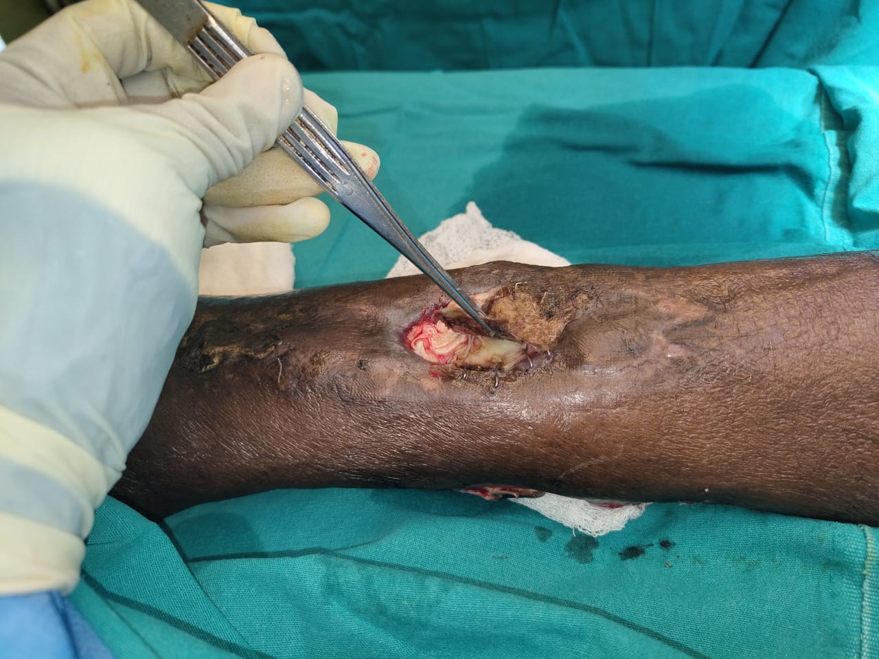
Figure 1: Wound with exposed bone
Implant was exposed in the middle of the wound. After serial debridement of necrotic tissues, exposed tendons, soft tissues are prepared for skin cover, Tensor Fascia Lata(TFL) graft done over the exposed bone (Figure 2,3).
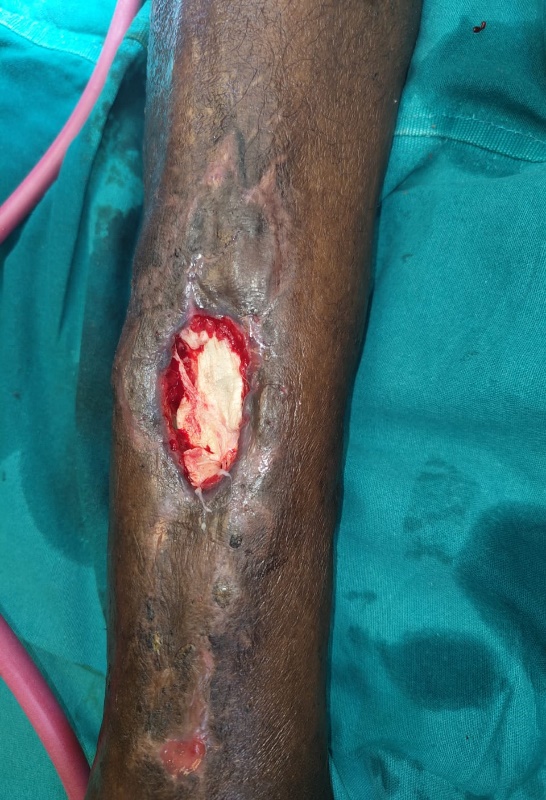
Figure 2: Tensor Fascia Lata graft applied over the wound bed
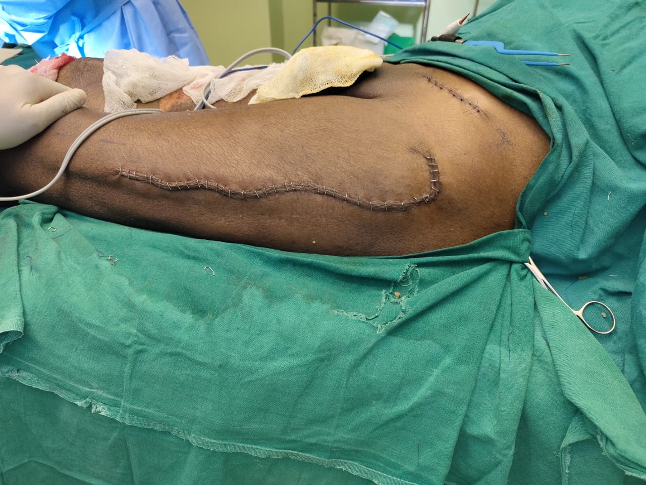
Figure 3: Tensor Fascia Lata donor site
Figure 4 : Limb at discharge
Full thickness raw area with exposed bone is present over the right leg. Split this skin graft was applied over the raw area once bone was covered with TFL.
Results
The use of Tensor fascia lata is helpful in preparing wound bed preparation in raw area in a lower leg middle third defect with exposed bone. Granulation tissue with soft tissue cover developed in the wound bed when wound was examined in postoperative day 7. Split thickness graft uptake was good. (Figure 4) Patient was discharged and referred to orthopedics department for further management.
Discussion
Successful wound closure and healing are a matter of concern for today's clinician. Determining if the wound will progress or not depends upon a comprehensive assessment, recognition of wound characteristics that will promote or impede the healing process and preparing the wound bed such that pathological features are removed allowing the healing cascade to occur. [2]The current study showed the versatile use of tensor fascia lata autograft for wound closure. Although we don’t have a pathological confirmation of how implanted tensor fascia lata allograft is incorporated into the wound, the fact that we have not encountered any wound complications is one of the most vital pieces of evidence of how this implantation technique is valuable and safe for soft tissue reconstructions. Human Fascia Lata Allograft (FLA) is a biodegradable low cost natural tissue with high elasticity and flexibility and therefore exhibits tensile strength and is easy to fit; furthermore it is biologically compatible, has a minimal risk of infection, lesser immunological response and is safe to use.[3].Therefore, when surgeons plan to close wounds with vascularized tissue, the addition of tensor fascia lata allografts can support the vascularized flap incorporated into the wound bed which is cost effective and minimises wound infection rates.[4].
Conclusion
Tensor fascia lata graft can be an excellent cost effective surgical option for wound closure, with high success rate and minimal complications.
References
- Frykberg RG, Banks J. (2015). Challenges in the treatment of chronic wounds. Advances in wound care.
View at Publisher | View at Google Scholar - Gokoo C. (2009). A primer on wound bed preparation. J Am Col Certif Wound Spec. 1(1):35-39. doi: 10.1016/j.jcws.2008.10.001. PMID: 24527107; PMCID: PMC3478922.
View at Publisher | View at Google Scholar - Park TH. (2023). The versatility of tensor fascia lata allografts for soft tissue reconstruction. Int Wound J. 20(3):784-791.
View at Publisher | View at Google Scholar - Zurek J, Dominiak M, Tomaszek K, Botzenhart U, Gedrange T. et al. (2016). Multiple gingival recession coverage with an allo-geneic biostatic fascia lata graft using the tunnel technique–ahistological assessment.Ann Anat.204:63-70
View at Publisher | View at Google Scholar
