

Research | DOI: https://doi.org/10.31579/2834-5126/119
Pathogenesis of CABG-Related Arrhythmias
- Maksimovich Yelizaveta *
Department of Propaedeutics of Internal Medicine, Grodno State Medical University, Grodno, Belarus.
*Corresponding Author: Maksimovich Yelizaveta, Department of Propaedeutics of Internal Medicine, Grodno State Medical University, Grodno, Belarus.
Citation: Maksimovich Yelizaveta, (2025), Pathogenesis of CABG-Related Arrhythmias, Clinical Trials and Clinical Research,4(6); DOI:10.31579/2834-5126/119
Copyright: © 2025, Maksimovich Yelizaveta. This is an open access article distributed under the creative commons’ attribution license, which permits unrestricted use, distribution, and reproduction in any medium, provided the original work is properly cited.
Received: 03 November 2025 | Accepted: 20 November 2025 | Published: 28 November 2025
Keywords: coronary artery bypass grafting; hemolysis; arrhythmias
Abstract
Further, the use of cardiopulmonary bypass (CPB) during CABG, necessary for maintaining blood circulation during the operation, is associated with potential red blood cell damage. This hemolysis may contribute to the development of cardiovascular complications in the postoperative period [14]. However, the specific relationship between intraoperative hemolysis (IOH) and the development of arrhythmias following CABG remains unclear in the existing literature.
Introduction
The pathogenesis of CABG-related arrhythmias is complex and not fully understood. Perioperative and early postoperative arrhythmias likely represent a reaction of the conduction system to the altered blood flow during the surgery, including the transition from cold cardioplegia to reperfusion. Coronary artery bypass surgery in patients with coronary heart disease (CHD) is frequently associated with postoperative complications, with arrhythmias being a significant concern [1, 2, 3, 4]. Potentially life-threatening arrhythmias, such as ventricular fibrillation and tachycardia, and third-degree atrioventricular block, as well as hemodynamically significant arrhythmias like atrial fibrillation (AF), severe bradycardia, and severe sinus tachycardia, are common. AF, a highly prevalent and dangerous postoperative arrhythmia, is frequently observed (25-65% of cases) [5, 6, 7, 8] and is associated with adverse outcomes, including increased risk of heart failure progression, thromboembolism, prolonged hospitalization, and mortality [ 9, 10, 11]. Patients experiencing AF after CABG have a higher risk of mortality related to cerebrovascular accidents and myocardial infarction [12]. Reoxygenation following the restoration of coronary blood flow can induce oxidative stress, metabolic disturbances, and electrical heterogeneity in the myocardium [13]. Further, the use of cardiopulmonary bypass (CPB) during CABG, necessary for maintaining blood circulation during the operation, is associated with potential red blood cell damage. This hemolysis may contribute to the development of cardiovascular complications in the postoperative period [14]. However, the specific relationship between intraoperative hemolysis (IOH) and the development of arrhythmias following CABG remains unclear in the existing literature. Purpose of the study. To establish the connection of intraoperative hemolysis (IOH) with the development of cardiac rhythm disturbances in patients with coronary artery disease after coronary shunting in conditions of cardiopulmonary bypass (CB).
Materials and methods
Was performed a prospective study of 123 patients with coronary heart disease undergoing CABG. The study was consistent with the Helsinki Declaration of the World Medical Association «Ethical Principles for Conducting Scientific Medical Research with Human Participation» and was approved by the ethics committees of the Grodno State Medical University and the Grodno Regional Clinical Cardiology Center healthcare institution. All patients underwent CB surgery in a planned manner under IR conditions. According to the level of free hemoglobin [Hb] in blood plasma, which is a marker of the degree of IOH, patients are divided into three groups: group 1 – without IOH (Hb⋜ 0.1 g/l), n=43, group 2 - with low IOH(lIOH) - with [Hb]>0.1g/l and <0 n=42, xss=removed>⋝0.5 g/l, n=38 [17]. The degree of IOH was assessed by the level of free hemoglobin [Hb] in the blood plasma at the beginning of the operation, immediately after connecting the patient to the artificial device and 15 minutes before removal from the artificial device (Fig. 1), using a HemoCue Plasma / Low Hb analyzer, Sweden [18, 19].
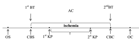
Figure 1: Diagram of the coronary bypass surgery operation
CB – cardiopulmonary bypass
CB – cardiopulmonary bypass start
CB – cardiopulmonary bypass completion
KP – cardioplegia
OS – operation start
OC – operation completion
1st BT – first blood test
2nd BT – second blood test
Patients of all groups are comparable by age and gender (Table 1).
Indicator
| Group 1 n=43 | Group 2 n=42 | Group 3 n=38 |
| Age, years | 60 (56; 63) | 64 (58; 66) | 66 (60; 68) |
| Gender (male), % | 36 (87,8%) | 32 (78,0%) | 31 (78,0%) |
| BMI (kg / m2) | 27,8(24,7; 29,2) | 27,7 (24,8; 29,2) | 29,1(25,9; 32,2) |
| Total protein (g / l) | 69 (62; 71) | 69(58; 68) | 66(57; 67) |
| Glucose, mmol /L | 5,0(4,5; 5,6) | 5,2(4,4; 6,1) | 5,3(4,5; 6,2) |
| Cholesterol, mmol /L | 4,1(3,3; 5,0) | 4,6(3,2; 5,7) | 5,0(4,6; 5,6) |
| Urea, mmol /L | 5,3(4,8; 5,6) | 6,0(5,5; 7,6) | 6,4(5,5; 7,2) |
| Creatinine, mmol /L | 99(89; 104) | 105(98; 110) | 106(99; 112) |
| CRP (mg / ml) | 1,2(0,8; 1,4) | 1,1(0,8; 1,3) | 1,0(0,6; 1,2) |
Table 1: Clinical characteristics of the subjects.
Notes: data are presented as Me [Q25; Q75], where Me is the median, Q25 is the value of the lower quartile; Q75 is the value of the upper quartile;
All patients underwent surgical intervention using a standard anesthetic protocol under normothermic artificial sirculation conditions with a hemodilution level of hematocrit of 25-30%.
The groups did not differ in the duration of artificial sirculation and the time of myocardial ischemia (p> 0.05), Table 2.
Indicator
| Group 1 n=43 | Group 2 n=42 | Group 3 n=38 |
| Ischemia-reperfusion time (min) | 69(65; 89) | 74 (68; 78) | 80 (75; 94) |
| Ischemia time (min) | 46(39; 64) | 58(56; 62) | 59(51; 68) |
Table 2: Duration of cardiopulmonary bypass (CB) and myocardial ischemia in patients with varying degrees of IOH during coronary artery bypass grafting.
Note: the data are presented in the form Me (Q25; Q75), where Me is the median of the indicator; Q25 - value of the lower quartile; Q75 is the value of the upper quartile.
Most patients (85%) underwent mammary-coronary bypass surgery in combination with aortic-coronary bypass surgery. Mammary-coronary bypass surgery was performed in 4% of patients (p <0> 0.05). More often, lesions of three or more coronary arteries, CA (63.1%) and significantly less often than one CA (7.1%) were revealed, Table 3.
| Number of shunts | Group 1 n=43 | Group 2 n=42 | Group 3 n=38 | p1-2 | p1-3 | p2-3 |
| 1 | 9,9 | 7,5 | 10,8 | 0,412 | 0,510 | 0,314 |
| 2 | 31,0 | 26,8 | 39,2 | 0,510 | 0,610 | 0,094 |
| 3 and more | 59,1 | 65,7 | 50,0 | 0,462 | 0,130 | 0,318 |
| Left anterior interventricular coronary artery | 87,8 | 100 | 100 | 0,21 | 0,31 | 0,31 |
| Left circumflex artery | 4,9 | 7,3 | 19,5 | 0,644 | 0,420 | 0,105 |
| Posterior interventricular branch of left circumflex artery | 14,6 | 39,0 | 61,0 | 0,210 | 0,310 | 0,406 |
| Left marginal artery | 56,1 | 65,9 | 80,5 | 0,172 | 0,22 | 0,324 |
| Right coronary artery | 24,4 | 58,5 | 61,0 | 0,231 | 0,341 | 0,821 |
| Right interventricular branch artery | 17,07 | 34,15 | 26,8 | 0,706 | 0,285 | 0,471 |
Table 3: Characterization of shunts in patients with coronary heart disease in groups with different levels of IOH.
Note: Accordingly, with myocardial revascularization, three or more coronary arteries (CA) were shunted more often - 56.9% of patients. The most common lesions were observed in the anterior interventricular branch of left coronary artery(p <0.05), posterior interventricular branch of the of left circumflex artery (p <0.05) and the left marginal artery (p <0.05).
Table 4 presents the nosological characteristics of patients.
Indicator
| Gr 1 n=43 | Gr 2 n=42 | Gr 3 n=38 |
| Ischemic heart disease duration | 8,5 (4,2; 11,4) | 8,9 (4,6; 10,8) | 9,5 (6,2; 12,1) |
| Duration of hypertension | 10 (6; 11) | 8 (5; 10) | 11,5 (9; 15) |
| Functional class II | 9 (20,1%) | 11 (26,2%) | 6 (15,8%) |
| Functional class III | 34 (79,9%) | 31(73,8%) | 32 (84,2%) |
| Postinfarction cardiosclerosis | 37 (86,1%) | 36 (85,7%) | 33 (86,8%) |
| The number of myocardial infarction (2 MI) in the history | 16(37,2%) | 18(42,8%) | 13(34,2%) |
| NYHAII | 36(83,7%) | 31(73,8%) | 33(86,8%) |
| NYHAIII | 7(16,3%) | 11(26,2%) | 5(13,2%) |
| Ischemic cardiomyopathy | 2 (0,86%) | 3 (1,26%) | 2 (0,76%) |
| History of arrhythmias | 7(16,3%) | 7(16,6%) | 6(15,4%) |
| Paroxysm of atrial fibrillation | 0 (9%) | 1(0,42) | 1 (0,38%) |
| supraventricular extrasystole | 4(1,72%) | 2 (0,84) | 2(0,76%) |
| ventricular extrasystole | 1(0,43%) | 1 (0,42%) | 1(0,38%) |
| right His bundle branch block | 1(0,43%) | 1 (0,42%) | 1(0,38%) |
| left His bundle branch block | 1(0,43%) | 2 (0,42%) | 2(0,76%) |
| blood hypertension | 36 (87,8%) | 38(90,2%) | 38(92,7%) |
| chronic bronchitis without exacerbation | 7(16,3%) | 9(21,4%) | 12(31,6%) |
| gastropathy | 18(41,9%) | 17(40,5%) | 20(52,6%) |
| urolithiasis | 6(13,9%) | 9(21,4%) | 7(18,4%) |
| osteoarthritis | 0(0%) | 3(7,1%) | 1(2,6%) |
| excess BMI and obesity | 36(83,7%) | 31(73,8%) | 33(86,8%) |
| excess BMI | 22(51,2%) | 18(42,9%) | 18(47,4%) |
| obesity | 14(32,6%) | 13(31%) | 15(39,5%) |
Table 4: Nosological characteristics of patients with coronary artery disease before coronary bypass surgery with varying degrees of intraoperative hemolysis (IOH).
Note: quantitative data are presented in the form Me [LQ; UQ], where Me is the median, LQ is the value of the lower quartile; UQ is the value of the upper quartile, and categorical – in the form of absolute and relative frequencies of signs; for all presented indicators, differences between the studied groups were absent (p> 0.05).
Most patients had one previously suffered myocardial infarction, MI. Patient groups were comparable in the number of MI (p> 0.05), the presence of ischemic cardiomyopathy (p> 0.05) and a history of cardiac arrhythmias (A), p> 0.05. Table 4 presents the frequency and structure of cardiac arrhythmias in patients with varying degrees of intraoperative hemolysis before coronary artery bypass surgery. As you can see, before surgery, cardiac arrhythmias were found in 22 people (17.89%). Among cardiac arrhythmias, AF paroxysms, supraventricular and ventricular extrasystoles, as well as blockade of the right and left legs of the bundle of His were found. At the same time, AF paroxysms were observed in 2 (1.63%) patients, extrasystoles were found in 11 (8.94%), including supraventricular extrasystoles, and in 3 (2,44%) – ventricular extrasystoles were noted. Dysfunction of the conduction function was noted in 8 people (6.5%), including blockade of the left leg of the bundle of His was noted in 5 people (4.07%), blockade of the right leg of the bundle of His - in 3 people (2.44%). The groups were comparable in the frequency and nature of cardiac arrhythmias in the anamnesis (p>0.05). Patients before CB (1-5 days) and after surgery (within 1-5 days) underwent daily ECG monitoring, as well as standard electrocardiography (ECG). In order to clarify the role of hemolysis in the development of postoperative arrhythmias in the studied groups of patients with different levels of IOH, we analyzed the incidence of cardiac arrhythmias in the perioperative (during the operation and during the first day after it) and in the early period (up to 1 month) and their structure [1]. The examined patients received standard therapy consisting of antiplatelet agents (79.7%), statins (76.4%), beta-blockers (84.6%), angiotensin-converting enzyme inhibitors (76.4%), antianginal drugs (79.7%), table 5. The drug treatment among patients of the studied groups did not differ in the administration of aspicard and clopidogrel (χ2 = 5.35; p = 0.069), β-blockers (χ2 = 3.18; p = 0.204), but it differed in the reception of statins (χ2 = 12.2; p = 0.006), inhibitors of the angiotensin-converting enzyme, ACE inhibitors (χ2 = 7.13; p = 0.028) and antianginal drugs (χ2 = 13.7; p<0.001). In particular, fewer patients in the third group took statins (57.9%, p <0.05), inhibitors (63.2%, p<0.001) and antianginal drugs (60.5%, p<0.001). Patients with a history of cardiac arrhythmias (paroxysmal AF) were treated with antiarrhythmic drugs 5-7 days before surgery.
| Index (%) | Gr 1 n=43 | Gr 2 n=42 | Gr 3 n=38 | Gr 1-3 n=123 | χ2 | p |
| beta blockers | 86,0 | 90,5 | 76,3 | 84,6 | 3,18 | 0,204 |
| ACE inhibitors | 88,4 | 76,2 | 63,2° | 76,4 | 7,13 | 0,028 |
| statins | 90,7 | 78,6 | 57,9°• | 76,4 | 12,2 | 0,006 |
| antianginal | 93,0 | 83,3 | 60,5°• | 79,7 | 13,7 | 0,0001 |
| antiplatelet agents | 88,4 | 81,0 | 68,4 | 79,7 | 5,35 | 0,069 |
Table 5: Characterization of drug therapy for examined patients with coronary artery disease before coronary artery bypass surgery with varying degrees of IOH.
Note: ° - p <0.05, °° - p <0.001– statistical differences with the group without IOH; • - p <0.05, •• - p <0.001 - statistical differences with group 2 (with lIOH)
To prevent arrhythmias during the operation, lidocaine was infused in a cardioplegic solution (1-1.5 mg / kg /min). After the operation, antiarrhythmic drugs were administered to arrest AF paroxysm (AF - 5 mg /kg intravenously dropwise for 60 min). Patients after CB took β-blockers (atenolol 25-50 mg / day, metoprolol at a dose of 25-50 mg / day, bisoprolol at a dose of 2.5-5 mg / day) depending on the level of blood pressure. In patients with atrial flutter and ventricular tachycardia in the perioperative period, temporary atrial pacemaker was performed, which was maintained for 72 hours with a frequency of 10 beats / min more than their own heart rate. Statistical data processing was carried out using the program Statistica 10.0 for Windows (StatSoft, Inc., USA). Given the abnormality of the distribution of attributes, nonparametric methods of descriptive statistics were used for processing: quantitative data are presented in the form Me [LQ; UQ], where Me is the median, LQ is the value of the lower quartile; UQ is the value of the upper quartile; categorical data are presented in the form of absolute and relative frequencies. When comparing the medians of quantitative variables of several independent groups, the Kruskell-Wallis test was used, to compare categorical data, the exact Fisher test, the χ2 criterion, with the Yeats correction at low frequencies were used. The strength of the relationship between the indicators was estimated using a correlation analysis based on the association coefficient (Kendall criterion) by its value (rs ⋜0.25 - weak; 0.25 <rs <0.75 - moderate and ⋝0.75 - strong). In order to check the dependence of the incidence of arrhythmias on the degree of IOG, determined by the level of free hemoglobin, a logistic regression analysis and ROC analysis were performed in the statistical program SPSS Statistics 21.0 (SPSS, USA). Differences were considered significant at p <0.05.
Results
Of the 123 examined with CB, 29 (23.6%; p<0> 0.05), in the early period - in 13 people (10.6%, p> 0.05). More often, in the operated patients, arrhythmias were revealed in 27 patients (21.95%, p <0 xss=removed xss=removed> Types of arrhythmias Arrhythmia frequency (%) p all PP EP n % n % n % Total arrhythmias 27 22,0 14 11,4 13 10,6 NS ventricular fibrillation 3 2,43 2 1,63 1 0,81 NS ventricular tachycardia 3 3,25 3 2,43 - - NS atrial fibrillation 7 5,70 3 2,43 4 3,25 NS atrial flutter 2 1,62 1 0,81 1 0,81 NS supraventricular tachycardia 1 0,81 - - 1 0,81 NS others: 11 8,9 5 4,07 4 3,25 NS AV block 1-2 degree 2 1,6 - - 2 1,6 NS ventricular extrasystole 5 4,1 4 3,25 3 2,43 NS supraventricular extrasystole 4 3,3 1 0,81 3 2,43 NS
Table 6: Frequency and structure of arrhythmias in patients with coronary artery disease after CBS.
Note: PP - perioperative period, EP - early period.
Data are presented in the form of absolute and relative frequencies of signs; AV block - atrio-ventricular block.
After surgery, arrhythmias developed in 27 (21.95%) patients. At the same time, 5 (4.9%) patients had life-threatening arrhythmias (ventricular fibrillation, ventricular tachycardia), 7 (5.7%) patients had hemodynamically significant arrhythmias – atrial fibrillation and flutter, supraventricular tachycardia. Atrial fibrillation was the most common type of postoperative arrhythmias (5.7%, p<0>
Most often, arrhythmias were observed in patients with a high degree of IOH (Table 7).
Types of complications | Group 1 n=43 | Group 2 n=42 | Group 3 n=38 | All n=123 | χ2 | p | ||||
n | % | n | % | n | % | n | % | |||
Arrhythmias | 2 | 4,7 | 5 | 11,9 | 20 | 52,6 | 27 | 22,0 | 21,95 | 0,000 |
Arrhythmias in the perioperative period | 2 | 4,7 | 2 | 4,8 | 10 | 26,3 | 14 | 11,4 | 14,8 | 0.0052 |
Arrhythmias in the early period | - | - | 3 | 7,14 | 10 | 26,3 | 13 | 10,6 | 15,6 | 0,0004 |
Table 7: The frequency of arrhythmias in patients with coronary heart disease after CB surgery with varying degrees of intraoperative hemolysis (IOH).
In the group with high IOH, the frequency of arrhythmias was 52.6%, which is higher than in the group with low IOH - 11.9%, p <0> Types of arrhythmias Gr 1 n=43 Gr 2 n=42 Gr 3 n=38 χ2 p n % n % n % all arrhythmias 2 4,65 5 11,9 20 52,6 39,54 0,0000 ventricular fibrillation - - 1 2,38 2 5,26 4,547 0.1020 ventricular tachycardia - - - - 3 7,89 6,878 0,032 atrial fibrillation 1 - 1 2,38 5 15,8 4,132 0.1260 atrial flutter - - 1 2,38 1 2,63 1,100 0.5760 supraventricular tachycardia - - - - 1 2,63 2,255 0,3238 other 1 2,32 2 4,80 8 23,7 10,06 0,0066
Table 8: The frequency and structure of arrhythmias in patients with coronary heart disease after CB with a different level of intraoperative hemolysis (IOH).
As can be seen from the table, in patients of the second (lIOH) and third (hIOH) groups, arrhythmias were more common than in the first group (without IOH), p<0>
arrhythmias and hemodynamically significant arrhythmias (13.2%). In 21.1% of patients with hIOH, other types of arrhythmias were noted (p <0>
Most often, arrhythmias were observed in patients with a high degree of IOH (Table 7).
Types of complications | Group 1 n=43 | Group 2 n=42 | Group 3 n=38 | All n=123 | χ2 | p | ||||
n | % | n | % | n | % | n | % | |||
Arrhythmias | 2 | 4,7 | 5 | 11,9 | 20 | 52,6 | 27 | 22,0 | 21,95 | 0,000 |
Arrhythmias in the perioperative period | 2 | 4,7 | 2 | 4,8 | 10 | 26,3 | 14 | 11,4 | 14,8 | 0.0052 |
Arrhythmias in the early period | - | - | 3 | 7,14 | 10 | 26,3 | 13 | 10,6 | 15,6 | 0,0004 |
Table 7: The frequency of arrhythmias in patients with coronary heart disease after CB surgery with varying degrees of intraoperative hemolysis (IOH).
In the group with high IOH, the frequency of arrhythmias was 52.6%, which is higher than in the group with low IOH - 11.9%, p <0> Types of arrhythmias Gr 1 n=43 Gr 2 n=42 Gr 3 n=38 χ2 p n % n % n % all arrhythmias 2 4,65 5 11,9 20 52,6 39,54 0,0000 ventricular fibrillation - - 1 2,38 2 5,26 4,547 0.1020 ventricular tachycardia - - - - 3 7,89 6,878 0,032 atrial fibrillation 1 - 1 2,38 5 15,8 4,132 0.1260 atrial flutter - - 1 2,38 1 2,63 1,100 0.5760 supraventricular tachycardia - - - - 1 2,63 2,255 0,3238 other 1 2,32 2 4,80 8 23,7 10,06 0,0066
Table 8: The frequency and structure of arrhythmias in patients with coronary heart disease after CB with a different level of intraoperative hemolysis (IOH).
As can be seen from the table, in patients of the second (lIOH) and third (hIOH) groups, arrhythmias were more common than in the first group (without IOH), p<0 xss=removed xss=removed xss=removed Rs=0.4167, p=0.000003). xss=removed xss=removed xss=removed xss=removed>
Based on logistic regression and ROC analysis, data were obtained that testify to the significance of the [Hb] indicator in assessing the likelihood of arrhythmias (Figure. 2).
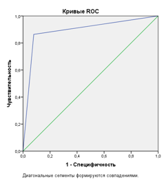
Figure 2: -ROC curve characterizing the sensitivity and specificity of the method for assessing the likelihood of developing postoperative arrhythmias by the concentration of free hemoglobin in blood plasma.
A high risk of developing arrhythmias in patients with coronary heart disease after CB was determined with a value of [Hb]>0.85 g/l (sensitivity - 86.4%, specificity - 92.7%, PPV (predictive value of a positive result) = 96.9 %, NPV (predictive value of a negative result) = 70.4%, area under the ROC-curve (AUC) = 0.892 (0.803-0.981), 95% confidence interval.
Discussion
As noted earlier, before the operation, cardiac arrhythmias occurred in 22 people (17.89%). Among arrhythmias, AF paroxysms, supraventricular and ventricular extrasystoles, as well as blockade of the right and left legs of the bundle of His were found. AF paroxysms were observed in 2 people (in 1 – in groups with lIOH and in 1 – in group with hIOH. The groups were comparable in history and frequency of arrhythmias in history (p>0.05). However, the incidence of arrhythmias in postoperative the period was highest in the third group with hIOH, and in the second group with lIOH it was greater than in the group without IOH According to the literature, the occurrence of arrhythmias associated with CB is caused by the restoration of blood flow in the ischemic zone, as a result of which the resumption of oxygenation initiates the development of oxidative stress. Action a reactive forms of oxygen and nitrogen leads to structural and metabolic disturbances, manifested by damage to cell membranes, electrolyte imbalance, forming a state of electrical myocardial heterogeneity, impaired excitability, pulse generation and conduction in the heart. Post-traumatic remodeling of heart chambers can contribute to the development of arrhythmias [6,7,9]. It was shown that not only the frequency of arrhythmias in groups after CB has
changed, but also the structure. Transformation of less life-threatening arrhythmias (extrasystole, blockade of the bundle of His) into arrhythmias was noted, which had more serious consequences for hemodynamics and posed a greater threat to the lives of patients (atrioventricular block of the 1st degree, atrial fibrillation and flutter, paroxysmal ventricular and supraventricular ventricles). Some authors have identified the relationship between arrhythmias and the features of surgical treatment (inadequate myocardial protection during surgery, due to the composition of the cardioplegic solution used, the direction of its administration, temperature, and the duration of cardioplegia). A positive correlation was revealed between the occurrence of arrhythmias and the duration of IR, the intensity
of inotropic support, blood transfusion, and the level of leukocytosis after surgery. Other authors have not found such a dependence on the duration of artificial circulation. CB has been shown to be most conducive to the development of arrhythmias in patients who had morphological changes in the heart (post-infarction cardiosclerosis) and a history of arrhythmias. Studies on the study of arrhythmias after CB using correlation, as well as logistic and ROC analysis revealed the dependence of the frequency of arrhythmias on the level of free hemoglobin as an indicator of the degree of intraoperative hemolysis. The destruction of red blood cells due to their mechanical damage in the artificial circuits of the exerts a pathogenic effect on the state of the rhythmogenic function of the cardiac conduction system and myocardial excitability, predisposing to the development of arrhythmias. The largest number of arrhythmias in the group with a high level of free hemoglobin, as well as the presence of correlation between the frequency of arrhythmias and [Hb] In the blood plasma at the end of the operation, as well as the results of the logistic and ROC analysis, indicate the important role of intraoperative hemolysis in their occurrence in perioperative and early periods. The pathogenetic role of free hemoglobin in the development of rhythm disturbances in CB, it is advisable to develop a set of perioperative preventive measures aimed at chelation of free iron, which reduce the activity of oxidative processes. Elimination of patient-dependent risk factors for increased hemolysis (smoking, alcohol consumption, normalization of blood pressure, body weight and cholesterol) is also important for the prevention of cardiac arrhythmias, as one of the most common complications of coronary artery bypass surgery.
Conclusions
- Postoperative arrhythmias occur in approximately 22% of patients undergoing CABG with cardiopulmonary bypass. A substantial portion of these arrhythmias are life-threatening or cause significant hemodynamic compromise and organ hypoperfusion.
- Analysis demonstrated a strong correlation (p<0>
- These findings suggest that monitoring free plasma hemoglobin levels during CABG procedures may serve as a crucial indicator for predicting and preventing postoperative arrhythmias, and potentially guiding interventions to correct any hemodynamic complications.
References
- Bokeriya, L.A. E.Z. Goluxova, B.G. Alekyani dr. (2011). Neposredstvenny`e rezul`taty` xirurgicheskogo i e`ndovaskulyarnogo lecheniya bol`ny`x ishemicheskoj bolezn`yu serdcza: perioperacionny`e oslozhneniya, faktory` riska, prognoz Kreativnaya kardiologiya,. – №1. – S. 41– 60.
View at Publisher | View at Google Scholar - Maksimovich E.N. Dementej A.I., Lavrinajt' V.V. i dr. (2017). Aritmii kak prichina letal'nogo iskhoda u pacientov posle operacii koronarnogo shuntirovaniya Sbornik tezisov XII mezhdunarodnoj (XXI Vserossijskoj) Pirogovskoj nauchnoj medicinskoj konferencii studentov i molodyh uchenyh. –, Moskva. – S.82.
View at Publisher | View at Google Scholar - Kim, L.K. P. Looser, R.V. Swaminathan, R.M. Minutello et al. (2016). Outcomes in patients undergoing coronary artery bypass graft surgery in the United States based on hospital volume, 2007 to 2011 J. Thorac. Cardiovasc. Surg. –. – V.151(6). – P.1686 –1692.
View at Publisher | View at Google Scholar - Maksimovich, E.N. T. P. Pron`ko, V.A. Snezhiczkij (2018). Aritmii u pacientov s IBS posle koronarnogo shunktirovaniya i raznoj spen`yu intraoperacionnogo gemoliza I s`ezd Evrazijskoj aritmologicheskoj associacii: sbornik materialov EURA Congress, 13-14 sentyabrya 2018 g. – Grodno, – S.46 – 47.
View at Publisher | View at Google Scholar - Gel'fand, I.M. M.N. Starkova, A.L. Syrkin. (1983). Prognosis of ventricular arrhythmias in myocardial infarct patients Kardiologiia. –. – V. 23, №5. – P. 9 –12.
View at Publisher | View at Google Scholar - Omer, S. L. Cornwell, A. (2016). Bakshi Incidence, predictors, and impact of postoperative atrial fibrillation after coronary artery bypass grafting in military veterans Tex. Heart Inst. J. , V. 43 (5). – P. 397 – 403.
View at Publisher | View at Google Scholar - Fengsrud, E. A. Englund, A. Ahlsson. (2017). Pre- and postoperative atrial fibrillation in CABG patients have similar prognostic impact Scand. Cardiovasc. J. –. – V.51 (1). – P.21 – 27.
View at Publisher | View at Google Scholar - Venetucci, L.A. A.W. Trafford, S.C.O'Neill, D.A. Eisner., (2008). The sarcoplasmic reticulum and arrhythmogenic calcium release Cardiovasc. Res. –. – V. 77 (2). – P 285 – 292.
View at Publisher | View at Google Scholar - Valeri, C.R. H. MacGregor, G. Ragno, N. Healey. (2006). Effects of centrifugal and roller pumps on survival of autologous red cells in cardiopulmonary bypass surgery Perfusion. – V. 21(5). – P.291 – 296.
View at Publisher | View at Google Scholar - Maksimovich, E.N. V.V. Vasilevich, D.D. Truxovskaya, Yu.A. Koshheev, V.V. Kruglik. (2018). Faktory` intraoperacionnogo gemoliza pri koronarnom shuntirovanii s ispol`zovaniem ickusstvennogo krovoobrashheniya Sbornik materialov konferencii studentov i molody`x ucheny`x, posvyashhennoj 60-letiyu uchrezhdeniya obrazovaniya
View at Publisher | View at Google Scholar - Vercaemst, L. (2008). Hemolysis in cardiac surgery patients undergo¬ing cardiopulmonary bypass: A review in search of a treatment algorithm J. of Extra. Corporeal. Technology. –. – V. 40, № 4. – P. 257 – 267.
View at Publisher | View at Google Scholar - Pan, K.C. D.P. McKenzie, V. Pellegrino, D. Murphy. (2016). The meaning of a high plasma free hemoglobin: retrospective review of the prevalence of hemolysis and circuit thrombosis in an adult ECMO centre over 5 years, Perfusion. –, V.31 (3). – P.223 – 231.
View at Publisher | View at Google Scholar - Svenmarker, S., E. Jansson, H. Stenlund, K. Engström (2000). Red blood cell trauma during cardiopulmonary bypass: Narrow pore filterability versus free hemoglobin Perfusion. –. – №15 (1). – P.33 – 40.
View at Publisher | View at Google Scholar - Maksimovich E.N., Vasilevich V.V., Koshheev Yu.A., Pron`ko T.P., Truсhovskaya D.D. (2018). Uroven` svobodnogo gemoglobina v plazme krovi pacientov s oslozhneniyami posle operacii koronarnogo shuntirovaniya. Mat. itogovoj nauchno-prakticheskoj konferencii «Aktual`ny`e problemy` mediciny`» 25 yanvarya 2019 g. – Grodno, – S. – 360 – 362.
View at Publisher | View at Google Scholar
